Review
. 2024 Jan 10;7(1):78.
doi: 10.1038/s42003-023-05691-4.

Architecture of the subthalamic nucleus

Affiliations
Review

Architecture of the subthalamic nucleus

Asheeta A Prasad et al. Commun Biol. .

Abstract

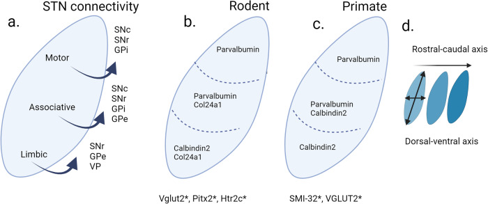
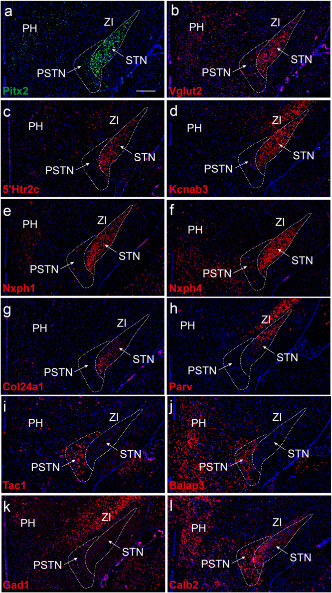
The subthalamic nucleus (STN) is a major neuromodulation target for the alleviation of neurological and neuropsychiatric symptoms using deep brain stimulation (DBS). STN-DBS is today applied as treatment in Parkinson´s disease, dystonia, essential tremor, and obsessive-compulsive disorder (OCD). STN-DBS also shows promise as a treatment for refractory Tourette syndrome. However, the internal organization of the STN has remained elusive and challenges researchers and clinicians: How can this small brain structure engage in the multitude of functions that renders it a key hub for therapeutic intervention of a variety of brain disorders ranging from motor to affective to cognitive? Based on recent gene expression studies of the STN, a comprehensive view of the anatomical and cellular organization, including revelations of spatio-molecular heterogeneity, is now possible to outline. In this review, we focus attention to the neurobiological architecture of the STN with specific emphasis on molecular patterns discovered within this complex brain area. Studies from human, non-human primate, and rodent brains now reveal anatomically defined distribution of specific molecular markers. Together their spatial patterns indicate a heterogeneous molecular architecture within the STN. Considering the translational capacity of targeting the STN in severe brain disorders, the addition of molecular profiling of the STN will allow for advancement in precision of clinical STN-based interventions.


Conflict of interest statement

The authors declare no competing interest.

Figures

Fig. 1
Fig. 1. The subthalamic nucleus (STN) outlined with connectivity and selected gene expression.
a Anatomical subdivisions are topologically organized with STN efferents based on published neural tracing studies by Carpenter et al., 1968, Parent et al., 1989, Nambu et al., 1996 and Karachi et al., 2004. b Selected gene expression (Parvalbumin, Calbindin 2, Col24a1: detected as mRNA or protein) in mouse (c) and primate based on Hardmann et al., 1997 (d). expression patterns are altered across rostral-caudal and the dorsal-ventral axis * indicates global STN gene expression. For details and full nomenclature, see the main text. Image created in Biorender.
Fig. 2
Fig. 2. Molecular heterogeneity of mouse STN as revealed by single nucleotide transcriptomics followed through with spatial mRNA mapping.
Serial mouse brain sections analyzed by fluorescent in situ hybridization (FISH) detects differential patterns and profiles of selected gene expression products; distribution within, and immediately surrounding, the STN structure: a Pitx2; b Vglut2; c Serotonin receptor subtype 2c, 5´Htr2c; d Kcnab3; e Nxph1; f Nxph4; g Col24a1; h PV i Tac1; j Baiap3; k Gad1; l Calb2. Note: Pitx2, Vglut2, Htr2c, Kcnab3, Nxph1 mRNAs (ae) are present throughout the entire STN structure; Nxph4 mRNA is similar but excluded from dorsolateral-most part of STN; Col24a1 primarily in medially and centrally located STN neurons and excluded dorsally with opposite pattern of PV mRNA, primarily in dorsally and centrally located STN neurons while excluded medially (compare g and h); Tac1 and Baiap3 mRNAs absent from STN (i and j), but Tac1 strong in para-STN (PSTN), Baiap3 in PSTN and also surrounding hypothalamus (lateral and posterior hypothalamus, PH, LH); Gad1 (k; GABA marker) largely absent from STN and PSTN (in accordance with their excitatory phenotype) but present in GABAergic structure zona incerta (ZI), Calb2 mRNA stronger in medial STN and para-STN (PSTN) than dorsal STN (l). For mRNA full name, see the main text. See original publication for details (Wallén-Mackenzie et al. ; published under Open access).
Fig. 3
Fig. 3. Tripartite model of primate STN presented next to a recent model of molecular organization within mouse STN.
a Tripartite model of primate STN; motor, associative and limbic territories based on multiple anatomical-functional studies (references in main text). Drawing based on original illustration in Benarrouch, 2008; here updated with information of afferent and efferent projections as outlined by Baunez et al., 2016. Abbreviations: GPe, globus pallidus externa; GPi, globus pallidus interna; SNr, substantia nigra pars reticulata; SNc, substantia nigra pars compacta; PPN, pedunculopontine nucleus; VTA, ventral tegmental area. b Gene expression patterns spatially mapped in brain sections using fluorescent in situ hybridization (FISH) (original data reprinted in Fig. 2) allowed the outlining of molecularly profiled domains within mouse STN: STNa (ventromedial STN), STNb (central STN), STNc (dorsal STN), STNds (dorsal strip of STN showing GABA-positive, rather than glutamatergic, mRNA profile). Gene expression (mRNAs) present throughout the STN structure listed in the text box; unique gene expression patterns distinct for each domain listed under the domain name. c Selected mRNAs, their distribution and extent of co-localization with STN marker Pitx2, as described in the mouse STN. b, c: For mRNA full name, see main text. Illustrations based on original publication (Wallén-Mackenzie et al. ; published under Open access).

References

    1. Stephenson-Jones M, Samuelsson E, Ericsson J, Robertson B, Grillner S. Evolutionary conservation of the basal ganglia as a common vertebrate mechanism for action selection. Curr. Biol. 2011;21:1081–1091. doi: 10.1016/j.cub.2011.05.001. - DOI - PubMed
    1. John MP. The subthalamic nucleus and Jules Bernard Luys (1828–97) J. Neurol., Neurosurg. Psychiatry. 2001;71:783. doi: 10.1136/jnnp.71.6.783. - DOI - PMC - PubMed
    1. Parent A. Jules Bernard Luys and the subthalamic nucleus. Mov. Disord. 2002;17:181–185. doi: 10.1002/mds.1251. - DOI - PubMed
    1. Hamada I, DeLong MR. Excitotoxic acid lesions of the primate subthalamic nucleus result in reduced pallidal neuronal activity during active holding. J. Neurophysiol. 1992;68:1859–1866. doi: 10.1152/jn.1992.68.5.1859. - DOI - PubMed
    1. Bergman H, Wichmann T, DeLong MR. Reversal of experimental Parkinsonism by lesions of the subthalamic nucleus. Science. 1990;249:1436–1438. doi: 10.1126/science.2402638. - DOI - PubMed

Publication types