

Metal-polyphenol networks-modified tantalum plate for craniomaxillofacial reconstruction
- PMID: 38200230
- PMCID: PMC10781789
- DOI: 10.1038/s41598-024-51640-4
Metal-polyphenol networks-modified tantalum plate for craniomaxillofacial reconstruction
Abstract
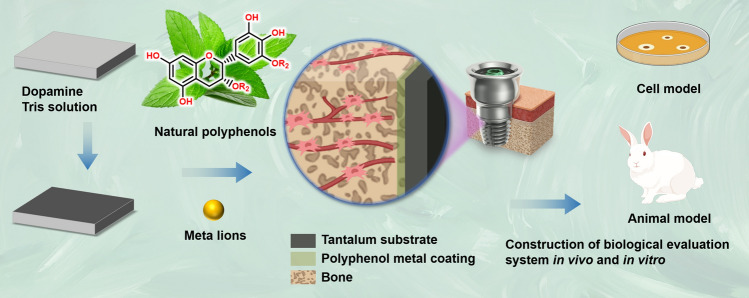
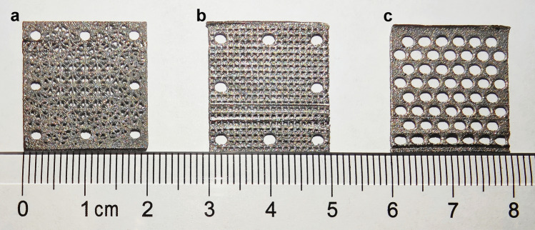
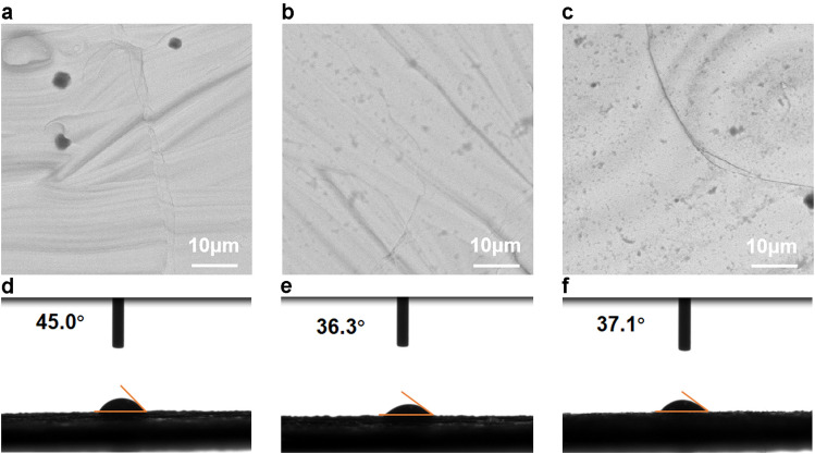
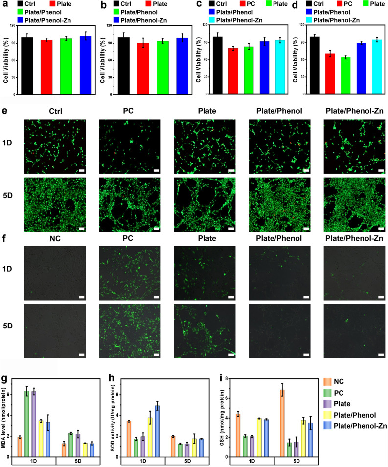
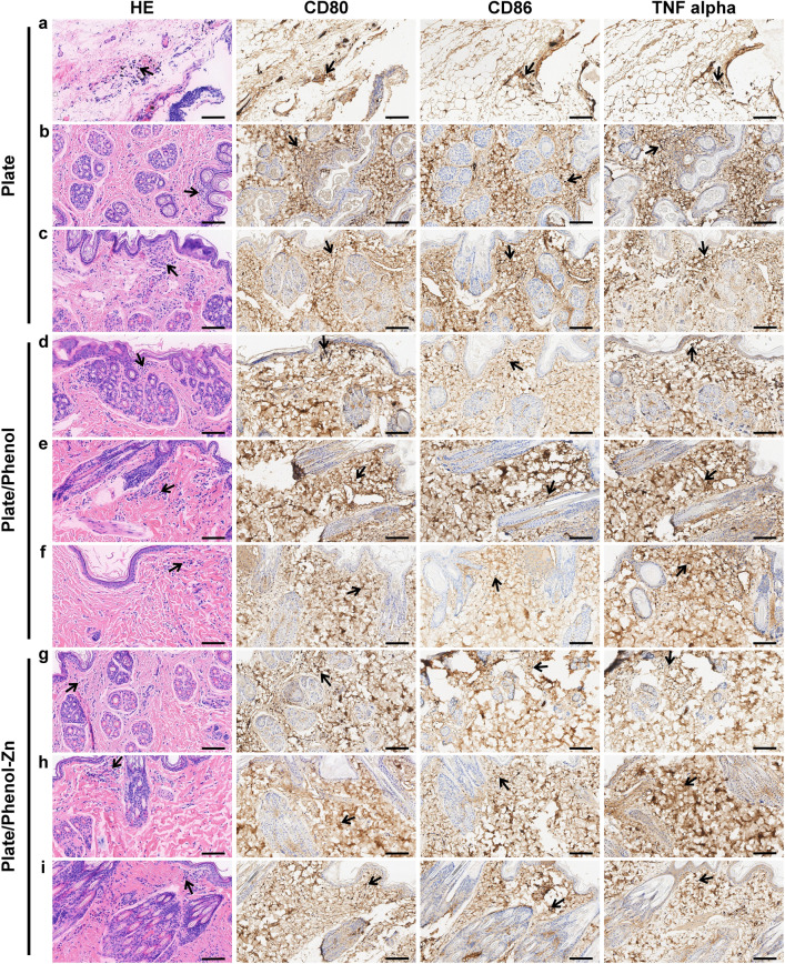
Using three-dimensional (3D) printing technology to make the porous tantalum plate and modify its surface. The physicochemical properties, cytocompatibility, antioxidant capacity, and histocompatibility of the modified materials were evaluated to prepare for the repair of craniomaxillofacial bone defects. The porous tantalum plates were 3D printed by selective laser melting technology. Tantalum plates were surface modified with a metal polyphenol network. The surface-modified plates were analyzed for cytocompatibility using thiazolyl blue tetrazolium bromide and live/dead cell staining. The antioxidant capacity of the surface-modified plates was assessed by measuring the levels of intracellular reactive oxygen species, reduced glutathione, superoxide dismutase, and malondialdehyde. The histocompatibility of the plates was evaluated by animal experiments. The results obtained that the tantalum plates with uniform small pores exhibited a high mechanical strength. The surface-modified plates had much better hydrophilicity. In vitro cell experiments showed that the surface-modified plates had higher cytocompatibility and antioxidant capacity than blank tantalum plates. Through subcutaneous implantation in rabbits, the surface-modified plates demonstrated good histocompatibility. Hence, surface-modified tantalum plates had the potential to be used as an implant material for the treatment of craniomaxillofacial bone defects.
© 2024. The Author(s).
Conflict of interest statement
The authors declare no competing interests.
Figures
Similar articles
-
Biocompatible Porous Tantalum Metal Plates in the Treatment of Tibial Fracture.Orthop Surg. 2019 Apr;11(2):325-329. doi: 10.1111/os.12432. Epub 2019 Mar 18. Orthop Surg. 2019. PMID: 30884151 Free PMC article.
-
The performance tests of three-dimensional printing titanium alloy craniomaxillofacial bone plate: A preliminary preclinical study.J Dent Sci. 2023 Oct;18(4):1756-1764. doi: 10.1016/j.jds.2021.11.005. Epub 2021 Dec 2. J Dent Sci. 2023. PMID: 37799913 Free PMC article.
-
Osteogenic differentiation of 3D-printed porous tantalum with nano-topographic modification for repairing craniofacial bone defects.Front Bioeng Biotechnol. 2023 Aug 21;11:1258030. doi: 10.3389/fbioe.2023.1258030. eCollection 2023. Front Bioeng Biotechnol. 2023. PMID: 37671184 Free PMC article.
-
Research progress and clinical translation of three-dimensional printed porous tantalum in orthopaedics.Biomater Transl. 2023 Sep 28;4(3):166-179. doi: 10.12336/biomatertransl.2023.03.005. eCollection 2023. Biomater Transl. 2023. PMID: 38283089 Free PMC article. Review.
-
Advances in surface modification of tantalum and porous tantalum for rapid osseointegration: A thematic review.Front Bioeng Biotechnol. 2022 Sep 13;10:983695. doi: 10.3389/fbioe.2022.983695. eCollection 2022. Front Bioeng Biotechnol. 2022. PMID: 36177183 Free PMC article. Review.
References
MeSH terms
Substances
Grants and funding
- No. LQ21H130001 and LY19H160014/Zhejiang Provincial Natural Science Foundation of China
- No.2022L005/Ningbo Clinical Research Center for Otolaryngology Head and Neck Disease
- No. PPXK2018-02/Ningbo Medical and Health Brand Discipline
- No. 2021KY307/Zhejiang Provincial Medical and Health Science Research Foundation
- No. 2021S171/Ningbo Public Science Research Foundation
LinkOut - more resources
Full Text Sources
