. 2024 Jan 10;25(1):23.
doi: 10.1186/s12931-023-02537-9.

Tobacco and menthol flavored nicotine-free electronic cigarettes induced inflammation and dysregulated repair in lung fibroblast and epithelium

Affiliations

Tobacco and menthol flavored nicotine-free electronic cigarettes induced inflammation and dysregulated repair in lung fibroblast and epithelium

Qixin Wang et al. Respir Res. .

Abstract

Background: Electronic cigarette (e-cig) vaping has increased in the past decade in the US, and e-cig use is misleadingly marketed as a safe cessation for quitting smoking. The main constituents in e-liquid are humectants, such as propylene glycol (PG) and vegetable glycerine (VG), but different flavoring chemicals are also used. However, the toxicology profile of flavored e-cigs in the pulmonary tract is lacking. We hypothesized that menthol and tobacco-flavored e-cig (nicotine-free) exposure results in inflammatory responses and dysregulated repair in lung fibroblast and epithelium.

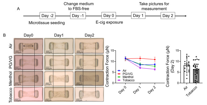
Method: We exposed lung fibroblast (HFL-1) and epithelium (BEAS-2B) to Air, PG/VG, menthol flavored, or tobacco-flavored e-cig, and determined the cytotoxicity, inflammation, and wound healing ability in 2D cells and 3D microtissue chip models.

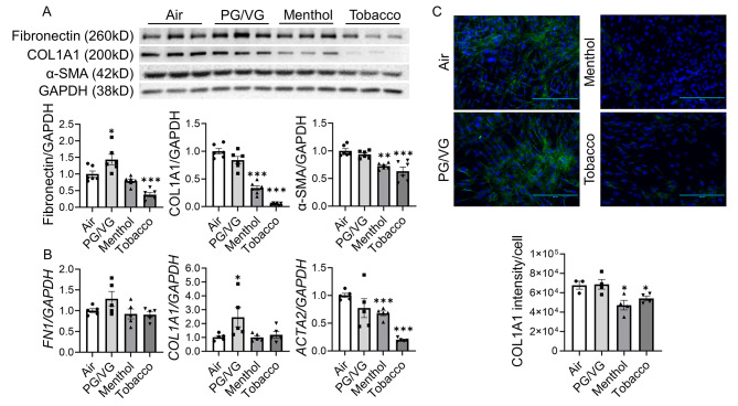
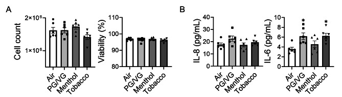
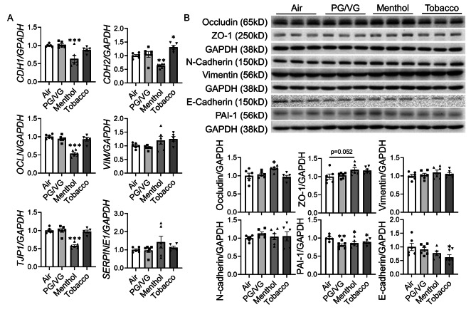
Results: After exposure, HFL-1 showed decreased cell number with increased IL-8 levels in the tobacco flavor group compared to air. BEAS-2B also showed increased IL-8 secretion after PG/VG and tobacco flavor exposure, while menthol flavor exposure showed no change. Both menthol and tobacco-flavored e-cig exposure showed decreased protein abundance of type 1 collagen α 1 (COL1A1), α-smooth-muscle actin (αSMA), and fibronectin as well as decreased gene expression level of αSMA (Acta2) in HFL-1. After tobacco flavor e-cig exposure, HFL-1 mediated wound healing and tissue contractility were inhibited. Furthermore, BEAS-2B exposed to menthol flavor showed significantly decreased tight junction gene expressions, such as CDH1, OCLN, and TJP1.

Conclusion: Overall, tobacco-flavored e-cig exposure induces inflammation in both epithelium and fibroblasts, and tobacco-flavored e-cig inhibits wound healing ability in fibroblasts.

Keywords: ENDS; Inflammation; Injury; Menthol; Nicotine-free; Repair; Tobacco.


Conflict of interest statement

The authors declare that they have no known competing financial interests or personal relationships that could have appeared to influence the work reported in this article.

Figures

Fig. 1
Fig. 1
Tobacco flavored e-cig induced inflammatory responses in lung fibroblast HFL-1 cells exposed to air, PG/VG, or tobacco flavored e-cig for 10 min, and then cultured for 2 days. (A). Representative pictures of HFL-1 cells were taken under 20x microscope. (B). Cell number and viability was measured by AO/PI staining. (C) Conditioned medium was collected for IL-6, IL-8 and TGF-β analysis. Data presented as mean ± SEM (n = 6–9. * P < 0.05, ** P < 0.01, vs. air)
Fig. 2
Fig. 2
Tobacco flavored e-cig inhibited fibroblast differentiation markers HFL-1 cells exposed to air, PG/VG, and tobacco flavored e-cig for 10 min, and then cultured for 2 days. (A) cells were lysed and protein was isolated for western blotting, fibronectin, COL1A1, and αSMA were analyzed, GAPDH was used as the endogenous control. (B). RNA was isolated from cells, and FN1, COL1A1, and ACTA2 were measured by qRT-PCR, GAPDH was used as the endogenous control. (C) Cells were fixed, and stained with COL1A1, the fluorescence intensity was measured by EVOMS, fluorescence intensity /cell was calculated via ImageJ. Data presented as mean ± SEM. (n = 3–6. * P < 0.05, ** P < 0.01, *** P < 0.001 vs. air). Scale bar = 200 μm
Fig. 3
Fig. 3
Tobacco flavored e-cigs inhibited wound healing mediated by lung fibroblast HFL-1 cells were scratched and then exposed to air, PG/VG, or tobacco flavored e-cig for 10 min, and then cultured for 2 days. The scratched wounds were monitored by taking pictures under the microscope daily. The same position has been selected by recording the coordinates from Cytation 5 imaging system. Data presented as mean ± SEM. (n = 11–12. * P < 0.05, ** P < 0.01, vs. air)
Fig. 4
Fig. 4
Tobacco flavored e-cig exposure decreased the contraction force of HFL-1 formed tissue (A). HFL-1 cells were used to form microtissue first, then exposed to air, PG/VG, and tobacco flavored e-cig for 10 min, and followed with 2 days culture. (B). The individual pictures of microtissue were taken by microscope for measurement of contraction force. Data presented as mean ± SEM. (n ≥ 10. * P < 0.05 vs. Air)
Fig. 5
Fig. 5
Unflavored and Tobacco flavored e-cig induced inflammatory responses in lung bronchial epithelium BEAS-2B cells exposed to air, PG/VG, and tobacco flavored e-cig for 10 min, and then cultured for 2 days. (A). Cell number and viability was measured by AO/PI staining/ (B) Conditioned medium was used for IL-6 and IL-8 analysis. Data presented as mean ± SEM (n = 6. * P < 0.05 vs. air)
Fig. 6
Fig. 6
Flavored e-cig dysregulated EMT in lung bronchial epithelium BEAS-2B cells exposed to air, PG/VG, and tobacco flavored e-cig for 10 min, and then cultured for 2 days. (A) Cells were lysed and RNA was isolated. The gene expression levels of CDH1, CDH2, OCLN, VIM, TJP1, and SERPINE1 were measured by qRT-PCR, and GAPDH was used as the endogenous control. (B) Protein was isolated and expression levels of Occludin, ZO-1, Vimentin, N-cadherin, PAI-1, and E-cadherin were measured by western blot. GAPDH was used as the endogenous control for both RNA and protein normalization. Data presented as mean ± SEM. (n = 5–6. * P < 0.05, ** P < 0.01, *** P < 0.001 vs. air)

Update of

References

    1. Farsalinos KE, Polosa R. Safety evaluation and risk assessment of electronic cigarettes as tobacco cigarette substitutes: a systematic review. Ther Adv Drug Saf. 2014;5:67–86. doi: 10.1177/2042098614524430. - DOI - PMC - PubMed
    1. Javed F, Kellesarian SV, Sundar IK, Romanos GE, Rahman I. Recent updates on electronic cigarette aerosol and inhaled nicotine effects on periodontal and pulmonary tissues. Oral Dis. 2017;23:1052–7. doi: 10.1111/odi.12652. - DOI - PMC - PubMed
    1. Grana R, Benowitz N, Glantz SA. E-cigarettes: a scientific review. Circulation. 2014;129:1972–86. doi: 10.1161/CIRCULATIONAHA.114.007667. - DOI - PMC - PubMed
    1. Cheng T. Chemical evaluation of electronic cigarettes. Tob Control. 2014;23(Suppl 2):ii11–17. doi: 10.1136/tobaccocontrol-2013-051482. - DOI - PMC - PubMed
    1. Kaur G, Muthumalage T, Rahman I. Mechanisms of toxicity and biomarkers of flavoring and flavor enhancing chemicals in emerging tobacco and non-tobacco products. Toxicol Lett. 2018;288:143–55. doi: 10.1016/j.toxlet.2018.02.025. - DOI - PMC - PubMed