Case Reports
. 2024 Jan 11;18(1):13.
doi: 10.1186/s13256-023-04311-3.

Conversion surgery for initially unresectable locally advanced pancreatic ductal adenocarcinoma after chemotherapy followed by carbon-ion radiotherapy: a case report

Affiliations
Case Reports

Conversion surgery for initially unresectable locally advanced pancreatic ductal adenocarcinoma after chemotherapy followed by carbon-ion radiotherapy: a case report

Yusuke Watanabe et al. J Med Case Rep. .

Abstract

Background: Recent advances in chemotherapy and chemoradiotherapy have enabled conversion surgery (CS) to be performed for selected patients with initially unresectable locally advanced (LA) pancreatic ductal adenocarcinoma (PDAC). Many studies indicate CS might extend the survival of patients with initially unresectable LA PDAC. However, several clinical questions concerning CS remain, such as the optimal preoperative treatment. Carbon-ion radiotherapy (CIRT) is a unique radiotherapy that offers higher biological effectiveness than conventional radiotherapy. Here, we report a long-term survival case with initially unresectable LA PDAC who underwent CS after chemotherapy followed by CIRT.

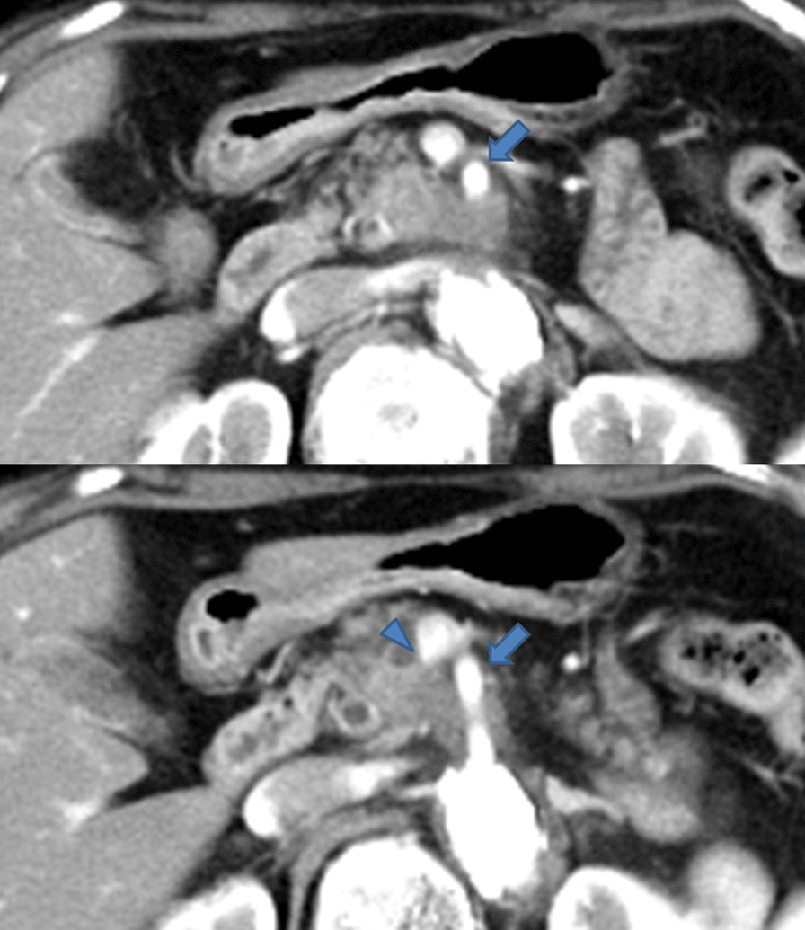
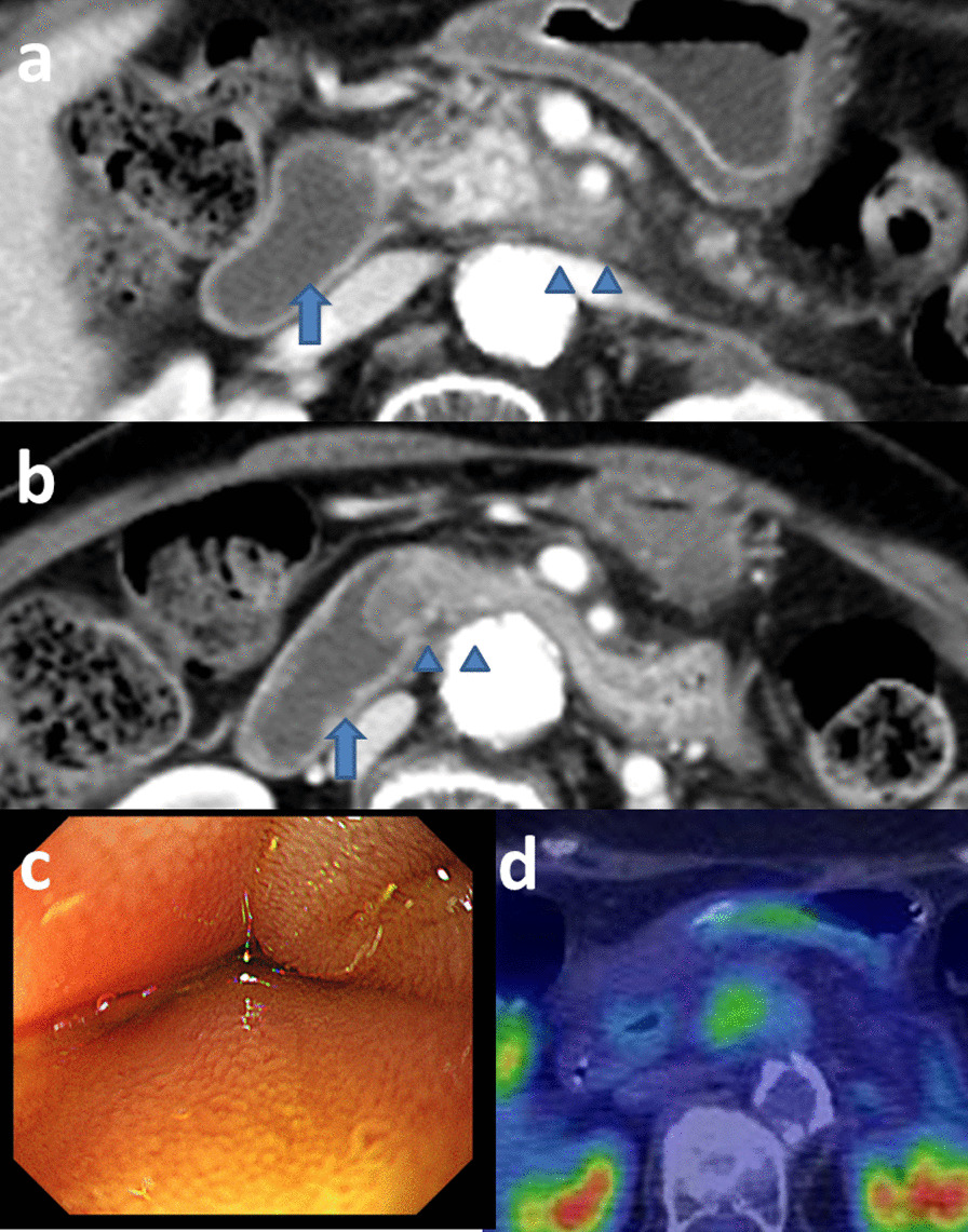
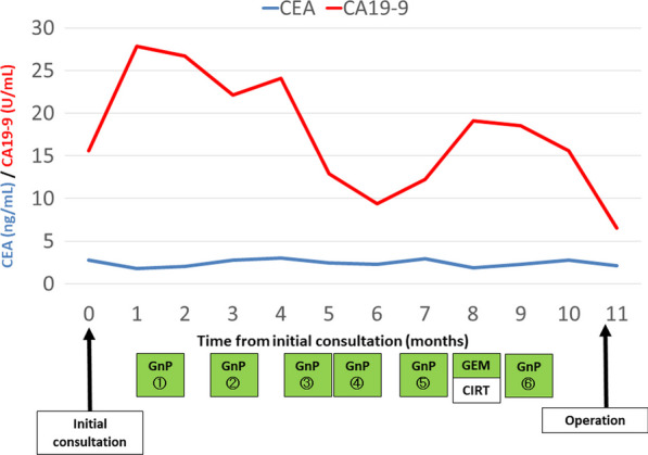
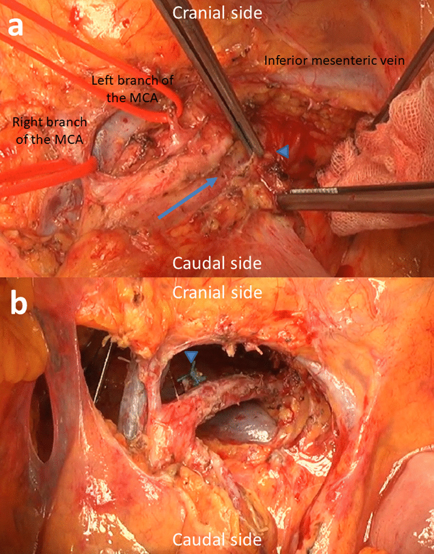
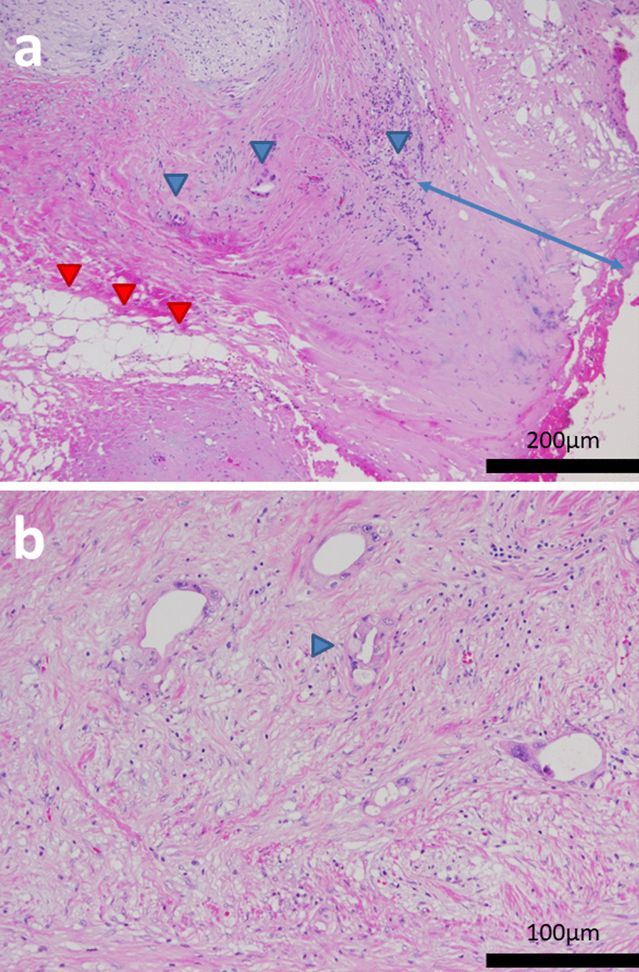
Case presentation: The patient was a 72-year-old Japanese woman with unresectable LA pancreatic head cancer with tumor contact to the superior mesenteric artery (SMA). She underwent four courses of chemotherapy (gemcitabine plus nanoparticle albumin-bound paclitaxel). However, the lesion did not shrink and tumor contact with the SMA did not improve after chemotherapy. Because the probability of achieving curative resection was judged to be low, she underwent radical dose CIRT, and chemotherapy was continued. She complained of vomiting 2 months after CIRT. Although imaging studies showed no tumor growth or metastasis, a duodenal obstruction which was speculated to be an adverse effect of CIRT was observed. She could not eat solid food and a trans-nasal feeding tube was inserted. Therapeutic intervention was required to enable enteral nutrition. We proposed several treatment options. She chose resection with the expectation of an anti-tumor effect of chemotherapy and CIRT rather than course observation with tube feeding or bypass surgery. Therefore, subtotal-stomach-preserving pancreatoduodenectomy with portal vein resection was performed as CS. Pathological examination of the resected specimen revealed an R0 resection with a histological response of Evans grade IIA. Postoperatively, she recovered uneventfully. Adjuvant chemotherapy with tegafur/gimeracil/oteracil (S1) was administrated. At the time of this report, 5 years have passed since the initial consultation and she has experienced no tumor recurrence.

Conclusions: The present case suggests that multidisciplinary treatment consisting of a combination of recent chemotherapy and CIRT may be beneficial for unresectable LA PDAC. However, further studies are required to assess the true efficacy of this treatment strategy.

Keywords: Carbon-ion radiotherapy; Conversion surgery; Pancreatic ductal adenocarcinoma.


Conflict of interest statement

The authors declare that they have no competing interests related to this manuscript.

Figures

Fig. 1
Fig. 1
Contrast-enhanced computed tomography findings. CECT revealed a hypovascular tumor in the uncinate process of the pancreas with tumor contact to the superior mesenteric artery (arrow) of more than 180° and portal vein involvement (arrowhead)
Fig. 2
Fig. 2
Three-dimensional constructed arterial imaging findings. The right gastroepiploic artery, left branch of the middle colic artery, first jejunal artery, and inferior pancreaticoduodenal artery formed a common trunk that branched from the right and slightly ventral side of the superior mesenteric artery
Fig. 3
Fig. 3
Findings of imaging studies after carbon-ion radiotherapy. a Contrast-enhanced computed tomography revealed no tumor growth (arrowheads). However, duodenal dilatation was observed (arrow). b CECT revealed a duodenal obstruction accompanied by wall thickening at the third portion of the duodenum (arrowheads). The oral duodenum was dilated (arrow). c Endoscopy revealed a duodenal compressive obstruction. The endoscope could not pass through this duodenal obstruction. d 18-fluoro-2-deoxy-glucose positron emission tomography/computed tomography after carbon-ion radiotherapy revealed slight FDG accumulation (standardized uptake value ranging from 3.14 to 3.20) in the pancreatic lesion without findings of distant or lymph nodal metastasis
Fig. 4
Fig. 4
The progress of preoperative treatment and changes in tumor markers. Tumor marker concentrations were within the normal limits during the preoperative period. CEA carcinoembryonic antigen, CA19-9 carbohydrate antigen 19-9, GnP gemcitabine plus nanoparticle albumin-bound paclitaxel, GEM gemcitabine, CIRT carbon-ion radiotherapy
Fig. 5
Fig. 5
Intraoperative findings. a After incising the left side of the nerve plexus around the superior mesenteric artery (SMA) (arrow indicates the incision line), part of the nerve plexus was assessed as a frozen tissue section to confirm the absence of tumor cells (arrowhead). Right and left branches of the middle colic arteries (MCAs) are taped. b View after resection via incised transverse mesocolon. Arrowhead indicates the stump of the common trunk of the right gastroepiploic artery, left branch of the MCA, first jejunal artery, and inferior pancreaticoduodenal artery. The dorsal nerve plexus around the SMA was dissected by approximately 240°. The left renal vein was observed on the dorsal side of the SMA
Fig. 6
Fig. 6
Microscopic findings of the resected specimen. a Viable tumor cells were observed in the replaced fibrotic tissues (blue arrowheads). Adipose degeneration was observed (red arrowheads). The distance of the dissected peripancreatic tissue margin was approximately 0.3 mm (blue arrow) and R0 resection was achieved. b Viable tumor cells with ductal formation were accompanied by vacuolar degeneration (arrowhead), and the effect grade was categorized as Evans grade IIA

Similar articles

Cited by

References

    1. Konstantinidis IT, Warshaw AL, Allen JN, et al. Pancreatic ductal adenocarcinoma: is there a survival difference for R1 resections versus locally advanced unresectable tumors? What is a “true” R0 resection? Ann Surg. 2013;257:731–736. doi: 10.1097/SLA.0b013e318263da2f. - DOI - PubMed
    1. Watanabe Y, Shinkawa T, Endo S, et al. Long-term outcomes after pancreatectomy for pancreatic ductal adenocarcinoma in elderly patients: special reference to postoperative adjuvant chemotherapy. World J Surg. 2018;42:2617–2626. doi: 10.1007/s00268-018-4496-y. - DOI - PubMed
    1. Yoshitomi H, Takano S, Furukawa K, et al. Conversion surgery for initially unresectable pancreatic cancer: current status and unresolved issues. Surg Today. 2019;49:894–906. doi: 10.1007/s00595-019-01804-x. - DOI - PubMed
    1. Furuse J, Shibahara J, Sugiyama M. Development of chemotherapy and significance of conversion surgery after chemotherapy in unresectable pancreatic cancer. J Hepatobiliary Pancreat Sci. 2018;25:261–268. doi: 10.1002/jhbp.547. - DOI - PubMed
    1. Satoi S, Yamaue H, Kato K, et al. Role of adjuvant surgery for patients with initially unresectable pancreatic cancer with a long-term favorable responses to non-surgical anti-cancer treatments: results of a project study for pancreatic surgery by the Japanese Society of Hepato-Biliary-Pancreatic Surgery. J Hepatobiliary Pancreat Sci. 2013;20:590–600. doi: 10.1007/s00534-013-0616-0. - DOI - PubMed

Publication types