. 2024 Jan 10;22(1):26.
doi: 10.1186/s12964-023-01399-4.

ALCAT1-mediated abnormal cardiolipin remodelling promotes mitochondrial injury in podocytes in diabetic kidney disease

Affiliations

ALCAT1-mediated abnormal cardiolipin remodelling promotes mitochondrial injury in podocytes in diabetic kidney disease

Yiqun Hao et al. Cell Commun Signal. .

Abstract

Background: Cardiolipin (CL) plays a critical role in maintaining mitochondrial membrane integrity and overall mitochondrial homeostasis. Recent studies have suggested that mitochondrial damage resulting from abnormal cardiolipin remodelling is associated with the pathogenesis of diabetic kidney disease (DKD). Acyl-coenzyme A:lyso-cardiolipin acyltransferase-1 (ALCAT1) was confirmed to be involved in the progression of Parkinson's disease, diet-induced obesity and other ageing-related diseases by regulating pathological cardiolipin remodelling. Thus, the purpose of this investigation was to determine the role of ALCAT1-mediated CL remodelling in DKD and to explore the potential underlying mechanism.

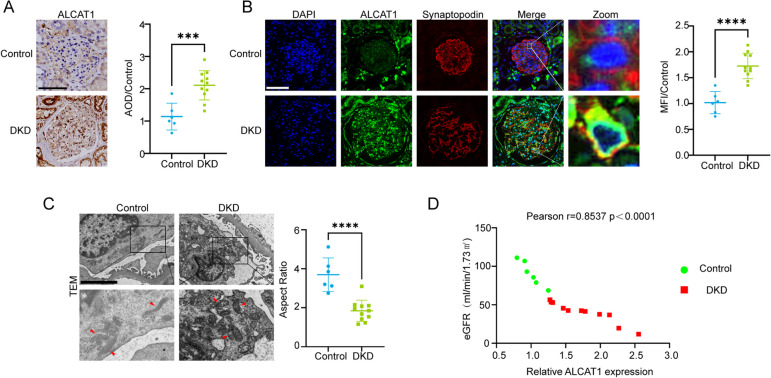
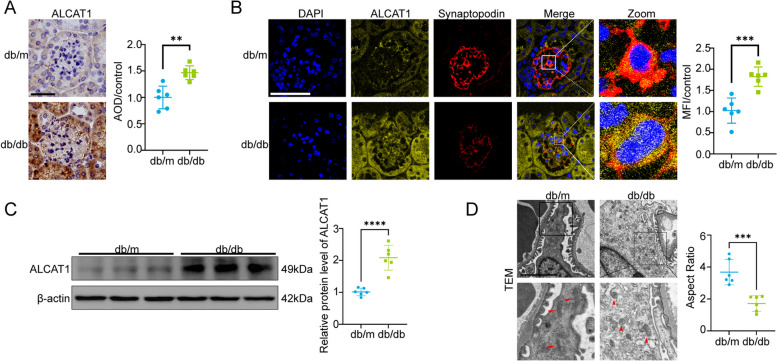
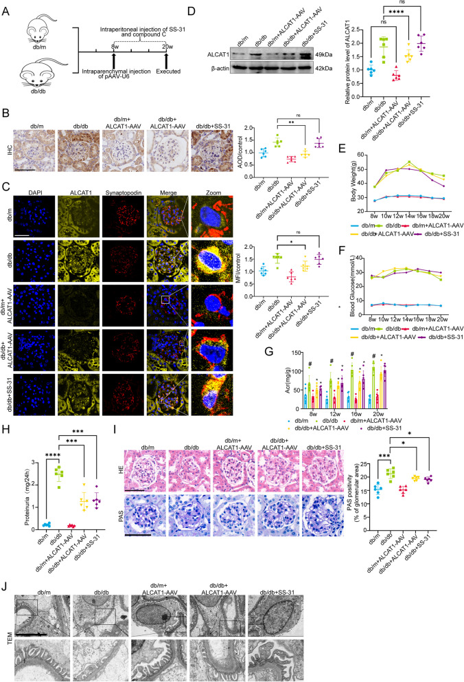
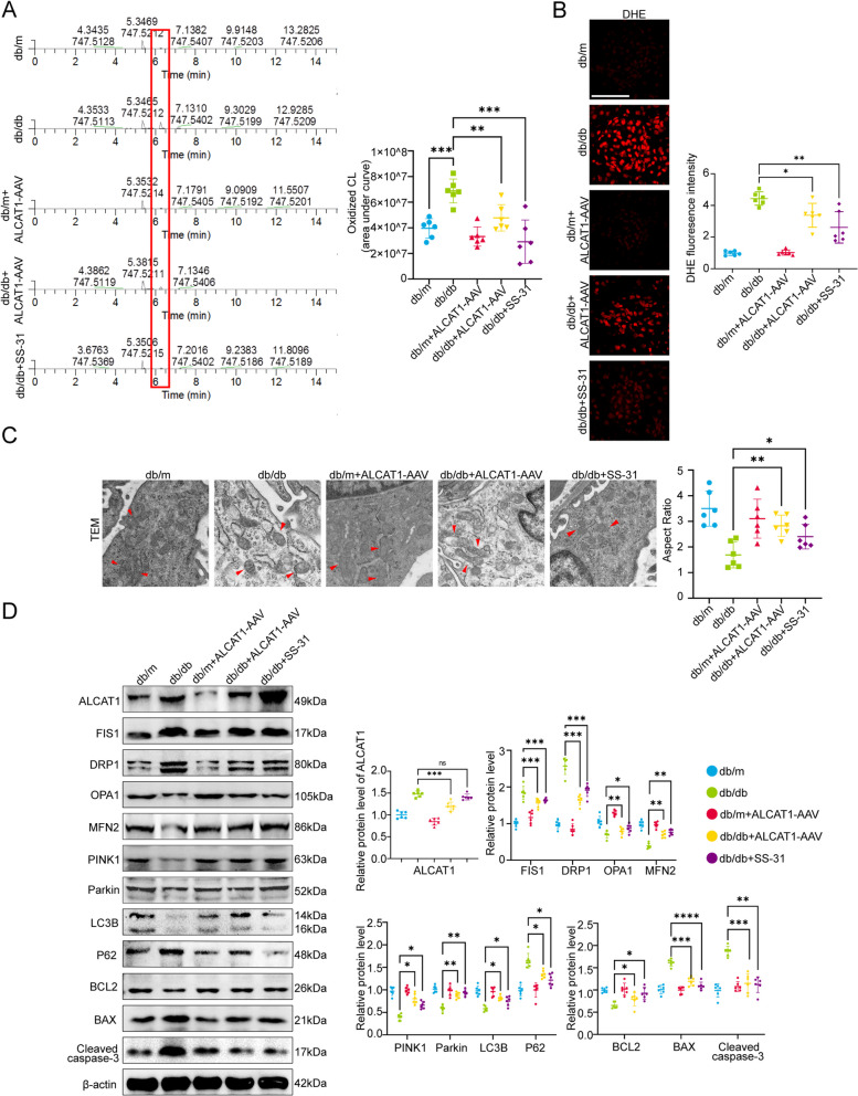
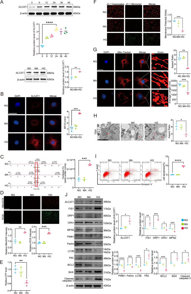
Methods: In vivo study, the mitochondrial structure was examined by transmission electron microscopy (TEM). The colocalization of ALCAT1 and synaptopodin was evaluated by double immunolabelling. Western blotting (WB) was performed to assess ALCAT1 expression in glomeruli. Lipidomics analysis was conducted to evaluate the composition of reconstructed cardiolipins. In vitro study, the lipidomics, TEM and WB analyses were similar to those in vivo. Mitochondrial function was evaluated by measuring the mitochondrial membrane potential (MMP) and the production of ATP and ROS.

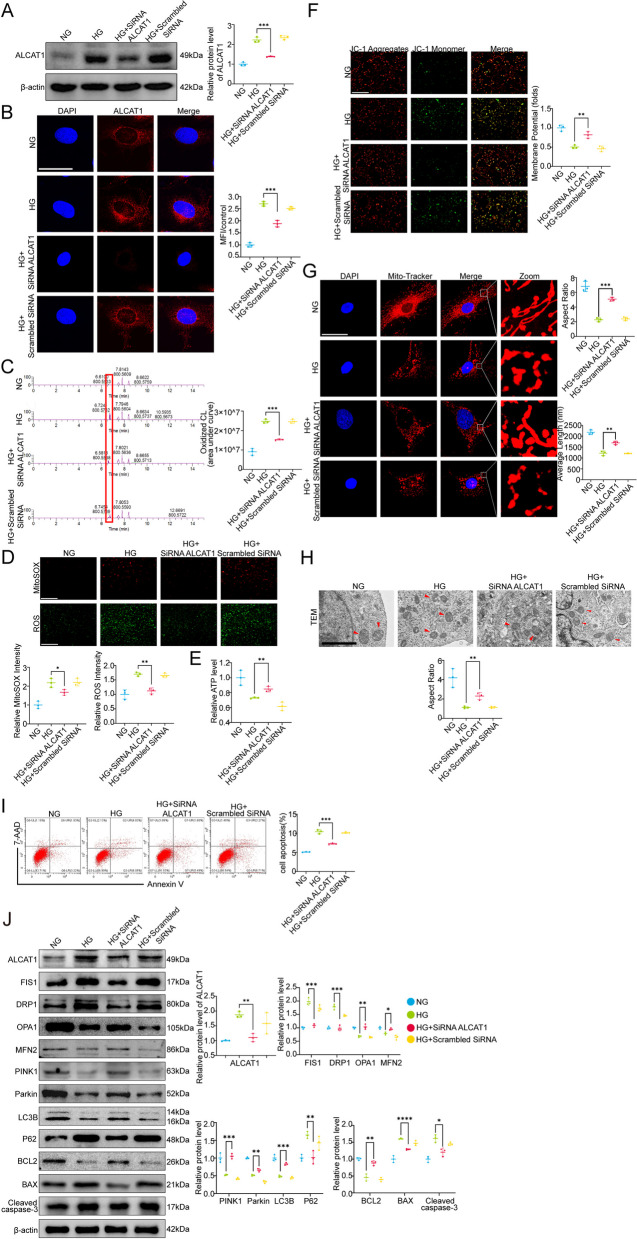
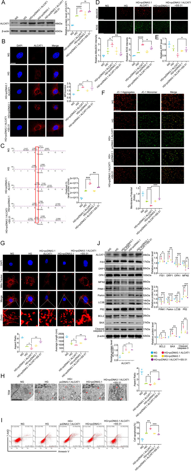
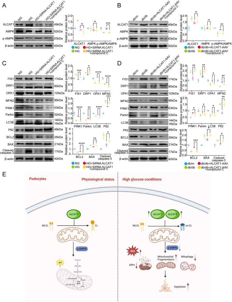
Results: Here, we showed that increased oxidized cardiolipin (ox-CL) and significant mitochondrial damage were accompanied by increased ALCAT1 expression in the glomeruli of patients with DKD. Similar results were found in db/db mouse kidneys and in cultured podocytes stimulated with high glucose (HG). ALCAT1 deficiency effectively prevented HG-induced ox-CL production and mitochondrial damage in podocytes. In contrast, ALCAT1 upregulation enhanced ox-CL levels and podocyte mitochondrial dysfunction. Moreover, treatment with the cardiolipin antioxidant SS-31 markedly inhibited mitochondrial dysfunction and cell injury, and SS-31 treatment partly reversed the damage mediated by ALCAT1 overexpression. We further found that ALCAT1 could mediate the key regulators of mitochondrial dynamics and mitophagy through the AMPK pathway.

Conclusions: Collectively, our studies demonstrated that ALCAT1-mediated cardiolipin remodelling played a crucial role in DKD, which might provide new insights for DKD treatment. Video Abstract.

Keywords: ALCAT1; Cardiolipin remodelling; Diabetic kidney disease; Mitochondrial dysfunction; Oxidized cardiolipin; Podocyte injury.


Conflict of interest statement

The authors declare no competing interests.

Figures

Fig. 1
Fig. 1
ALCAT1 levels were increased in glomerular podocytes along with mitochondrial injury in individuals with DKD. A Immunohistochemical staining and semiquantitative analysis of ALCAT1 in the glomeruli of patients with DKD (= 11) and control participants (n = 6) (***p<0.001, scale bars: 80 μm). B Immunofluorescence double staining and semiquantitative analysis of ALCAT1 and synaptopodin in glomeruli from patients with DKD (= 11) and control participants (= 6) (****p<0.0001, scale bars: 80 μm). C Transmission electron micrography (TEM) results of podocyte foot processes, glomerular basement membrane (GBM) and mitochondrial morphology from patients with DKD (= 11) and control participants (n = 6) (****p<0.0001, scale bars: 4 μm). D Correlation between ALCAT1 expression and eGFR (Control group: n = 6; DKD group: n = 11)
Fig. 2
Fig. 2
ALCAT1 was increased in the glomerular podocytes of db/db mice, accompanied by mitochondrial damage. A Immunohistochemical staining and semiquantitative analysis of ALCAT1 in glomeruli from each group (n = 6, ***p<0.001, scale bars: 40 μm). B Immunofluorescence double staining and semiquantitative analysis of ALCAT1 and synaptopodin in glomeruli in each group (n = 6, ***p<0.001, scale bars: 60 μm). C Western blots and relative expression of ALCAT1 (n = 6, ****p<0.0001). D Representative transmission electron micrographs of capillary loops and mitochondrial morphology in the glomeruli from each group (n = 6, ***p<0.001, scale bars: 2 μm)
Fig. 3
Fig. 3
Knockdown of ALCAT1 or inhibition of cardiolipin oxidation ameliorated kidney injury. A Illustration of the Animal Model Construction Pathway. B Immunohistochemical staining and semiquantitative analysis of ALCAT1 in glomeruli from each group (n = 6, **p<0.01, ns p>0.05, scale bars: 40 μm). C Immunofluorescence double staining and semiquantitative analysis of ALCAT1 and synaptopodin in glomeruli in each group (n = 6, *p<0.05, ns p>0.05, scale bars: 40 μm). D ALCAT1 protein levels in each group evaluated by Western blot (n = 6, *p<0.05, ****p<0.0001, ns p>0.05). E-G Body weight, blood sugar and urinary albumin creatinine ratio (UACR) from each group (n = 6, *p<0.05, #p<0.0001). H Results of 24-hour urine protein quantification (n = 6, ***p<0.001, ****p<0.0001). I Pathological damage detection by HE and PAS staining of kidney sections and quantification of the extent of mesangial expansion (n = 6, *p<0.05, ***p<0.001, scale bars: 30 μm). J Ultrastructure of capillary loops according to transmission electron microscopy in each group (n = 6, scale bars: 6 μm)
Fig. 4
Fig. 4
ALCAT1 inhibition or SS-31 treatment attenuated abnormal cardiolipin remodelling and podocyte mitochondrial damage in diabetic mice. A Relative content of oxidized cardiolipin in the renal cortex (n = 6, **p<0.01, ***p<0.001). B Micrographs and semiquantitative DHE staining in different groups (n = 6, *p<0.05, **p<0.01, scale bars: 30 μm). C Representative electron microscopy images of the ultrastructure of podocyte mitochondria from different groups (n = 6, *p<0.05, **p<0.01, scale bars: 1 μm). D Western blotting in the glomerulus of ALCAT1, mitochondrial fusion/fission-related proteins (FIS1, DRP1, OPA1, MFN2), autophagy-related proteins (PINK1, LC3B, P62), and apoptosis-related proteins (BCL2, BAX, cleaved caspase-3) (n = 6, *p<0.05, **p<0.01, ***p<0.001, ****p<0.0001, ns p>0.05)
Fig. 5
Fig. 5
ALCAT1 was increased in HG-treated podocytes, accompanied by abnormal cardiolipin remodelling and mitochondrial dysfunction. Western blotting was used to measure the variations in ALCAT1 levels in cultured podocytes subjected to glucose stimulation with time gradients (0, 6, 12, 24, 36, and 48 h) (n = 3, **p<0.01, ****p<0.0001). B Representative immunofluorescence staining for ALCAT1 (red) and semiquantitative analysis of these results in each group (n = 3, ***p<0.001, scale bars: 40 μm). C Relative content of oxidized cardiolipin (n = 3, ***p<0.001). D Cellular ROS and mitochondrial ROS production were assessed by DCFH-DA fluorescent probe and MitoSox Red fluorescence staining in different groups (n = 3, **p<0.01, ***p<0.001, scale bars: 200 μm). E Determination of relative ATP content in each group (n = 3, **p<0.01). F Relative quantification of mitochondrial membrane potential by JC-1 staining in podocytes (n = 3, ***p<0.001, scale bars: 200 μm). G Microscopy images of mitochondrial fission and fusion by MitoTracker Red staining and semiquantitative analysis of the average mitochondrial length and aspect ratio (n = 3, **p<0.01, ***p<0.001, scale bars: 40 μm). H TEM results of mitochondrial structure damage in podocytes (n = 3, *p<0.05, scale bars: 1 μm). I Apoptosis detection by flow cytometry in podocytes (n = 3, ****p<0.0001). J Western blotting of mitochondrial fusion/fission-related proteins (FIS1, DRP1, OPA1, MFN2), autophagy-related proteins (PINK1, LC3B, P62), and apoptosis-related proteins (BCL2, BAX, cleaved caspase-3) (n = 3, *p<0.05, **p<0.01, ***p<0.001, ****p<0.0001)
Fig. 6
Fig. 6
ALCAT1 deficiency improved abnormal cardiolipin remodelling and suppressed mitochondrial damage in high glucose-treated podocytes. A Verification of the knockdown effect of ALCAT1 siRNA by Western blotting (n = 3, ***p<0.001). B Immunofluorescence staining of ALCAT1 (red) in podocytes from each group (n = 3, ***p<0.001, scale bars: 40 μm). C Relative content of oxidized cardiolipin in podocytes measured by lipidomic analysis (n = 3, ***p<0.001). D Cellular ROS and mitochondrial ROS production were evaluated by DCFH-DA fluorescent probe and MitoSox Red fluorescence staining (n = 3, *p<0.05, **p<0.01, scale bars: 200 μm). E Relative ATP content and semiquantitative analysis of each group (n = 3, **p<0.01). F Relative quantification of mitochondrial membrane potential detected by JC-1 staining (n = 3, **p<0.01, scale bars: 200 μm). G Microscopy images of mitochondrial fission and fusion in podocytes and semiquantitative analysis of the average mitochondrial length and aspect ratio (n = 3, **p<0.01, ***p<0.001, scale bars: 60 μm). H Electron microscopy observation of the ultrastructure of mitochondria in podocytes (n = 3, **p<0.01, scale bars: 2 μm). I Apoptosis detection in each group by flow cytometry (n = 3, ***p<0.001). J Western blot analysis of mitochondrial fusion/fission-related proteins (FIS1, DRP1, OPA1, MFN2), autophagy-related proteins (PINK1, LC3B, P62), and apoptosis-related proteins (BCL2, BAX, cleaved caspase-3) (n = 3, *p<0.05, **p<0.01, ***p<0.001, ****p<0.0001)
Fig. 7
Fig. 7
Inhibition of cardiolipin oxidation by SS-31 reversed ALCAT1 upregulation-mediated abnormal cardiolipin remodelling and mitochondrial damage in HG-induced podocytes. A The effects of overexpression plasmid transfection and SS-31 treatment on ALCAT1 expression were detected by Western blotting (n = 3, ****p<0.0001, ns p>0.05). B Immunofluorescence staining of ALCAT1 (red) in podocytes from each group (n = 3, ***p<0.001, ns p>0.05, scale bars: 20 μm). C Relative oxidized cardiolipin content in podocytes measured by lipidomic analysis (n = 3, **p<0.01, ***p<0.001). D Cellular ROS and mitochondrial ROS production were evaluated by DCFH-DA fluorescent probe and MitoSox Red fluorescence staining (n = 3, *p<0.05, **p<0.01, ****p<0.0001, scale bars: 200 μm). E Relative ATP content and semiquantitative analysis of each group (n = 3, *p<0.05). F Relative quantification of mitochondrial membrane potential detected by JC-1 staining (n = 3, ***p<0.001, scale bars: 200 μm). G Microscopy images of mitochondrial fission and fusion in podocytes by MitoTracker Red staining and semiquantitative analysis of the average mitochondrial length and aspect ratio (n = 3, *p<0.05, **p<0.01, scale bars: 40 μm). H Electron microscopy observation of the ultrastructure of mitochondria in podocytes (n = 3, *p<0.05, ***p<0.001, scale bars: 2 μm). I Apoptosis detection in each group by flow cytometry (n = 3, **p<0.01, ***p<0.001). J Western blot analysis of mitochondrial fusion/fission-related proteins (FIS1, DRP1, OPA1, MFN2), autophagy-related proteins (PINK1, LC3B, P62), and apoptosis-related proteins (BCL2, BAX, cleaved caspase-3) (n = 3, *p<0.05, **p<0.01, ***p<0.001, ****p<0.0001, ns p>0.05)
Fig. 8
Fig. 8
Aberrant cardiolipin remodelling mediated by ALCAT1 promoted mitochondrial malfunction in podocytes by deactivating the AMPK pathway. A Western blotting analysis of ALCAT1, AMPK and p-AMPK in vitro, as well as semi-quantitative of pAMPK/AMPK ratios (n = 3, ***p<0.001, ns p>0.05). B Western blotting analysis of ALCAT1, AMPK and p-AMPK in vivo, as well as semi-quantitative of pAMPK/AMPK ratios (n = 3, **p<0.01, ns p>0.05). C Western blot analysis of mitochondrial fusion/fission-related proteins (FIS1, DRP1, OPA1, MFN2), autophagy-related proteins (PINK1, LC3B, P62), and apoptosis-related proteins (BCL2, BAX, cleaved caspase-3) in vitro (n = 3, *p<0.05, **p<0.01, ***p<0.001, ****p<0.0001). D Western blot analysis of mitochondrial fusion/fission-related proteins (FIS1, DRP1, OPA1, MFN2), autophagy-related proteins (PINK1, LC3B, P62), and apoptosis-related proteins (BCL2, BAX, cleaved caspase-3) in vivo (n = 3, *p<0.05, **p<0.01, ***p<0.001). Schematic of the molecular model proposed in this study

Similar articles

Cited by

References

    1. Zhang L, Long J, Jiang W, Shi Y, He X, Zhou Z, Li Y, Yeung RO, Wang J, Matsushita K, et al. Trends in chronic Kidney Disease in China. N Engl J Med. 2016;375:905–6. - PubMed
    1. Ma RCW. Epidemiology of Diabetes and diabetic Complications in China. Diabetologia. 2018;61:1249–60. - PubMed
    1. DeFronzo RA, Reeves WB, Awad AS. Pathophysiology of diabetic Kidney Disease: impact of SGLT2 inhibitors. Nat Rev Nephrol. 2021;17:319–34. - PubMed
    1. Guedes M, Pecoits-Filho R. Can we cure diabetic Kidney Disease? Present and future perspectives from a nephrologist’s point of view. J Intern Med. 2022;291:165–80. - PubMed
    1. Tao Y, Young-Stubbs C, Yazdizadeh Shotorbani P, Su DM, Mathis KW, Ma R. Sex and strain differences in renal hemodynamics in mice. Physiol Rep. 2023;11:e15644. - PMC - PubMed

Publication types