. 2024 Jan 10;22(1):43.
doi: 10.1186/s12967-023-04830-z.

Targeting the SphK1/S1P/PFKFB3 axis suppresses hepatocellular carcinoma progression by disrupting glycolytic energy supply that drives tumor angiogenesis

Affiliations

Targeting the SphK1/S1P/PFKFB3 axis suppresses hepatocellular carcinoma progression by disrupting glycolytic energy supply that drives tumor angiogenesis

Xin Tracy Liu et al. J Transl Med. .

Abstract

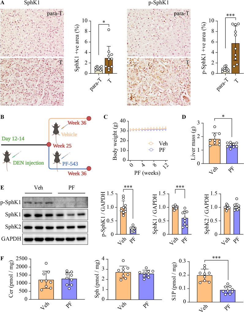
Background: Hepatocellular carcinoma (HCC) remains a leading life-threatening health challenge worldwide, with pressing needs for novel therapeutic strategies. Sphingosine kinase 1 (SphK1), a well-established pro-cancer enzyme, is aberrantly overexpressed in a multitude of malignancies, including HCC. Our previous research has shown that genetic ablation of Sphk1 mitigates HCC progression in mice. Therefore, the development of PF-543, a highly selective SphK1 inhibitor, opens a new avenue for HCC treatment. However, the anti-cancer efficacy of PF-543 has not yet been investigated in primary cancer models in vivo, thereby limiting its further translation.

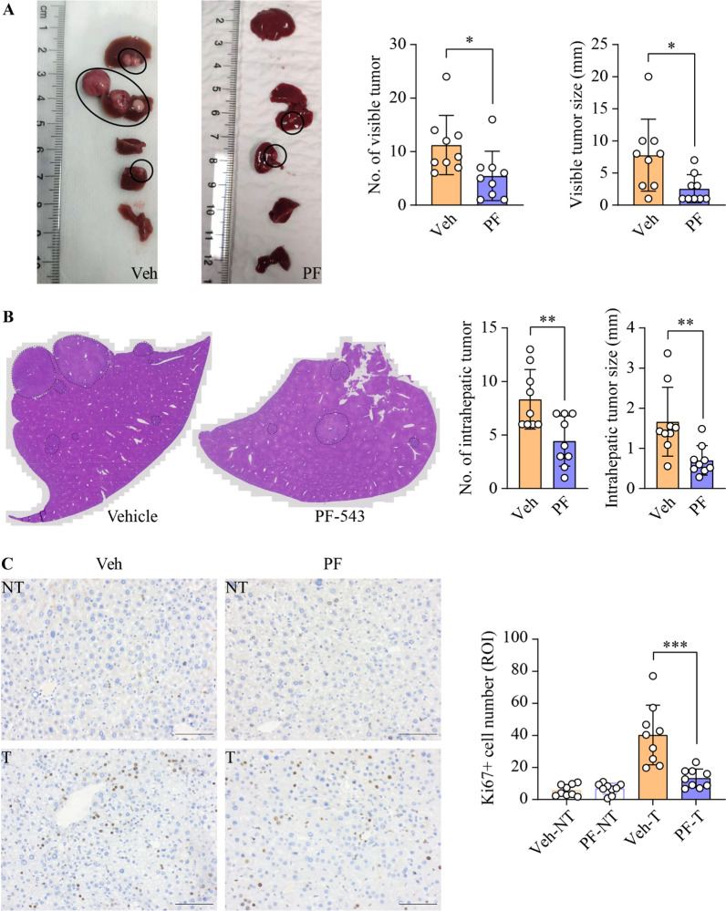
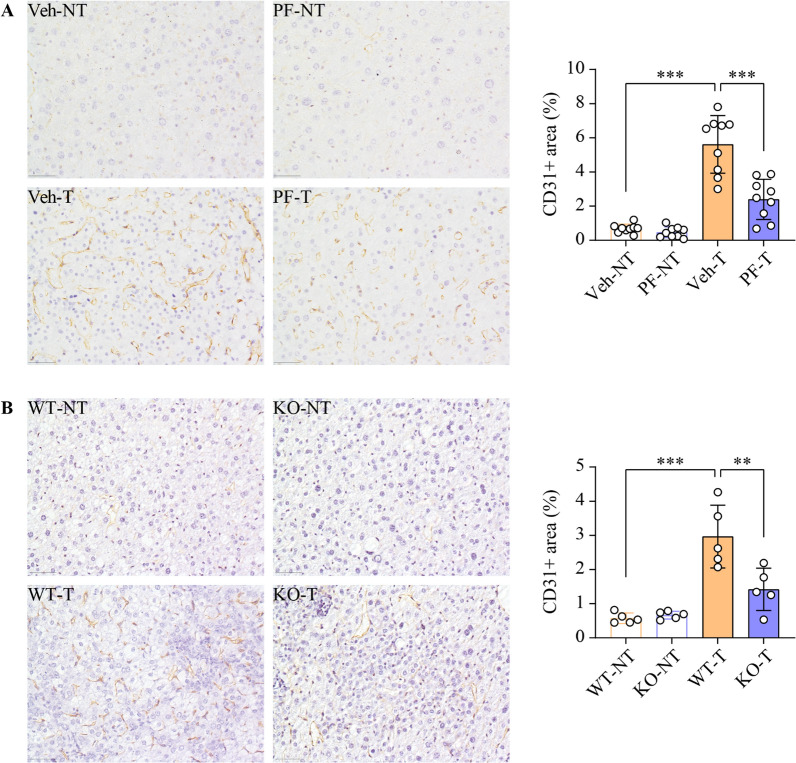
Methods: Building upon the identification of the active form of SphK1 as a viable therapeutic target in human HCC specimens, we assessed the capacity of PF-543 in suppressing tumor progression using a diethylnitrosamine-induced mouse model of primary HCC. We further delineated its underlying mechanisms in both HCC and endothelial cells. Key findings were validated in Sphk1 knockout mice and lentiviral-mediated SphK1 knockdown cells.

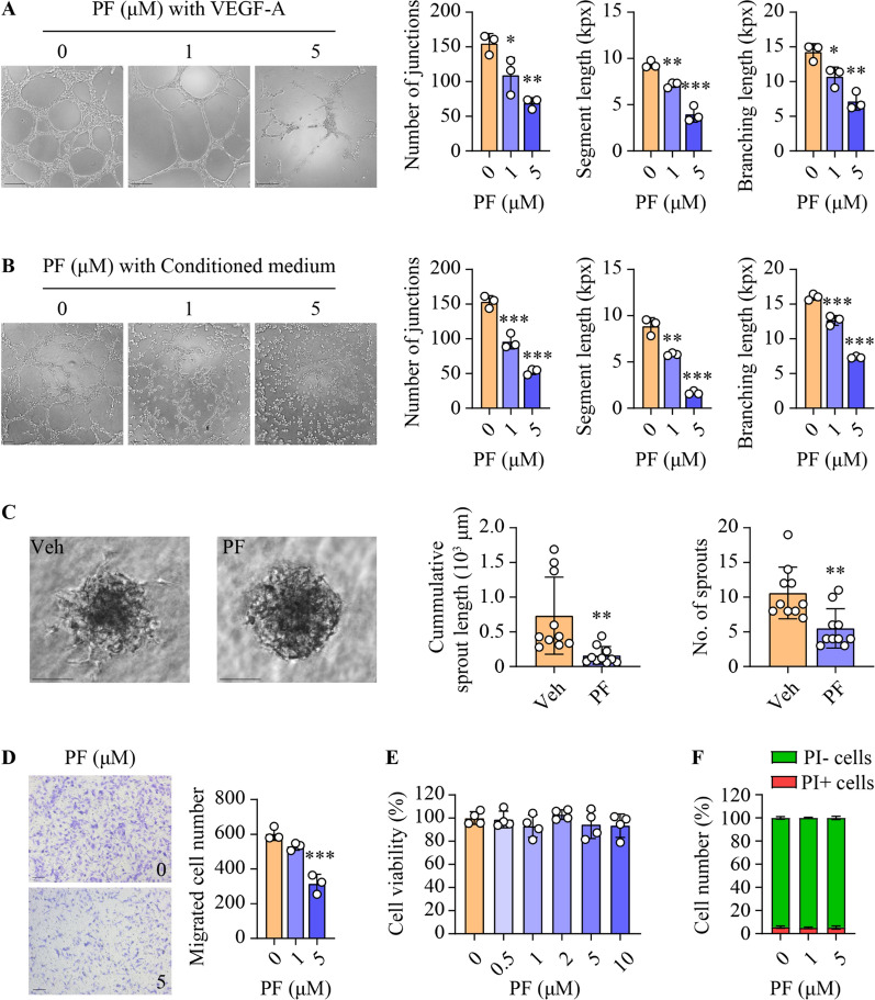
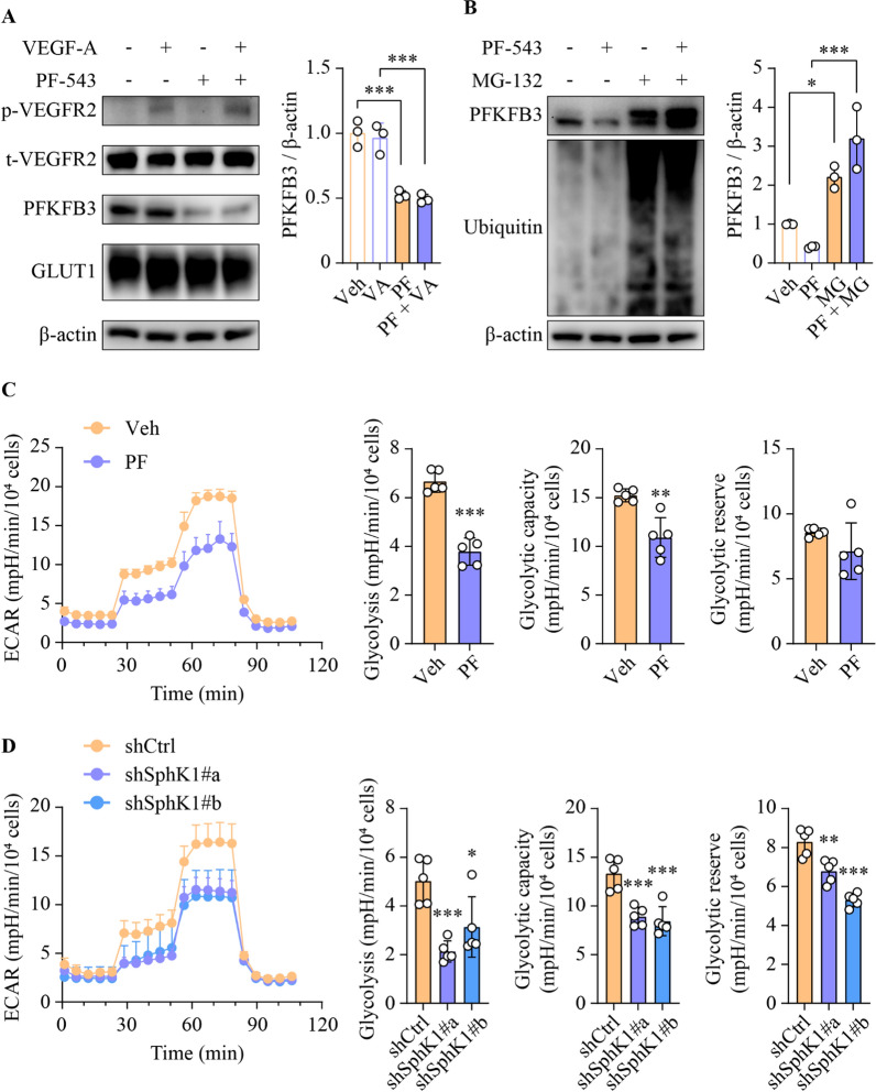
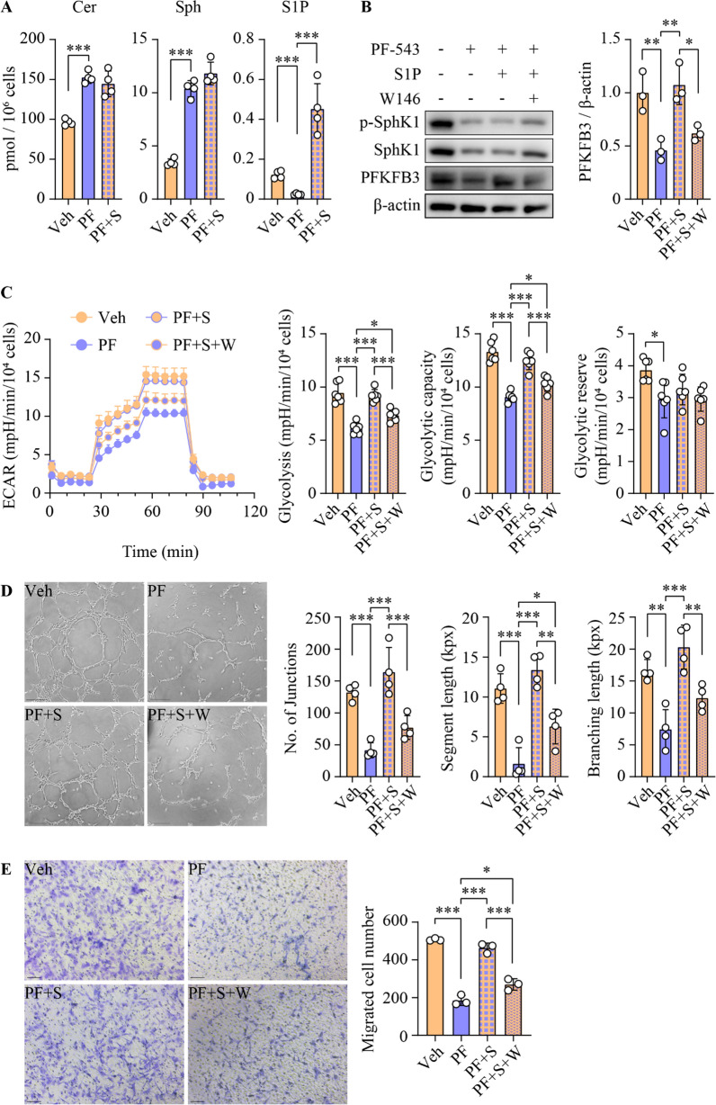
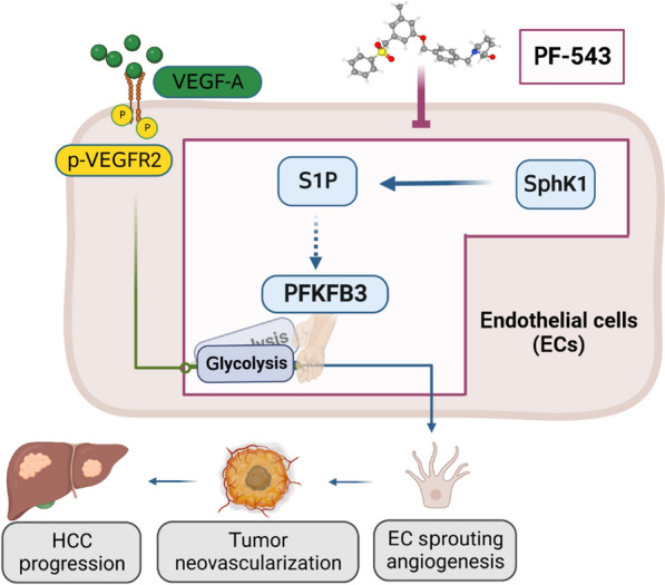
Results: SphK1 activity was found to be elevated in human HCC tissues. Administration of PF-543 effectively abrogated hepatic SphK1 activity and significantly suppressed HCC progression in diethylnitrosamine-treated mice. The primary mechanism of action was through the inhibition of tumor neovascularization, as PF-543 disrupted endothelial cell angiogenesis even in a pro-angiogenic milieu. Mechanistically, PF-543 induced proteasomal degradation of the critical glycolytic enzyme 6-phosphofructo-2-kinase/fructose-2,6-biphosphatase 3, thus restricting the energy supply essential for tumor angiogenesis. These effects of PF-543 could be reversed upon S1P supplementation in an S1P receptor-dependent manner.

Conclusions: This study provides the first in vivo evidence supporting the potential of PF-543 as an effective anti-HCC agent. It also uncovers previously undescribed links between the pro-cancer, pro-angiogenic and pro-glycolytic roles of the SphK1/S1P/S1P receptor axis. Importantly, unlike conventional anti-HCC drugs that target individual pro-angiogenic drivers, PF-543 impairs the PFKFB3-dictated glycolytic energy engine that fuels tumor angiogenesis, representing a novel and potentially safer therapeutic strategy for HCC.

Keywords: Angiogenesis; Glycolysis; Hepatocellular carcinoma; PF-543; PFKFB3; Sphingosine kinase.


Conflict of interest statement

The authors declare no potential conflicts of interest.

Figures

Fig. 1
Fig. 1
PF-543 inhibits SphK1 activity in DEN-treated mice. A Total and phospho-SphK1 levels were examined using immunohistochemical staining in tumorous (T) and para-tumorous (para-T) tissues of human HCC specimens; scale bar = 50 μm; n = 10. B Schematic illustration of treatment schedules for in vivo studies, created with BioRender.com. C Body weight over 12 weeks of vehicle (veh) or PF-543 (PF) treatment. D Liver mass was weighed. E phospho(p)-SphK1 SphK1 and SphK2 protein levels in liver tissues were determined using Western blotting. F Hepatic levels of ceramide (Cer), sphingosine (Sph), and S1P were analyzed using lipidomics. C-F n = 9. Data are expressed as mean ± SD. *p < 0.05; ***p < 0.001
Fig. 2
Fig. 2
Inhibition of SphK1 suppresses HCC progression in DEN-treated mice. DEN-injected mice were treated with vehicle (veh) or PF-543 (PF) for 12 weeks. A The number and maximal diameter of visible liver tumors were quantified from macroscopic images. B The number and maximal diameter of intrahepatic liver tumors were quantified from the scanning of the entire H&E-stained liver tissue sections. C Cell proliferation in non-tumorous (NT) and tumorous (T) liver tissues was stained and quantified using Ki67 immunohistochemistry; scale bar = 50 μm. Data are expressed as mean ± SD. n = 9. *p < 0.05; **p < 0.01; ***p < 0.001
Fig. 3
Fig. 3
SphK1 inhibition or ablation reduces vessel density in HCC tumors. A DEN-injected mice were treated with vehicle (veh) or PF-543 (PF) for 12 weeks. Blood vessels in non-tumorous (NT) and tumorous (T) liver tissues were stained and quantified by CD31 immunohistochemistry; scale bar = 50 μm; n = 9. B Blood vessels in DEN-injected wild-type (WT) and Sphk1 knockout (KO) mice were examined by CD31 immunohistochemistry; scale bar = 50 μm; n = 5. Data are expressed as mean ± SD. **p < 0.01; ***p < 0.001
Fig. 4
Fig. 4
Inhibition of SphK1 impairs angiogenesis in HUVECs. Primary HUVECs were treated with PF-543 at the indicated concentrations for 16 h. A and B Tube formation was induced by 50 ng/ml VEGF-A (A) or conditioned medium collected from Huh7 HCC cell culture (B). Quantification of tube formation is presented as the number of junctions, segment length, and total branching length. kpx, 1000 pixels; scale bar = 200 μm (A) or 100 μm (B); n = 3. C Sprouting assays were performed in three-dimensional spheroids. Quantitation of the sprouting is presented as cumulative sprout length and number of sprouts per spheroid; scale bar = 100 μm; n = 10. D Cell migration was determined by transwell assay, and migrated cells were stained with crystal violet; scale bar = 100 μm; n = 3. E Cell viability was determined using MTS assay; n = 4. F Cell death was assessed using flow cytometry with propidium iodide (PI) staining; PI-, living cells (green); PI+, dead cells (red); n = 3. Data are expressed as mean ± SD. *p < 0.05; **p < 0.01; ***p < 0.001
Fig. 5
Fig. 5
SphK1 promotes angiogenesis by regulating the glycolytic modulator PFKFB3. A Primary HUVECs were treated with 5 µM PF-543 for 16 h, prior to stimulation with 50 ng/ml VEGF-A for 15 min. B Following 1 h pre-treatment with 10 µM MG-132, primary HUVECs were treated with PF-543 at 5 µM for 16 h. A and B Western blotting analyses of the indicated proteins; n = 3. C and D Glycolytic rate, capacity and reserve were determined using Seahorse real-time glycolytic stress assay in PF-543-treated (C) or shRNA-mediated SphK1 knockdown (D) primary HUVECs. ECAR, extracellular acidification rate; n = 5. Data are expressed as mean ± SD. *p < 0.05; **p < 0.01; ***p < 0.001
Fig. 6
Fig. 6
S1P is required for glycolysis and angiogenesis in an S1P1-dependent manner. Primary HUVECs were treated with 5 µM PF-543 (PF) and 0.5 µM S1P (S) for 16 h, with 2 µM S1P1 antagonist W146 added 1 h prior to these treatments. A Levels of ceramide (Cer), sphingosine (Sph) and S1P were analyzed using lipidomics; n = 4. B phospho(p)-SphK1, SphK1 and PFKFB3 protein levels were determined using Western blotting; n = 3. C Glycolytic rate, capacity and reserve were examined using Seahorse real-time glycolytic stress assay. ECAR, extracellular acidification rate; n = 6. D Tube formation was quantified as the number of junctions, segment length, and total branching length. kpx, 1000 pixels; scale bar = 200 μm; n = 4. E Cell migration was determined by transwell assay, and migrated cells were stained with crystal violet; scale bar = 100 μm; n = 3. Data are expressed as mean ± SD. *p < 0.05; **p < 0.01; ***p < 0.001
Fig. 7
Fig. 7
Model depicting the anti-HCC actions of PF-543. Elevated pro-angiogenic factors, exemplified by VEGF-A, act through their receptors to drive sprouting angiogenesis in tumor endothelial cells (EC), promoting tumor neovascularization and HCC progression. PFKFB3 serves as a molecular switch in this process, dictating the glycolytic energy supply that is essential for sprouting angiogenesis. The selective SphK1 inhibitor PF-543 abrogates S1P production, subsequently turning off the PFKFB3-mediated glycolytic switch, which leads to the inhibition of sprouting angiogenesis and eventually suppression of HCC progression. The graphical abstract was created with BioRender.com. . The 3D conformer of PF-543 was adapted from PubChem, pubchem.ncbi.nlm.nih.gov, CID 66,577,038

References

    1. Rumgay H, Arnold M, Ferlay J, Lesi O, Cabasag CJ, Vignat J, et al. Global burden of primary liver cancer in 2020 and predictions to 2040. J Hepatol. 2022;77:1598. doi: 10.1016/j.jhep.2022.08.021. - DOI - PMC - PubMed
    1. Zhu AX, Duda DG, Sahani DV, Jain RK. HCC and angiogenesis: possible targets and future directions. Nat Rev Clin Oncol. 2011;8:292–301. doi: 10.1038/nrclinonc.2011.30. - DOI - PMC - PubMed
    1. Qin S, Li A, Yi M, Yu S, Zhang M, Wu K. Recent advances on anti-angiogenesis receptor tyrosine kinase inhibitors in cancer therapy. J Hematol Oncol. 2019;12:27. doi: 10.1186/s13045-019-0718-5. - DOI - PMC - PubMed
    1. Calderaro J, Ziol M, Paradis V, Zucman-Rossi J. Molecular and histological correlations in liver cancer. J Hepatol. 2019;71:616–630. doi: 10.1016/j.jhep.2019.06.001. - DOI - PubMed
    1. Moawad AW, Szklaruk J, Lall C, Blair KJ, Kaseb AO, Kamath A, et al. Angiogenesis in hepatocellular carcinoma; pathophysiology, targeted therapy, and role of imaging. J Hepatocell Carcinoma. 2020;7:77–89. doi: 10.2147/JHC.S224471. - DOI - PMC - PubMed

Publication types