. 2023 Dec 21;14(1):36.
doi: 10.3390/ani14010036.

Evaluation of the Effectiveness of Medical-Grade Honey and Hypericum Perforatum Ointment on Second-Intention Healing of Full-Thickness Skin Wounds in Cats

Affiliations

Evaluation of the Effectiveness of Medical-Grade Honey and Hypericum Perforatum Ointment on Second-Intention Healing of Full-Thickness Skin Wounds in Cats

Kyriakos Chatzimisios et al. Animals (Basel). .

Abstract

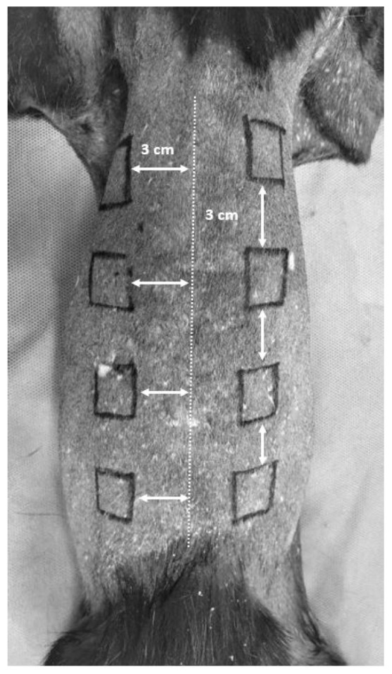
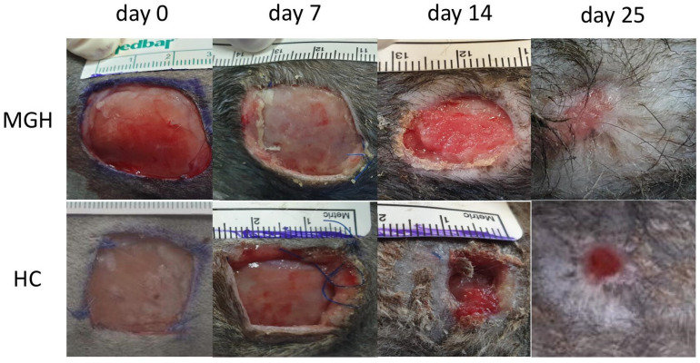
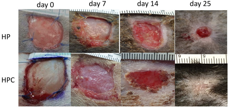
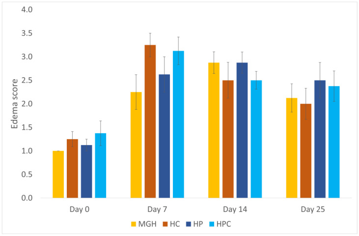
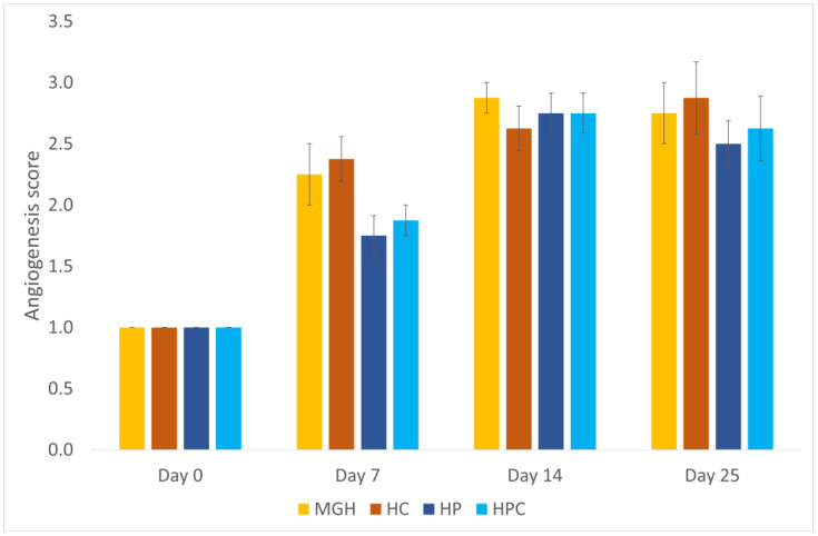
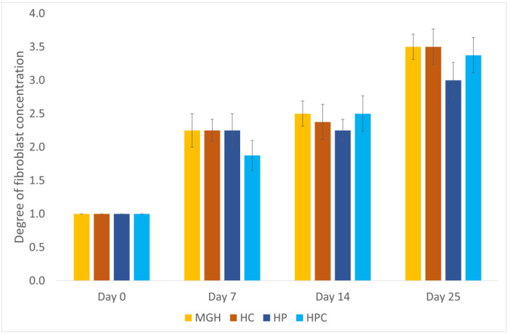
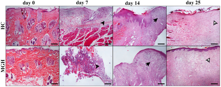
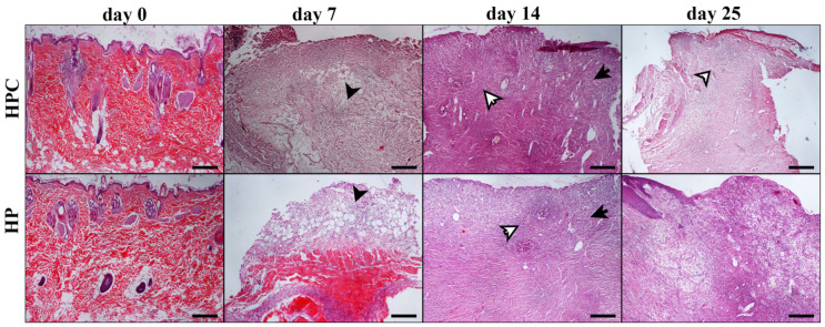
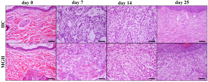
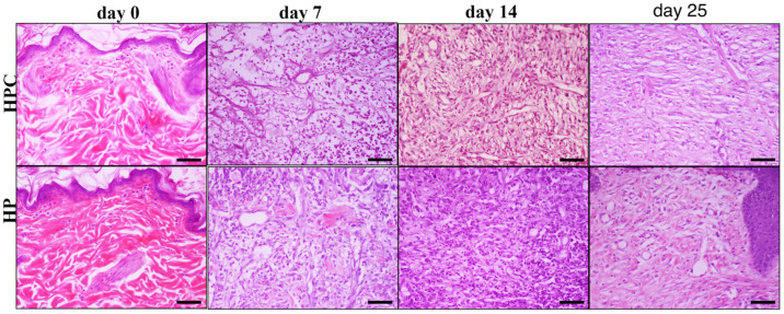
This study aimed to determine the effects of two topical treatments on second-intention wound healing in cats. Eight 2 × 2 cm full-thickness wounds were created, four on each side of the dorsal midline of eight laboratory cats, to receive either medical-grade honey ointment (MGH) and its control (HC), or Hypericum-based ointment (HP) and its control (HPC). MGH or HP ointment was applied to four wounds on the same side, while the remaining four were used as controls, chosen at random. Planimetry, laser Doppler flowmetry, daily physical examinations, and histologic examinations on days 0, 7, 14, and 25 were used to assess the healing of wounds. Tissue perfusion was better in the MGH-treated (2.14 ± 0.18 mm/s) and HP-treated wounds (2.02 ± 0.13 mm/s) than in the untreated controls HC (1.59 ± 0.11 mm/s) and HPC (1.60 ± 0.05 mm/s), respectively (p = 0.001). Histopathology revealed that the median edema score was lower in the MGH-treated (2; range 1-4) compared to the HC-treated wounds (3; range 2-4) on day 7 (p < 0.05). The median angiogenesis score was higher on day 7 in the MGH-treated (2; range 1-3) compared to the HP-treated wounds (2; range 1-2) (p = 0.046). The fibroblast concentration was increased in the MGH-treated wounds (3.5; range 3-4) compared to the HP-treated wounds (3; range 2-4) on day 25 (p = 0.046). MGH and HP increased tissue perfusion compared to the untreated controls. The MGH-treated wounds had histologic parameters superior to the HP-treated wounds regarding angiogenesis and fibroblast concentration in cutaneous wound healing in cats. Topical application of MGH and HP did not accelerate the healing process of feline cutaneous wounds.

Keywords: Hypericum; cat; honey; wound healing.


Conflict of interest statement

Niels Cremers works as Head of Research at Triticum Exploitatie BV, the manufacturer of the honey-based product used in this study. He was involved in manuscript preparation and editing. There are no relevant financial or non-financial competing interests to report. All other authors declare no conflicts of interest.

Figures

Figure 1
Figure 1
Eight 2 × 2 cm squares were drawn, four on either side of the dorsal midline. The squares were 3 cm away from each other and 3 cm away from the dorsal midline.
Figure 2
Figure 2
MGH-treated and HC wounds at different measurement times (days 0, 7, 14, 25).
Figure 3
Figure 3
HP-treated and HPC wounds at different measurement times (days 0, 7, 14, 25).
Figure 4
Figure 4
Edema score of honey-treated wounds (MGH) compared to Hypericum-treated wounds (HP), and their controls (HC and HPC). Honey-treated wounds showed a significantly lower edema score than HC on day 7. Bars represent the mean ± standard deviation (yellow = MGH, orange = HC, dark blue = HP, light blue = HPC).
Figure 5
Figure 5
Mean angiogenesis score of honey-treated wounds (MGH) compared to Hypericum-treated wounds (HP), and their controls (HC and HPC). Honey-treated wounds showed a significantly greater score than HP-treated wounds on day 7. Bars represent the mean ± standard deviation (yellow = MGH, orange = HC, dark blue = HP, light blue = HPC).
Figure 6
Figure 6
Mean fibroblast concentration scores of honey-treated wounds (MGH) compared to Hypericum-treated wounds (HP) to their control wounds (HC and HPC). Honey-treated wounds had significantly increased fibroblast concentration scores compared to HP-treated wounds on day 25. Bars represent the mean ± standard deviation (yellow = MGH, orange = HC, dark blue = HP, light blue = HPC).
Figure 7
Figure 7
Representative images of tissue sections from honey-treated (MGH) wounds and their controls (HC) at different measurement times (days 0, 7, 14, 25): On day 0, the excised skin in both images appears normal. On day 7, the edema of the HC wound compared to the MGH-treated wound is more intense, showing prominent separation of cells and collagen by acellular material (black arrowheads). On day 14, no significant differences are depicted between the two groups concerning the degrees of inflammatory cell infiltration, edema, fibroblast, and blood vessel aggregation, as well as collagen density (black arrows). Finally, on day 25, in MGH-treated wounds, a little lower collagen density and fibroblast accumulation and relatively increased capillary aggregation, relative to HC wounds, were noticed (white arrows). The formation of the epidermis is characteristic in both images, although in MGH it seems closer to the normal architecture. MGH and HC/days 0, 7, 14, 25: H-E; Hematoxylin and eosin (H&E) staining, magnification 100×, bar = 100 μm.
Figure 8
Figure 8
Representative images of tissue sections from hypericum-treated (HP) and HPC wounds at different measurement times (days 0, 7, 14, 25). On day 0, images show normal skin histology. On day 7, both HPC and HP-treated wounds present inflammatory cell infiltration and edema, with the former showing a little higher intensity than the latter (black arrowheads). The opposite is noticed regarding fibroblast aggregation and collagen density. On day 14, mild edema is shown in both groups, accompanied by pronounced fibroblast aggregation of a little higher intensity in HP-treated wounds relative to HPC (black arrows). The opposite is observed for inflammatory cell infiltration (white arrows). Finally, on day 25, intense collagen density and fibroblast aggregation is noticed in the HPC wound (white arrowhead). The wounds exhibit incomplete epithelial coverage. HPC and HP/days 0, 7, 14, 25: H-E; Hematoxylin and eosin (H&E) staining, magnification 100×, bar = 100 μm.
Figure 9
Figure 9
Representative images of tissue sections from honey-treated (MGH) wounds and their controls (HC) at different measurement times (days 0, 7, 14, 25): On day 0, the excised skin in both images appears normal. On day 7, the edema of the HC wound compared to the MGH-treated wound is more intense, showing prominent separation of cells and collagen by acellular material. On day 14, no significant differences are depicted between the two groups concerning the degrees of inflammatory cell infiltration, edema, fibroblast, and blood vessel aggregation, as well as collagen density. Finally, on day 25, in MGH-treated wounds a little lower collagen density and fibroblast accumulation and relatively increased capillary aggregation, relative to HC wounds were noticed. MGH and HC/days 0, 7, 14, 25: H-E; Hematoxylin and eosin (H&E) staining, magnification 200×, bar = 50 μm.
Figure 10
Figure 10
Representative images of tissue sections from hypericum-treated (HP) and HPC wounds at different measurement times (days 0, 7, 14, 25). On day 0, images show normal skin histology. On day 7 both HPC and HP-treated wounds, present inflammatory cell infiltration and edema, with the former showing a little higher intensity than the latter. The opposite is noticed regarding fibroblast aggregation and collagen density. On day 14, mild edema is shown in both groups, accompanied by pronounced fibroblast aggregation of a little higher intensity in HP-treated wounds related to HPC. The opposite is observed for inflammatory cell infiltration. Finally, on day 25, increased collagen density and fibroblast aggregation is mostly noticed in the HPC-treated wound. HPC and HP/days 0, 7, 14, 25: H-E; Hematoxylin and eosin (H&E) staining, magnification 200×, bar = 50 μm.

Similar articles

Cited by

References

    1. Bohling M.W., Henderson R.A., Swaim S.F., Kincaid S.A., Wright J.C. Cutaneous wound healing in the cat: A macroscopic description and comparison with cutaneous wound healing in the dog. Vet. Surg. 2004;33:579–587. doi: 10.1111/j.1532-950X.2004.04081.x. - DOI - PubMed
    1. Bohling M.W., Henderson R.A. Differences in cutaneous wound healing between dogs and cats. Vet. Clin. Small Anim. 2006;36:687–692. doi: 10.1016/j.cvsm.2006.02.001. - DOI - PubMed
    1. Volk S.W., Bohling M.W. Comparative wound healing-Are the small animal veterinarian’s clinical patients an improved trans-lational model for human wound healing research? Wound Rep. Reg. 2013;21:372–381. doi: 10.1111/wrr.12049. - DOI - PubMed
    1. Karayannopoulou M., Loukopoulos P., Papazoglou L.G., Tsioli V., Anagnostou T.L., Assaloumidis N., Constantinidis T.C., Assimopoulou A.N., Kaldrymidou E., Papageorgiou V.P. Naturally occurring isohexenylnapthazarins and wound healing: An experimental study in dogs. J. Cutan. Med. Surg. 2010;14:62–70. doi: 10.2310/7750.2010.09024. - DOI - PubMed
    1. Karayannopoulou M., Tsioli V., Loukopoulos P., Anagnostou T.L., Giannakas N., Savvas I., Papazoglou L.G., Kaldrymidou E. Evaluation of the effectiveness of an ointment based on Alkannins/Shikonins on second intention wound healing in the dog. Can. J. Vet. Res. 2011;75:42–48. - PMC - PubMed