. 2023 Dec 19;13(1):2.
doi: 10.3390/cells13010002.

Biomarkers of Affective Dysregulation Associated with In Utero Exposure to EtOH

Affiliations

Biomarkers of Affective Dysregulation Associated with In Utero Exposure to EtOH

Nune Darbinian et al. Cells. .

Abstract

Introduction: Children with fetal alcohol spectrum disorders (FASD) exhibit behavioral and affective dysregulation, including hyperactivity and depression. The mechanisms are not known, but they could conceivably be due to postnatal social or environmental factors. However, we postulate that, more likely, the affective dysregulation is associated with the effects of EtOH exposure on the development of fetal serotonergic (5-HT) and/or dopaminergic (DA) pathways, i.e., pathways that in postnatal life are believed to regulate mood. Many women who use alcohol (ethanol, EtOH) during pregnancy suffer from depression and take selective serotonin reuptake inhibitors (SSRIs), which might influence these monoaminergic pathways in the fetus. Alternatively, monoaminergic pathway abnormalities might reflect a direct effect of EtOH on the fetal brain. To distinguish between these possibilities, we measured their expressions in fetal brains and in fetal brain-derived exosomes (FB-Es) isolated from the mothers' blood. We hypothesized that maternal use of EtOH and/or SSRIs during pregnancy would be associated with impaired fetal neural development, detectable as abnormal levels of monoaminergic and apoptotic biomarkers in FB-Es.

Methods: Fetal brain tissues and maternal blood were collected at 9-23 weeks of pregnancy. EtOH groups were compared with unexposed controls matched for gestational age (GA). The expression of 84 genes associated with the DA and 5-HT pathways was analyzed by quantitative reverse transcription polymerase chain reaction (qRT-PCR) on microarrays. FB-Es also were assayed for serotonin transporter protein (SERT) and brain-derived neurotrophic factor (BDNF) by enzyme-linked immunosorbent assay (ELISA).

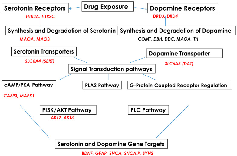
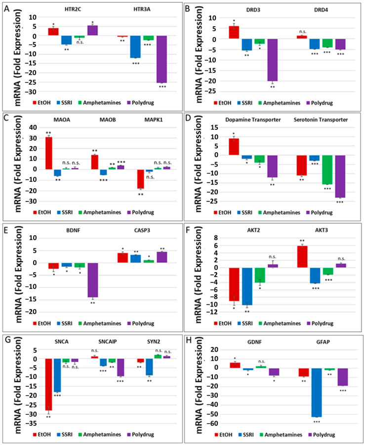
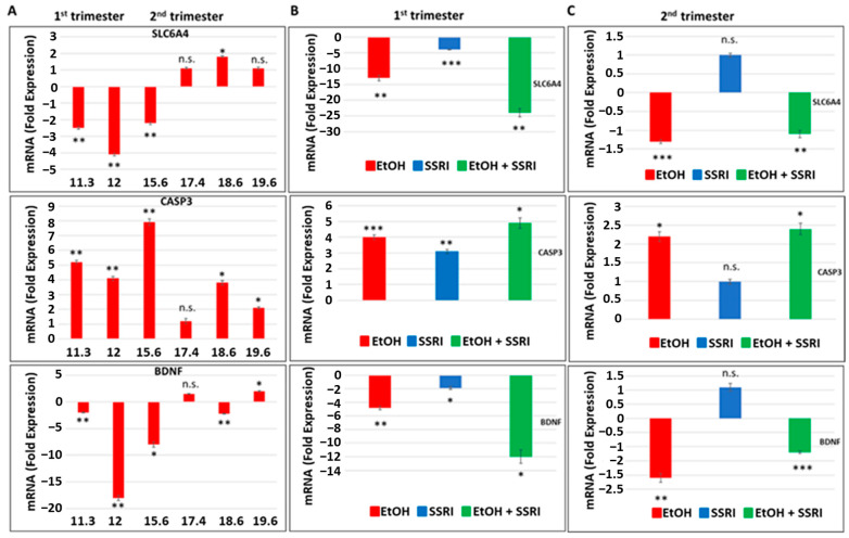
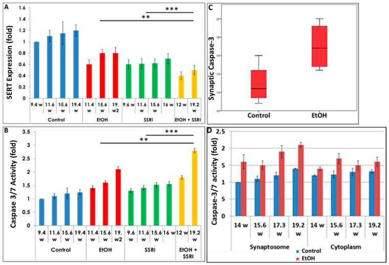
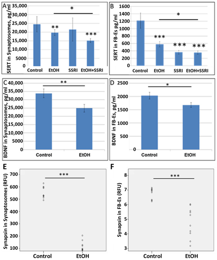
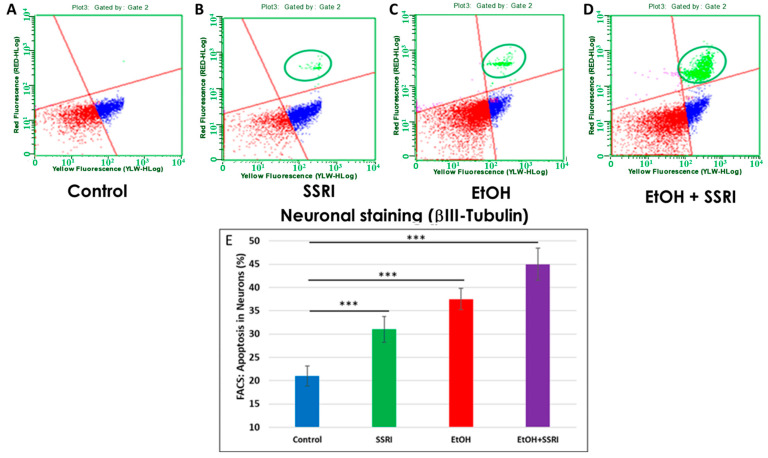
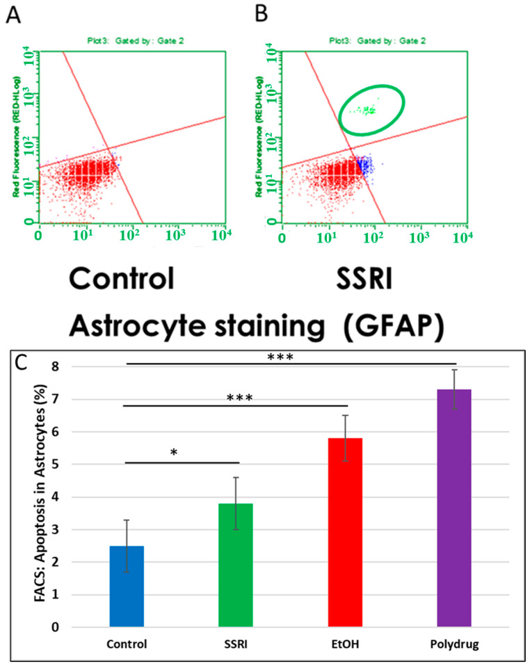
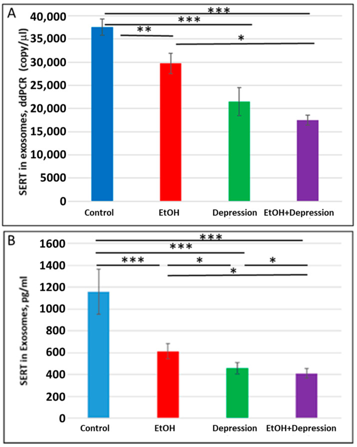
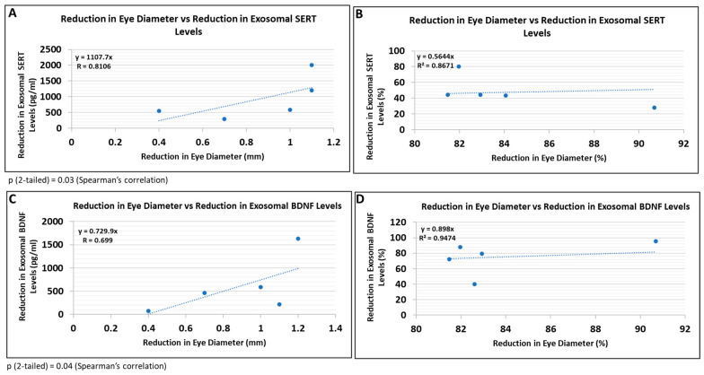
Results: Six EtOH-exposed human fetal brain samples were compared to SSRI- or polydrug-exposed samples and to unexposed controls. EtOH exposure was associated with significant upregulation of DA receptor D3 and 5-HT receptor HTR2C, while HTR3A was downregulated. Monoamine oxidase A (MAOA), MAOB, the serine/threonine kinase AKT3, and caspase-3 were upregulated, while mitogen-activated protein kinase 1 (MAPK1) and AKT2 were downregulated. ETOH was associated with significant upregulation of the DA transporter gene, while SERT was downregulated. There were significant correlations between EtOH exposure and (a) caspase-3 activation, (b) reduced SERT protein levels, and (c) reduced BDNF levels. SSRI exposure independently increased caspase-3 activity and downregulated SERT and BDNF. Early exposure to EtOH and SSRI together was associated synergistically with a significant upregulation of caspase-3 and a significant downregulation of SERT and BDNF. Reduced SERT and BDNF levels were strongly correlated with a reduction in eye diameter, a somatic manifestation of FASD.

Conclusions: Maternal use of EtOH and SSRI during pregnancy each was associated with changes in fetal brain monoamine pathways, consistent with potential mechanisms for the affective dysregulation associated with FASD.

Keywords: FAS; FASD; biomarkers; brain development; depression; dopamine receptors; exosomes; serotonin receptors.


Conflict of interest statement

The authors declare no competing financial interest.

Figures

Figure 1
Figure 1
Serotonin and dopamine pathways and upstream and downstream target genes in fetal brains, studied with RNA arrays.
Figure 2
Figure 2
Prenatal EtOH, SSRI, and polydrug exposures disrupt 5-HT and DA pathways in fetal brain. Brains from fetuses with exposure to EtOH (n = 6), SSRIs (n = 5), EtOH + SSRIs (n = 3) amphetamines (n = 2) or polydrugs (n = 6) were compared with unexposed controls (n = 12), as enumerated in Table 1. The brains were assayed for expression of molecules in the 5-HT and DA pathways by qRT-PCR on micro-arrays. Downregulation for most genes was greatest in the cases with EtOH and SSRI exposure (graphs show means from triplicate assays +/− SD). The p-values shown in this figure and in Figure 3 are only for the comparison between the indicated exposure group and controls. In several cases, there are significant differences between exposure groups, and these are not shown here to avoid clutter but are commented on in the text. * p < 0.05, ** p < 0.01, and *** p < 0.001; n.s.: not significant. Values are shown in fold change in expression, normalized to the housekeeping gene for actin. (A). RNA levels for the 5-HT receptor HTR3A were downregulated in EtOH- and SSRI-exposed fetal brains. (B). Maternal EtOH use increased mRNA for DA receptor DRD3 in fetal brain, while SSRI and polydrug use downregulated DRD3. (C). Effect of EtOH and poly drug use on MAOA, MAOB, and MAPK1 in 5-HT and DA pathways in fetal brain. (D). EtOH exposure increased DAT and reduced SERT mRNAs in fetal brain. All other drug exposures showed downregulation of both. (E). All drug exposures inhibited mRNA expression of BDNF, which is downstream of the 5-HT/DA/NE pathways, and upregulated caspase-3 in fetal brain. (F). EtOH exposure downregulated RNA expression of AKT2 and upregulated AKT3 in fetal brain. SSRIs and amphetamines downregulated both, while polydrug exposure showed no significant effects. (G). Downregulation of RNA expression for some synaptic genes in fetal brain by exposure to EtOH, SSRI, and amphetamines. (H). SSRI and polydrug exposure inhibited mRNA expression of GDNF and GFAP, which are downstream genes in the 5-HT and DA pathways.
Figure 3
Figure 3
Effects of early or late exposure to EtOH and SSRI on gene expression for the 5-HT transporter SLC6A4, BDNF, and caspase-3. RNAs from the brain of fetuses with early (1st trimester) or late (2nd trimester) exposure to EtOH, SSRIs, or both, and from non-exposed controls, were studied for SLC6A4 (the gene coding for SERT), caspase-3 and BDNF gene expression by qRT-PCR. The numbers of fetuses studied were as in Table 1: EtOH-exposed (n = 6), SSRI (n = 5), EtOH + SSRI (n = 3) and unexposed controls (n = 12). Values for qRT-PCR are shown in fold change of expression normalized to actin. Error bars are averaged values for the means of triplicate assays +/− SD. * p < 0.05, ** p < 0.01, and *** p < 0.001. n.s.: not significant. (A). Early EtOH exposure was associated with downregulation of SLC6A4 (top), upregulation of caspase-3 (middle), and downregulation of BDNF (bottom). Late exposure showed mainly up-regulation of caspase-3, while SLC6A4 and BDNF showed only inconsistent or small changes. (B). Synergistic effects of early EtOH and SSRI exposure on mRNA expression of SLC6A4 (top), caspase-3 (middle panel), and BDNF (bottom). Early exposure to EtOH or SSRIs induced downregulation of SLC6A4 and BDNF, and upregulation of caspase-3. In each case, the effects of EtOH + SSRIs were greater than those of either exposure alone. (C). Similarly, late exposure to EtOH was associated with downregulation of SLC6A4 (top) and BDNF (bottom) genes, while caspase-3 was upregulated (middle), but the changes were less pronounced than in the 1st trimester. Moreover, the effects of late exposure to SSRIs produced no significant effects on these three markers, and the combination of EtOH + SSRI was not significantly greater than either alone (n = 12 controls, n = 6 EtOH, n = 3 SSRI, and n = 3 EtOH + SSRI).
Figure 4
Figure 4
Effects of gestational age, EtOH, and SSRIs on fetal synaptosome SERT protein expression and on neuronal injury. Synaptic and cytoplasmic extracts were prepared and SERT levels were analyzed by qWestern blot assays using the anti-serotonin transporter antibody AB10514P (Millipore). Apoptosis was assessed in fetal brain tissue by analysis of caspase-3 activation, as described in Methods. (A). Exposure to either EtOH or SSRIs was associated with reduction in SERT levels in fetal brain synaptosomes. The effect of EtOH + SSRI was greater than that of either alone. (B). Activation of caspase-3 in the same cases. Exposure to either EtOH or SSRIs increased activated caspase-3 levels in fetal brain synaptosomes. The effect of EtOH + SSRI was significantly greater than that of either exposure alone. (C). EtOH exposure was associated with increased caspase-3 activation in fetal brain synaptosomes (n = 10 EtOH vs. 10 controls; p < 0.05). (D). In 4 EtOH-exposed fetal brains, caspase-3 activity was significantly increased (p < 0.05) in both synaptosomes and, to a lesser degree, in cytoplasm, compared to their GA- and sex-matched controls. Each bar represents the mean and SE of three determinations in one fetus. ** p < 0.01, and *** p < 0.001.
Figure 5
Figure 5
EtOH exposure inhibits expression of SERT and synaptic proteins in both synaptosomes and exosomes. (A). SERT and BDNF levels were quantified by ELISA and values expressed in pg/mL, after normalization to the exosomal marker protein CD81. EtOH and SSRI each reduced SERT levels (measured by ELISA; n = 6/group) in synaptosomes, and the effects were additive. (B). EtOH and SSRI each reduced SERT levels in FB-Es (ELISA n = 6/group), and the effect of combined EtOH + SSRI was about the same as that of SSRI alone. (C). Exposure to EtOH reduced BDNF levels in synaptosomes (ELISA; n = 10/group). (D). BDNF levels were reduced in FB-Es of EtOH-exposed cases (p < 0.05; n = 10/group). Graphs show means from triplicate assays +/− SD. (E). Exposure to EtOH reduced synapsin levels compared to controls (n = 10 cases/group) in synaptosomes and in exosomes (F), as assayed by quantitative Western blot, and shown in relative fluorescence units (RFU). * p < 0.05, ** p < 0.01, and *** p < 0.001.
Figure 6
Figure 6
Neuronal apoptosis in EtOH- and SSRI-exposed fetal brains. Neuronal viability in human fetal brain was assessed by FACS analysis, using multiple fluorescent dyes to sort cell types. Representative dot-plots of the flow cytometry to measure membrane changes associated with apoptosis, using Annexin V-PE and 7-AAD to identify dead cells, together with the neuronal marker βIII-tubulin. Three neuronal populations are identified with the Guava Nexin Reagent and analyzed with the Guava Nexin Software (5.3.1): The lower left quadrant represents the live cells 7-AAD and annexin-V-PE negative (in red); the lower right quadrant represents cells with early signs of injury or enhanced vulnerability (apoptotic cells 7-AAD negative and Annexin-V-PE positive; in blue). The upper right quadrant represents cells with signs of progressive cell injury and cell death (necrotic cells 7-AAD and annexin-V-PE positive; in green). (A). Representative images from flow cytometry analysis in 6 control fetal brains in the late 1st through mid-2nd trimester. (B). Same as (A) for 5 fetuses exposed to SSRI. (C). Same as (A) for 6 fetuses exposed to EtOH. (D). Same as (A) for 3 fetuses exposed to both EtOH + SSRI. Late injury and cell death were detected in SSRI-exposed neuronal cells, more so in EtOH-exposed fetuses, and most in a combined EtOH + SSRI exposure. (E). Apoptosis in neurons (% of neuronal cells expressing activated caspase-3). Graphs represent average data from flow cytometry measures in control brains (n = 6), SSRI (n = 5), EtOH (n = 6), and EtOH + SSRI (n = 3). *** for p < 0.001.
Figure 7
Figure 7
Increased apoptosis in astrocytes after SSRI exposure. GFAP was used as an astrocytic marker to assess glial viability by FACS analysis (n = 6 control and 5 SSRI-exposed fetal brains). Annexin V was used as a nexin assay for early apoptosis, and 7-AAD for late apoptosis. (A). No apoptotic cells were detected in control astrocytic cells (all cells in the left lower quadrant; red). (B). SSRI exposure increased apoptosis of astrocytic cells. As in Figure 6, the upper right quadrant represents cells with signs of progressive injury and death (green). The lower right quadrant represents cells with early signs of injury (blue). (C). Apoptosis (% of astrocytic cells expressing activated caspase-3). Graphs represent average data from flow cytometry measures in control brains (n = 6), SSRI (n = 5), EtOH (n = 6), and polydrug (n = 3). * p < 0.05, *** p < 0.001.
Figure 8
Figure 8
Maternal use of EtOH is associated with downregulation of SERT mRNA and protein in FB-Es. FB-Es isolated from the plasma of mothers who drank EtOH (n = 10) or did not drink EtOH (n = 10) and who did or did not suffer from depression (n = 10 each group) during pregnancy were isolated from maternal blood and assayed for SERT mRNA by ddPCR and for SERT protein by ELISA. For absolute quantitation of SERT mRNA in exosomes by ddPCR, values are shown in copies/μL. For quantitation of SERT protein by ELISA, values are shown in pg/mL (normalized to CD81). (A). EtOH and depression each were associated with downregulation of SERT mRNA levels in FB-Es. Downregulation was greatest in the cases with EtOH + depression, although the difference between the combined effect compared with that of depression alone was not statistically significant. (B). Downregulation of SERT protein levels in FB-Es by EtOH and depression. The same FB-E preparations were studied by ELISA for SERT protein levels. EtOH and depression each reduced SERT levels, and the effect of the combination of EtOH + depression was significantly stronger than that of EtOH exposure or depression alone. Graphs show means from triplicate assays +/− SD (n = 10 fetuses/group); all comparison differences were significant at * p < 0.05 and ** p < 0.01, *** p < 0.001, or less.
Figure 9
Figure 9
Reductions in FB-E SERT and BDNF levels correlate with reductions in eye diameter in fetuses exposed to EtOH + SSRIs. Eye diameters were measured in histological sections of human fetuses that had been exposed to both EtOH and SSRI. FB-E SERT and BDNF levels were measured by ELISA. Each of five EtOH + SSRI-exposed fetal eyes from 1st- and 2nd-trimester pregnancies was paired with a GA- and sex-matched unexposed control. Their matching maternal blood samples were obtained at the time of voluntary pregnancy termination. Assays were performed in triplicate on contents of FB-Es isolated from the maternal blood. Correlation between reduction in eye size (difference between EtOH- or SSRI-exposed fetus and its paired control) and reduction in exosomal SERT (A,B) and BDNF (C,D) levels is presented as scatter plots. Data in (B,D) are presented in %. Calculations are based on Spearman’s correlation on exact two-tailed probabilities critical and p-values for N > 2 <= 18.

Similar articles

Cited by

References

    1. Ornoy A., Ergaz Z. Alcohol abuse in pregnant women: Effects on the fetus and newborn, mode of action and maternal treatment. Int. J. Environ. Res. Public Health. 2010;7:364–379. doi: 10.3390/ijerph7020364. - DOI - PMC - PubMed
    1. Tan C.H., Denny C.H., Cheal N.E., Sniezek J.E., Kanny D. Alcohol use and binge drinking among women of childbearing age—United States, 2011–2013. MMWR Morb. Mortal. Wkly. Rep. 2015;64:1042–1046. doi: 10.15585/mmwr.mm6437a3. - DOI - PubMed
    1. Mutch R.C., Jones H.M., Bower C., Watkins R.E. Fetal Alcohol Spectrum Disorders: Using Knowledge, Attitudes and Practice of Justice Professionals to Support their Educational Needs. J. Popul. Ther. Clin. Pharmacol. 2016;23:e77–e89. - PubMed
    1. May P.A., Chambers C.D., Kalberg W.O., Zellner J., Feldman H., Buckley D., Kopald D., Hasken J.M., Xu R., Honerkamp-Smith G., et al. Prevalence of Fetal Alcohol Spectrum Disorders in 4 US Communities. JAMA. 2018;319:474–482. doi: 10.1001/jama.2017.21896. - DOI - PMC - PubMed
    1. Sampson P.D., Streissguth A.P., Bookstein F.L., Little R.E., Clarren S.K., Dehaene P., Hanson J.W., Graham J.M., Jr. Incidence of fetal alcohol syndrome and prevalence of alcohol-related neurodevelopmental disorder. Teratology. 1997;56:317–326. doi: 10.1002/(SICI)1096-9926(199711)56:5<317::AID-TERA5>3.0.CO;2-U. - DOI - PubMed

Publication types