

A Microfluidics Approach for Ovarian Cancer Immune Monitoring in an Outpatient Setting
- PMID: 38201211
- PMCID: PMC10778191
- DOI: 10.3390/cells13010007
A Microfluidics Approach for Ovarian Cancer Immune Monitoring in an Outpatient Setting
Abstract
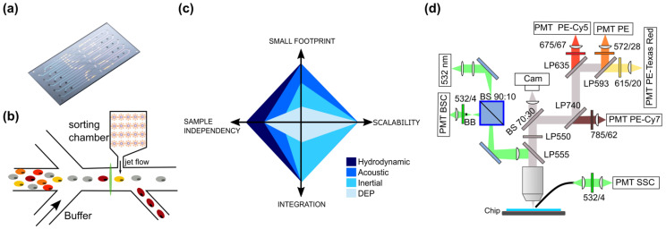
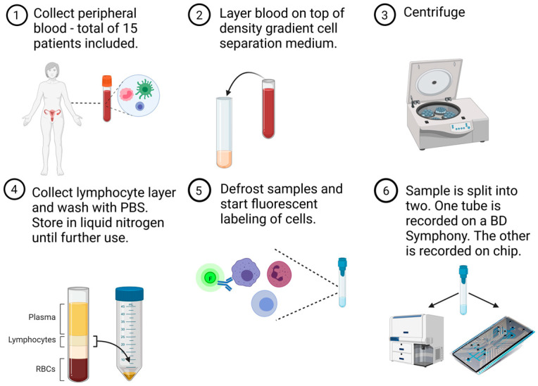
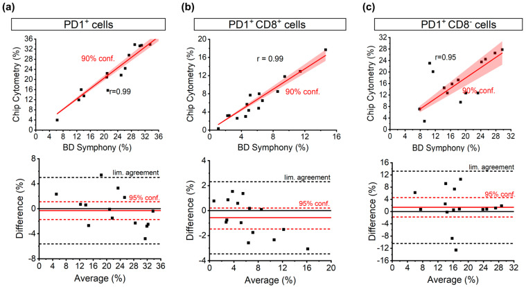
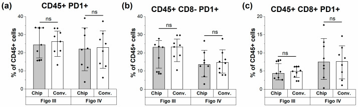
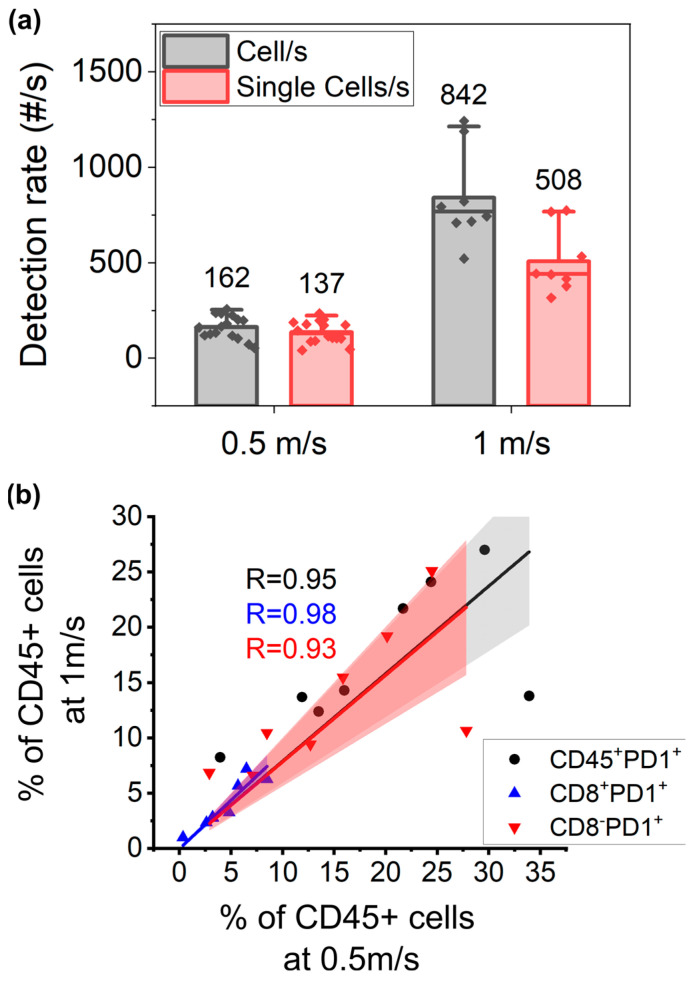
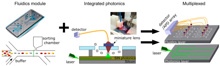
Among cancer diagnoses in women, ovarian cancer has the fifth-highest mortality rate. Current treatments are unsatisfactory, and new therapies are highly needed. Immunotherapies show great promise but have not reached their full potential in ovarian cancer patients. Implementation of an immune readout could offer better guidance and development of immunotherapies. However, immune profiling is often performed using a flow cytometer, which is bulky, complex, and expensive. This equipment is centralized and operated by highly trained personnel, making it cumbersome and time-consuming. We aim to develop a disposable microfluidic chip capable of performing an immune readout with the sensitivity needed to guide diagnostic decision making as close as possible to the patient. As a proof of concept of the fluidics module of this concept, acquisition of a limited immune panel based on CD45, CD8, programmed cell death protein 1 (PD1), and a live/dead marker was compared to a conventional flow cytometer (BD FACSymphony). Based on a dataset of peripheral blood mononuclear cells of 15 patients with ovarian cancer across different stages of treatment, we obtained a 99% correlation coefficient for the detection of CD8+PD1+ T cells relative to the total amount of CD45+ white blood cells. Upon further system development comprising further miniaturization of optics, this microfluidics chip could enable immune monitoring in an outpatient setting, facilitating rapid acquisition of data without the need for highly trained staff.
Keywords: flow cytometry; immune monitoring; immunotherapy; lab on chip; ovarian cancer.
Conflict of interest statement
There are no conflicts of interest to report regarding this study. However, for full disclosure, we report that A.C. is a contracted researcher for Oncoinvent AS and Novocure and a consultant for Sotio a.s. and Epics Therapeutics SA. T.B. has received travel fees from MSD and Tesaro/GSK, is chair holder of an endowed chair from Roche, and is a consultant for Tesaro/GSK. T.V.G. has received honoraria for advisory boards from Eisai (Inst), OncXerna Therapeutics (Inst), AstraZeneca (Inst), GSK (Inst), MSD (Inst), Seagen (Inst), Tubulis (Inst), and ImmunoGen (Inst); research funding from Amgen (Inst), Roche (Inst), and AstraZeneca (Inst); and expense reimbursements from MSD, Immunogen, PharmaMar, and AstraZeneca.
Figures
Similar articles
-
Handheld Microflow Cytometer Based on a Motorized Smart Pipette, a Microfluidic Cell Concentrator, and a Miniaturized Fluorescence Microscope.Sensors (Basel). 2019 Jun 19;19(12):2761. doi: 10.3390/s19122761. Sensors (Basel). 2019. PMID: 31248214 Free PMC article.
-
A multiplexable, microfluidic platform for the rapid quantitation of a biomarker panel for early ovarian cancer detection at the point-of-care.Cancer Prev Res (Phila). 2015 Jan;8(1):37-48. doi: 10.1158/1940-6207.CAPR-14-0248. Epub 2014 Nov 11. Cancer Prev Res (Phila). 2015. PMID: 25388014 Free PMC article.
-
Pembrolizumab with low-dose carboplatin for recurrent platinum-resistant ovarian, fallopian tube, and primary peritoneal cancer: survival and immune correlates.J Immunother Cancer. 2021 Sep;9(9):e003122. doi: 10.1136/jitc-2021-003122. J Immunother Cancer. 2021. PMID: 34531249 Free PMC article.
-
New Approaches for Immune Directed Treatment for Ovarian Cancer.Curr Treat Options Oncol. 2016 Mar;17(3):14. doi: 10.1007/s11864-016-0389-1. Curr Treat Options Oncol. 2016. PMID: 26942589 Review.
-
Microfluidics and materials for smart water monitoring: A review.Anal Chim Acta. 2021 Nov 22;1186:338392. doi: 10.1016/j.aca.2021.338392. Epub 2021 Mar 13. Anal Chim Acta. 2021. PMID: 34756264 Review.
References
-
- Globocan. [(accessed on 26 January 2023)]. Available online: https://gco.iarc.fr/survival/survmark/visualizations/viz1/?groupby=%22ca....
-
- SEER Cancer Statistics. [(accessed on 26 January 2023)]; Available online: https://seer.cancer.gov/statfacts/html/ovary.html.
-
- Schadendorf D., Hodi F.S., Robert C., Weber J.S., Margolin K., Hamid O., Patt D., Chen T.T., Berman D.M., Wolchok J.D. Pooled Analysis of Long-Term Survival Data from Phase II and Phase III Trials of Ipilimumab in Unresectable or Metastatic Melanoma. J. Clin. Oncol. 2015;33:1889–1894. doi: 10.1200/JCO.2014.56.2736. - DOI - PMC - PubMed
Publication types
MeSH terms
Grants and funding
LinkOut - more resources
Full Text Sources
Medical
Research Materials
Miscellaneous
