. 2023 Dec 20;13(1):9.
doi: 10.3390/cells13010009.

WAVE2 Is a Vital Regulator in Myogenic Differentiation of Progenitor Cells through the Mechanosensitive MRTFA-SRF Axis

Affiliations

WAVE2 Is a Vital Regulator in Myogenic Differentiation of Progenitor Cells through the Mechanosensitive MRTFA-SRF Axis

Mai Thi Nguyen et al. Cells. .

Abstract

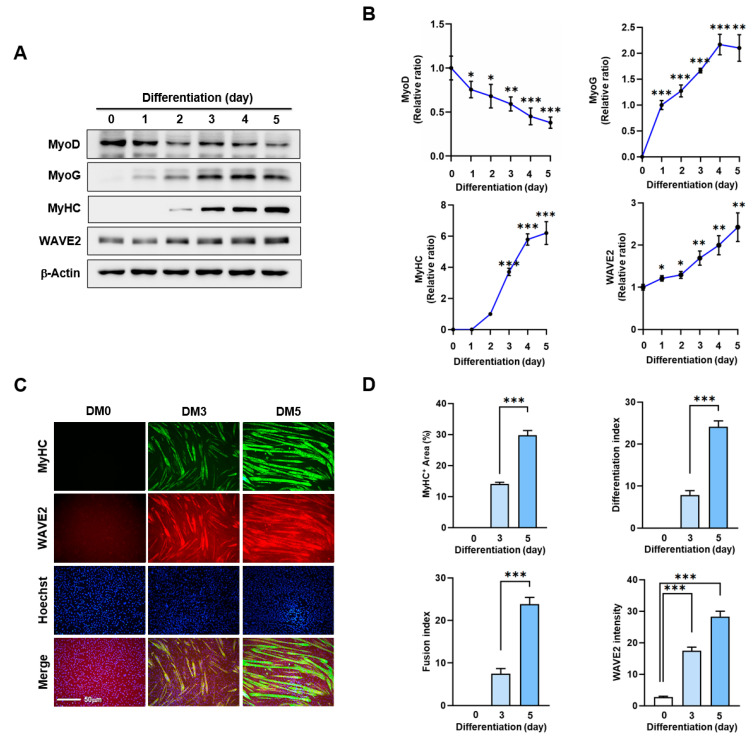
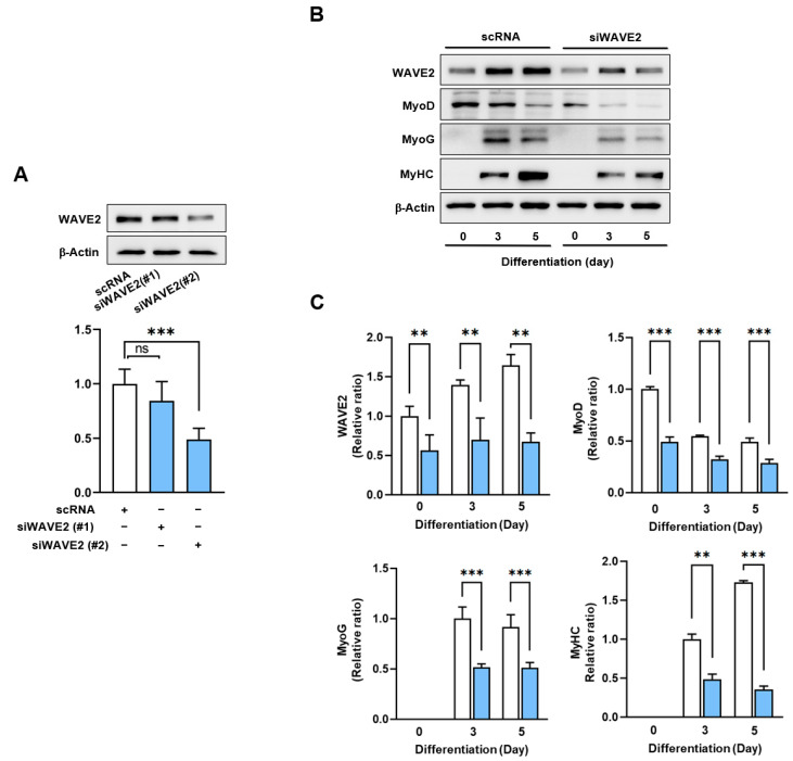
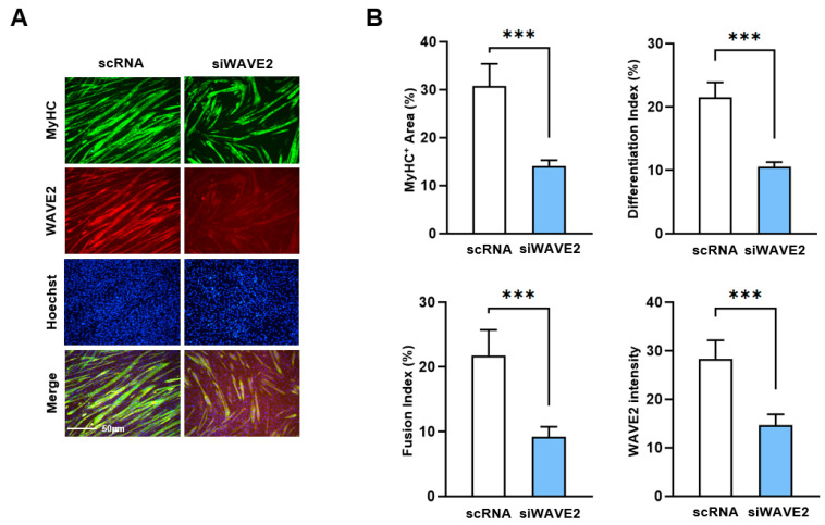
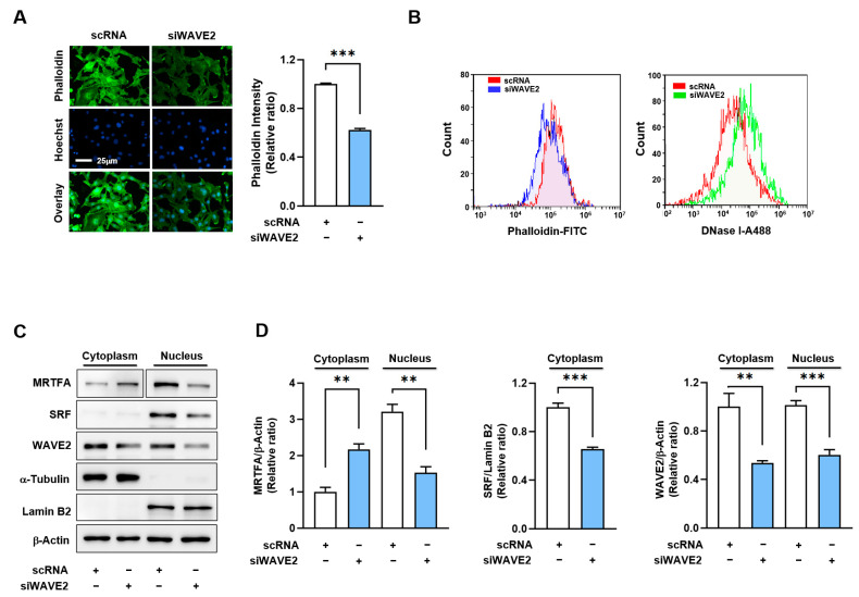
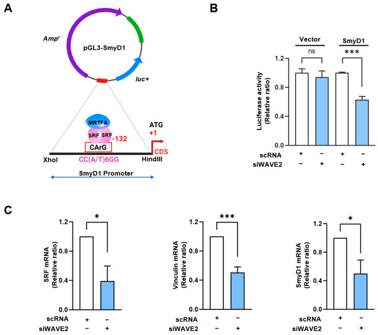
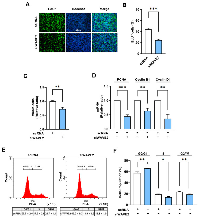
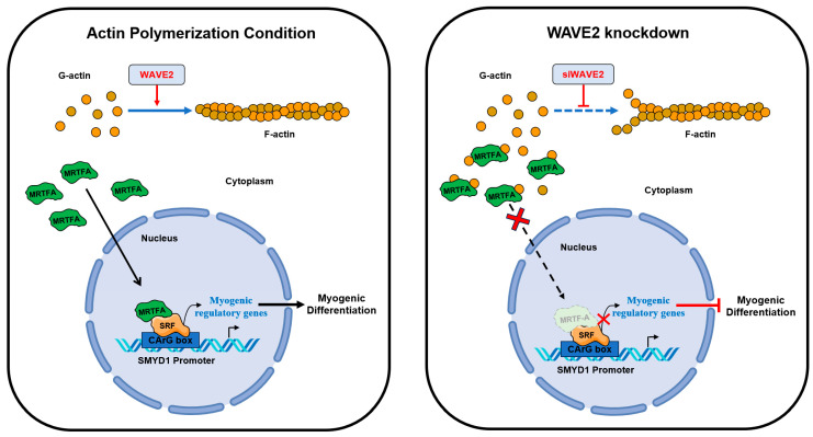
Skeletal myogenesis is an intricate process involving the differentiation of progenitor cells into myofibers, which is regulated by actin cytoskeletal dynamics and myogenic transcription factors. Although recent studies have demonstrated the pivotal roles of actin-binding proteins (ABPs) as mechanosensors and signal transducers, the biological significance of WAVE2 (Wiskott-Aldrich syndrome protein family member 2), an ABP essential for actin polymerization, in myogenic differentiation of progenitor cells has not been investigated. Our study provides important insights into the regulatory roles played by WAVE2 in the myocardin-related transcription factor A (MRTFA)-serum response factor (SRF) signaling axis and differentiation of myoblasts. We demonstrate that WAVE2 expression is induced during myogenic differentiation and plays a pivotal role in actin cytoskeletal remodeling in C2C12 myoblasts. Knockdown of WAVE2 in C2C12 cells reduced filamentous actin levels, increased globular actin accumulation, and impaired the nuclear translocation of MRTFA. Furthermore, WAVE2 depletion in myoblasts inhibited the expression and transcriptional activity of SRF and suppressed cell proliferation in myoblasts. Consequently, WAVE2 knockdown suppressed myogenic regulatory factors (i.e., MyoD, MyoG, and SMYD1) expressions, thereby hindering the differentiation of myoblasts. Thus, this study suggests that WAVE2 is essential for myogenic differentiation of progenitor cells by modulating the mechanosensitive MRTFA-SRF axis.

Keywords: MRTFA; SRF; WAVE2; differentiation; myogenesis; proliferation.


Conflict of interest statement

The authors declare no conflict of interest.

Figures

Figure 1
Figure 1
WAVE2 expression is induced during myoblast differentiation. C2C12 cells were allowed to differentiate for up to five days. (A) MyoD, MyoG, MyHC, and WAVE2 expressions were determined by immunoblotting. (B) Protein levels in (A) were measured using densitometry and normalized against β-Actin. Results are shown as relative ratios, with the degree of differentiation on day 0 (MyoD, WAVE2, and MyoG) or day 2 (MyHC) set to one. (C) Cells were subjected to immunostaining with anti-MyHC and anti-WAVE2 antibodies, followed by Alexa 488 antibody (green) and Hoechst 34,452 (blue) nuclear counterstain after three and five days of differentiation. Scale bar: 50 μm. (D) Myotubes are quantified using MyHC-positive cells, WAVE2 intensities, differentiation and fusion indexes, and myotube widths. Results are presented as means ± standard errors (n > 3). Significance levels are represented by *, p < 0.05; **, p < 0.01; and ***, p < 0.001.
Figure 2
Figure 2
Depletion of WAVE2 suppresses myogenic regulatory factors. (A) WAVE2 in C2C12 cells was determined by immunoblotting 24 h after transfection with 200 nM of scRNA control, siWAVE2-1 or -2. (B) C2C12 myoblasts were differentiated for three or five days after transfecting with scRNA or siWAVE2 (200 nM). WAVE2, MyHC, MyoD, and MyoG expressions were determined by immunoblotting. (C) Protein levels in (B) were measured using densitometry and normalized against β-Actin. Results are shown as relative ratios, with the degree of differentiation on day 0 (WAVE2 and MyoD) or day 3 (MyoG and MyHC) set to one. Results are presented as means ± standard errors (n > 3). Statistical significance is indicated by **, p < 0.01; ***, p < 0.001 vs. scRNA. ns: not significant.
Figure 3
Figure 3
Depletion of WAVE2 inhibits differentiation and fusion of myoblasts. C2C12 myoblasts were differentiated after transfecting with scRNA or siWAVE2 (200 nM). (A) After five days of differentiation, cells were subjected to immunostaining with anti-MyHC and anti-WAVE2 antibodies, and Hoechst 34,452 (blue) was used for nucleus staining. Scale bar: 50 μm. (B) Myotubes are quantified using MyHC-positive cells, WAVE2 intensities, differentiation and fusion indexes, and myotube widths. Results are presented as means ± standard errors (n > 3). Statistical significance is indicated by ***, p < 0.001 vs. scRNA.
Figure 4
Figure 4
WAVE2 knockdown reduces F-actin and nuclear MRTFA levels. (A) Representative images showing FITC-phalloidin staining (green) for F-actin and Hoechst 34,452 (blue) for the nucleus staining. Scale bar: 25 μm. (B) F- and G-actin levels in cells stained with FITC-phalloidin and DNase I, respectively, were determined by flow cytometry. (C,D) Immunoblots showed MRTFA, SRF, and WAVE2 protein expressions in cytoplasmic and nuclear fractions of C2C12 cells after transfection with scRNA or siWAVE2. For MRTFA detection, different exposure times were required because it is distributed differently in the cytoplasm and nucleus. α-Tubulin and Lamin B2 were used as markers for cytoplasm and nucleus, respectively. Immunoblot intensities are presented as relative ratios and normalized against β-Actin or Lamin B2. Results are presented as means ± standard errors (n > 3), and significance levels are represented as **, p < 0.01; ***, p < 0.001 vs. scRNA.
Figure 5
Figure 5
WAVE2 knockdown inhibits the transcriptional activity of SRF. (A) Luciferase reporter assays of truncated SMYD1 promoter constructs, including the CArG box sequence for immediate early sites upstream of the SMYD1 gene. (B) Luciferase activity was determined 24 h after transfection with 200 nM of scRNA or siWAVE2 and normalized against pRLSV40 activity. (C) Relative expression levels of SRF, Vinculin, and SMYD1 transcripts in C2C12 cells were analyzed 24 h after transfection by qRT-PCR, normalized against U6. Results are presented as means ± standard errors (n > 3), and significance levels are represented as *, p < 0.05; ***, p < 0.001 vs. scRNA. ns: not significant.
Figure 6
Figure 6
Depletion of WAVE2 negatively regulates cell proliferation. C2C12 cells were transfected with scRNA or siWAVE2 (200 nM), and the following analyses were performed 24 h after transfection. (A) EdU (green) was used for cell proliferation analysis to label cells undergoing DNA replication, and Hoechst 33,342 (blue) was applied to nuclear staining. Scale bar: 50 μm. (B) Proportions of EdU-positive cells were determined using ImageJ software. (C) Cell viability analysis. (D) The relative levels of PCNA, cyclin B1, and cyclin D1 in C2C12 cells were determined by qRT-PCR and normalized against U6. (E,F) Cell cycle analysis using flow cytometry. qRT-PCR and immunoblot intensities are presented as relative ratios against scRNA controls. Results are presented as means ± standard errors (n > 3). Significance levels are represented as *, p < 0.05; **, p < 0.01; ***, p < 0.001 vs. scRNA.
Figure 7
Figure 7
Roles of WAVE2 in myogenic differentiation.

Similar articles

Cited by

References

    1. Frontera W.R., Ochala J. Skeletal muscle: A brief review of structure and function. Calcif. Tissue Int. 2015;96:183–195. doi: 10.1007/s00223-014-9915-y. - DOI - PubMed
    1. Baskin K.K., Winders B.R., Olson E.N. Muscle as a “mediator” of systemic metabolism. Cell Metab. 2015;21:237–248. doi: 10.1016/j.cmet.2014.12.021. - DOI - PMC - PubMed
    1. Chal J., Pourquie O. Making muscle: Skeletal myogenesis in vivo and in vitro. Development. 2017;144:2104–2122. doi: 10.1242/dev.151035. - DOI - PubMed
    1. Sartori R., Romanello V., Sandri M. Mechanisms of muscle atrophy and hypertrophy: Implications in health and disease. Nat. Commun. 2021;12:330. doi: 10.1038/s41467-020-20123-1. - DOI - PMC - PubMed
    1. Benarroch L., Bonne G., Rivier F., Hamroun D. The 2020 version of the gene table of neuromuscular disorders (nuclear genome) Neuromuscul. Disord. 2019;29:980–1018. doi: 10.1016/j.nmd.2019.10.010. - DOI - PubMed

Publication types

LinkOut - more resources