

Microfibre-Functionalised Silk Hydrogels
- PMID: 38201214
- PMCID: PMC10777932
- DOI: 10.3390/cells13010010
Microfibre-Functionalised Silk Hydrogels
Abstract
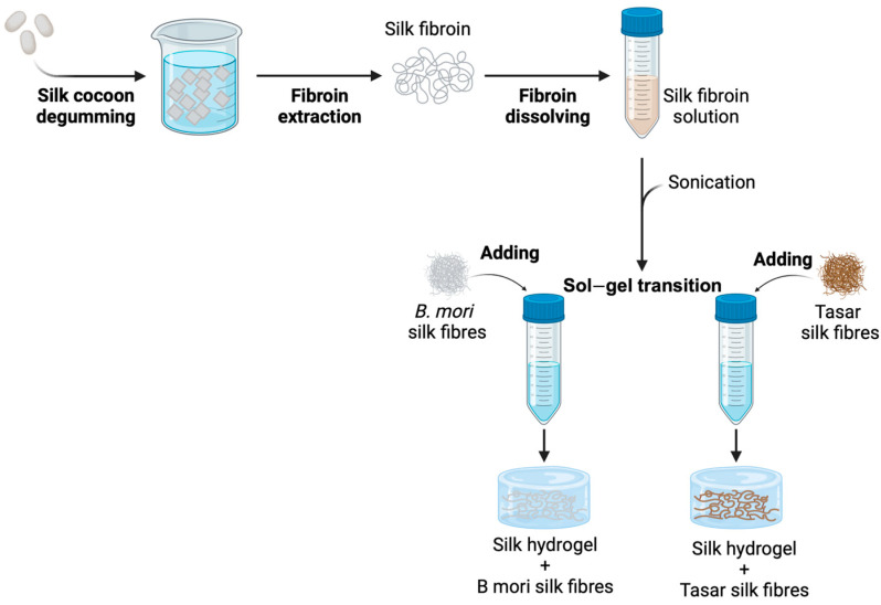
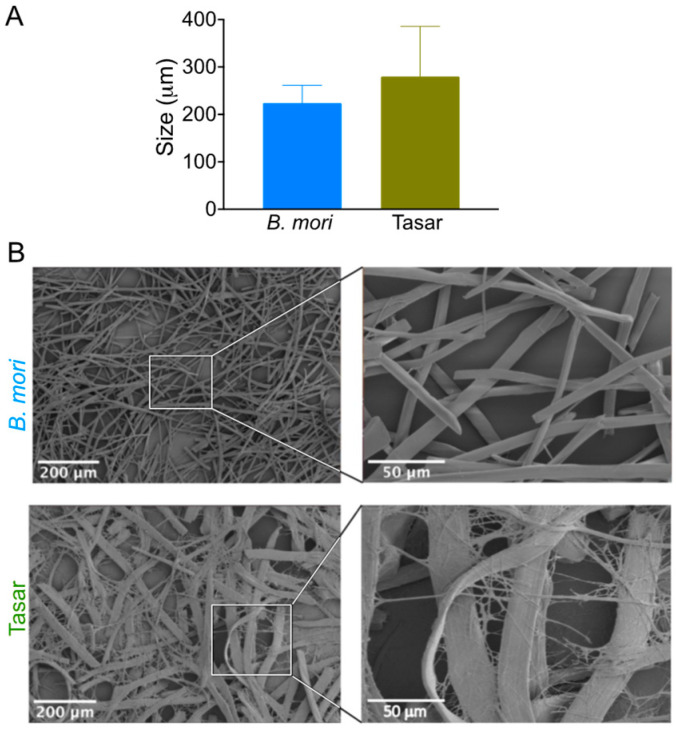
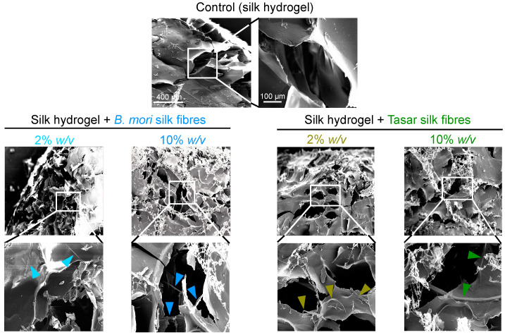
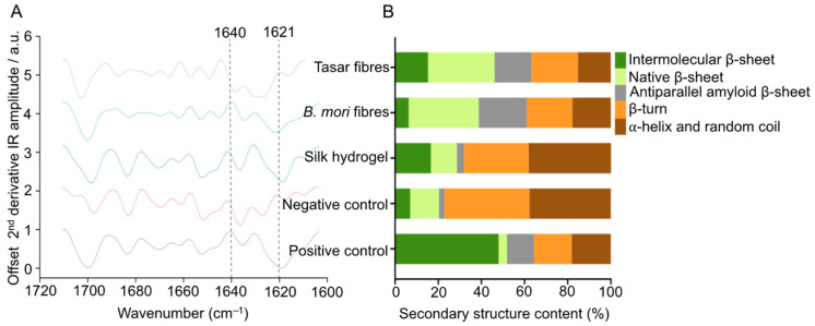
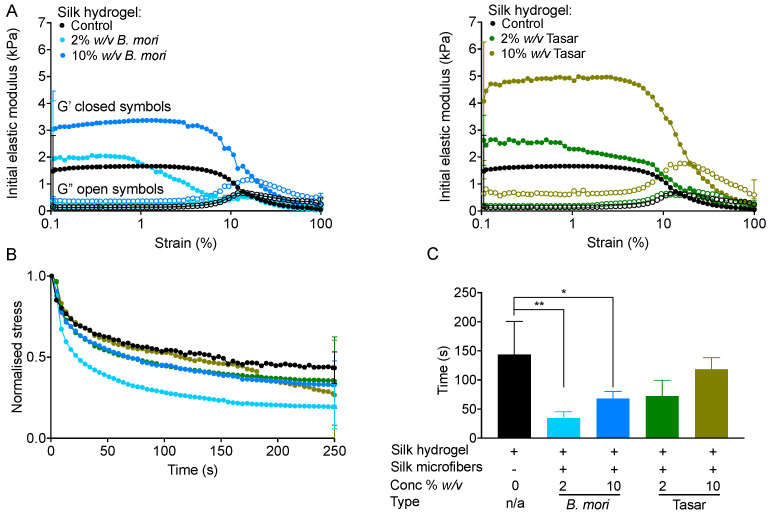
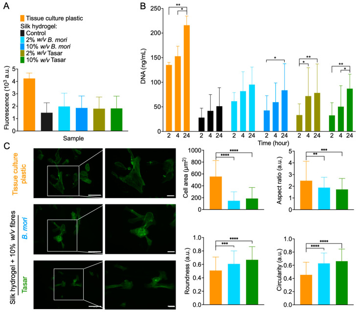
Silk hydrogels have shown potential for tissue engineering applications, but several gaps and challenges, such as a restricted ability to form hydrogels with tuned mechanics and structural features, still limit their utilisation. Here, Bombyx mori and Antheraea mylitta (Tasar) silk microfibres were embedded within self-assembling B. mori silk hydrogels to modify the bulk hydrogel mechanical properties. This approach is particularly attractive because it creates structured silk hydrogels. First, B. mori and Tasar microfibres were prepared with lengths between 250 and 500 μm. Secondary structure analyses showed high beta-sheet contents of 61% and 63% for B. mori and Tasar microfibres, respectively. Mixing either microfibre type, at either 2% or 10% (w/v) concentrations, into 3% (w/v) silk solutions during the solution-gel transition increased the initial stiffness of the resulting silk hydrogels, with the 10% (w/v) addition giving a greater increase. Microfibre addition also altered hydrogel stress relaxation, with the fastest stress relaxation observed with a rank order of 2% (w/v) > 10% (w/v) > unmodified hydrogels for either fibre type, although B. mori fibres showed a greater effect. The resulting data sets are interesting because they suggest that the presence of microfibres provided potential 'flow points' within these hydrogels. Assessment of the biological responses by monitoring cell attachment onto these two-dimensional hydrogel substrates revealed greater numbers of human induced pluripotent stem cell-derived mesenchymal stem cells (iPSC-MSCs) attached to the hydrogels containing 10% (w/v) B. mori microfibres as well as 2% (w/v) and 10% (w/v) Tasar microfibres at 24 h after seeding. Cytoskeleton staining revealed a more elongated and stretched morphology for the cells growing on hydrogels containing Tasar microfibres. Overall, these findings illustrate that hydrogel stiffness, stress relaxation and the iPSC-MSC responses towards silk hydrogels can be tuned using microfibres.
Keywords: fibre; gel; iPSC; mechanics; silk fibroin; stem cells; tissue engineering.
Conflict of interest statement
The authors declare no conflicts of interest.
Figures
Similar articles
-
Functionalising silk hydrogels with hetero- and homotypic nanoparticles.RSC Adv. 2024 Jan 22;14(5):3525-3535. doi: 10.1039/d3ra07634b. eCollection 2024 Jan 17. RSC Adv. 2024. PMID: 38259992 Free PMC article.
-
Silk fibroin/collagen protein hybrid cell-encapsulating hydrogels with tunable gelation and improved physical and biological properties.Acta Biomater. 2018 Mar 15;69:218-233. doi: 10.1016/j.actbio.2017.12.026. Epub 2018 Feb 2. Acta Biomater. 2018. PMID: 29410166
-
Silk Hydrogel Substrate Stress Relaxation Primes Mesenchymal Stem Cell Behavior in 2D.ACS Appl Mater Interfaces. 2021 Jul 7;13(26):30420-30433. doi: 10.1021/acsami.1c09071. Epub 2021 Jun 25. ACS Appl Mater Interfaces. 2021. PMID: 34170674 Free PMC article.
-
Nonmulberry silk proteins: multipurpose ingredient in bio-functional assembly.Biomed Mater. 2021 Sep 22;16(6). doi: 10.1088/1748-605X/ac20a0. Biomed Mater. 2021. PMID: 34428758 Review.
-
Reverse-engineered silk hydrogels for cell and drug delivery.Ther Deliv. 2018 May 1;9(6):469-487. doi: 10.4155/tde-2018-0016. Ther Deliv. 2018. PMID: 29722634 Review.
References
-
- Chen S., Guo Y., Liu R., Wu S., Fang J., Huang B., Li Z., Chen Z., Chen Z. Tuning surface properties of bone biomaterials to manipulate osteoblastic cell adhesion and the signaling pathways for the enhancement of early osseointegration. Colloids Surf. B Biointerfaces. 2018;164:58–69. doi: 10.1016/j.colsurfb.2018.01.022. - DOI - PubMed
-
- Tian Y., Liu H., Sheldon B.W., Webster T.J., Yang S., Yang H., Yang L. Surface energy-mediated fibronectin adsorption and osteoblast responses on nanostructured diamond. J. Mater. Sci. Technol. 2019;35:817–823. doi: 10.1016/j.jmst.2018.11.009. - DOI
Publication types
MeSH terms
Substances
LinkOut - more resources
Full Text Sources
