. 2023 Dec 20;13(1):16.
doi: 10.3390/cells13010016.

Absent in Melanoma (AIM)2 Promotes the Outcome of Islet Transplantation by Repressing Ischemia-Induced Interferon (IFN) Signaling

Affiliations

Absent in Melanoma (AIM)2 Promotes the Outcome of Islet Transplantation by Repressing Ischemia-Induced Interferon (IFN) Signaling

Selina Wrublewsky et al. Cells. .

Abstract

Clinical islet transplantation is limited by ischemia-induced islet cell death. Recently, it has been reported that the absent in melanoma (AIM)2 inflammasome is upregulated by ischemic cell death due to recognition of aberrant cytoplasmic self-dsDNA. However, it is unknown whether AIM2 determines the outcome of islet transplantation. To investigate this, isolated wild type (WT) and AIM2-deficient (AIM2-/-) islets were exposed to oxygen-glucose deprivation to mimic ischemia, and their viability, endocrine function, and interferon (IFN) signaling were assessed. Moreover, the revascularization and endocrine function of grafted WT and AIM2-/- islets were analyzed in the mouse dorsal skinfold chamber model and the diabetic kidney capsule model. Ischemic WT and AIM2-/- islets did not differ in their viability. However, AIM2-/- islets exhibited a higher protein level of p202, a transcriptional regulator of IFN-β and IFN-γ gene expression. Accordingly, these cytokines were upregulated in AIM2-/- islets, resulting in a suppressed gene expression and secretion of insulin. Moreover, the revascularization of AIM2-/- islet grafts was deteriorated when compared to WT controls. Furthermore, transplantation of AIM2-/- islets in diabetic mice failed to restore physiological blood glucose levels. These findings indicate that AIM2 crucially determines the engraftment and endocrine function of transplanted islets by repressing IFN signaling.

Keywords: AIM2; inflammasome; interferon; islet transplantation; revascularization.


Conflict of interest statement

The authors declare no conflicts of interest.

Figures

Figure 1
Figure 1
AIM2 deficiency does not affect the cellular composition of isolated islets under normoxia. (a) Double stainings of insulin/glucagon, insulin/somatostatin, and insulin/CD31 in isolated WT and AIM2−/− islets. Hoechst 33342 (blue) was used to stain cell nuclei. Scale bar: 50 µm. (b) Insulin- (β-cells), glucagon- (α-cells), somatostatin- (δ-cells), and CD31- (endothelial) positive cells (in % of all islet cells) in isolated WT and AIM2−/− islets were quantitatively analyzed (n = 20 each). Mean ± SEM. (c) Stainings of MPO-, CD68- and CD3-positive cells in isolated WT and AIM2−/− islets. Scale bar: 50 µm. (d) MPO-, CD68- and CD3-positive cells (in % of all islet cells) in isolated WT and AIM2−/− islets were quantitatively analyzed (n = 20 each). Mean ± SEM.
Figure 2
Figure 2
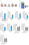
AIM2 deficiency reduces insulin expression in an IFN-dependent manner. (a) Schematic illustration of the experimental in vitro setting. Freshly isolated islets (left: bright field microscopic image; scale bar: 75 µm) were exposed to hypoxia (5% O2) and low glucose (LG) medium (1 g/L glucose) for 48 h to induce central cell death within the islets (right: bright field microscopic image; scale bar: 75 µm). These ischemic islets were used for further experiments. (b) Propidium iodide/annexin V-stained cells (in % of total cell number) were quantitatively analyzed in ischemic WT and AIM2−/− islets subdivided in necrotic, necroptotic, apoptotic, and vital cells (n = 3 each). (c) Representative Western blot of p202 expression from extracts of ischemic WT and AIM2−/− islets (lower panels). Quantitative analysis of p202 expression (upper panel). β-actin was used as loading control. Data are expressed in % of WT (n = 3 each). Mean ± SEM. * p < 0.05 vs. WT. (d,e) IFN-β (d) and IFN-γ (e) mRNA expression in ischemic WT and AIM2−/− islets were quantitatively analyzed. Data are expressed in % of WT (n = 3 each). Mean ± SEM. * p < 0.05 vs. WT. (f) Quantitative analysis of insulin secretion (µU/mL) from ischemic WT and AIM2−/− islets (n = 8 each). Mean ± SEM. * p < 0.05 vs. WT. (g,h) Ins1 (g) and Ins2 (h) mRNA expression in ischemic WT and AIM2−/− islets were quantitatively analyzed. Data are expressed in % of WT (n = 3 each). Mean ± SEM. * p < 0.05 vs. WT. (i) Quantitative analysis of insulin secretion (µU/mL) from ischemic WT islets exposed to vehicle, IFN-β, or IFN-γ (n = 8 each). Mean ± SEM. * p < 0.05 vs. WT + Vehicle; + p < 0.05 vs. WT + IFN-β. (j,k) Quantitative analysis of Ins1 (j) and Ins2 (k) mRNA expression in ischemic WT islets exposed to vehicle, IFN-β, or IFN-γ. Data are expressed in % of WT + Vehicle (n = 3 each). Mean ± SEM. * p < 0.05 vs. WT + Vehicle.
Figure 3
Figure 3
AIM2 deficiency impairs the graft revascularization. (a) Experimental in vivo setting. On day -2, we implanted the dorsal skinfold chambers. WT and AIM2−/− islets were transplanted on day 0. Intravital fluorescence microscopy was performed on day 0, 3, 6, 10, and 14 after islet transplantation. (b) Take rate of WT and AIM2−/− islets (% of transplanted islets; n = 8 each) on day 14. Mean ± SEM. * p < 0.05 vs. WT. (c) Intravital fluorescent microscopic images of transplanted WT and AIM2−/− islets within the dorsal skinfold chamber. Blood-perfused microvessels are stained with FITC-labeled dextran. The border of the grafts is marked by broken lines. Scale bar: 100 μm. (d) The revascularized area (mm2) of WT and AIM2−/− islets was quantitatively analyzed in (n = 8 each). Mean ± SEM. * p < 0.05 vs. WT. (e) The functional microvessel density (cm/cm2) of WT and AIM2−/− islets was quantitatively analyzed in (n = 8 each). Mean ± SEM. * p < 0.05 vs. WT. (f) Intravital fluorescent microscopic images of transplanted WT and AIM2−/− islets within the dorsal skinfold chamber. We used Rhodamine 6G to stain the perfusion of the endocrine tissue (bright signals). Broken lines mark the border of the transplants. Scale bar: 100 µm. (g) Quantitative analysis of the rhodamine 6G-positive area (% of islet size) within transplanted WT and AIM2−/− islets (n = 8 each). Mean ± SEM. * p < 0.05 vs. WT. (h) Tube formation assays were performed with HUVEC exposed to vehicle, IFN-β, or IFN-γ. The formation of vessel-like structures was analyzed 5 h and 7 h after seeding. Scale bar: 750 µm. (i) Quantitative analysis of the number of tube meshes (% of vehicle) after 5 h and 7 h (n = 3 each). Mean ± SEM. * p < 0.05 vs. Vehicle; + p < 0.05 vs. IFN-β.
Figure 4
Figure 4
AIM2 deficiency in transplanted islets impairs restoration of normoglycemia in diabetic mice. (a) Experimental in vivo setting. Eight days prior to islet transplantation, we induced a diabetic phenotype by STZ injection. On day 0, 400 islets were transplanted under the kidney capsule of diabetic recipient mice. Subsequently, we measured body weights and blood glucose levels twice a week. On day 28, we performed an IPGTT and excised the kidneys to determine the insulin content of the transplants. (b) Blood glucose levels (mg/mL) of diabetic recipient mice transplanted with WT and AIM2−/− islets from day -8 to day 28 (n = 6 each). Nondiabetic animals were used as negative control (n = 6 each). Mean ± SEM. * p < 0.05 vs. WT; + p < 0.05 vs. AIM2−/−. (c) AUC of the blood glucose levels from (b) (n = 6 each). Mean ± SEM. * p < 0.05 vs. WT; + p < 0.05 vs. AIM2−/−. (d) Quantitative analysis of blood glucose levels (mg/dL) on day 28 according to the IPGTT of diabetic recipient mice transplanted with WT and AIM2−/− islets (n = 6 each). Nondiabetic animals were used as negative control (n = 6 each). Mean ± SEM. * p < 0.05 vs. WT; + p < 0.05 vs. AIM2−/−. (e) AUC of IPGTT from (d) (n = 6 each). Mean ± SEM. * p < 0.05 vs. WT; + p < 0.05 vs. AIM2−/−. (f) The fraction of animals achieving physiological blood glucose levels (n = 6 each). (g) Insulin content (µU/mL) of the removed grafts from diabetic mice transplanted with WT and AIM2−/− islets (n = 6 each). Mean ± SEM. * p < 0.05 vs. WT.

Similar articles

References

    1. Kobayashi N. The current status of islet transplantation and its perspectives. Rev. Diabet. Stud. 2008;5:136–143. doi: 10.1900/RDS.2008.5.136. - DOI - PMC - PubMed
    1. Nyqvist D., Speier S., Rodriguez-Diaz R., Molano R.D., Lipovsek S., Rupnik M., Dicker A., Ilegems E., Zahr-Akrawi E., Molina J., et al. Donor islet endothelial cells in pancreatic islet revascularization. Diabetes. 2011;60:2571–2577. doi: 10.2337/db10-1711. - DOI - PMC - PubMed
    1. Brissova M., Fowler M., Wiebe P., Shostak A., Shiota M., Radhika A., Lin P.C., Gannon M., Powers A.C. Intraislet endothelial cells contribute to revascularization of transplanted pancreatic islets. Diabetes. 2004;53:1318–1325. doi: 10.2337/diabetes.53.5.1318. - DOI - PubMed
    1. Emamaullee J.A., Shapiro A.M.J. Factors Influencing the Loss of beta-Cell Mass in Islet Transplantation. Cell Transplant. 2007;16:1–8. doi: 10.3727/000000007783464461. - DOI - PubMed
    1. Kim G., Lee H.S., Oh B.J., Kwon Y., Kim H., Ha S., Jin S.M., Kim J.H. Protective effect of a novel clinical-grade small molecule necrosis inhibitor against oxidative stress and inflammation during islet transplantation. Am. J. Transplant. 2020;21:1440–1452. doi: 10.1111/ajt.16323. - DOI - PubMed

Publication types