. 2023 Dec 21;13(1):20.
doi: 10.3390/cells13010020.

Fully Human Herpesvirus-Specific Neutralizing IgG Antibodies Generated by EBV Immortalization of Splenocytes-Derived from Immunized Humanized Mice

Affiliations

Fully Human Herpesvirus-Specific Neutralizing IgG Antibodies Generated by EBV Immortalization of Splenocytes-Derived from Immunized Humanized Mice

Sebastian J Theobald et al. Cells. .

Abstract

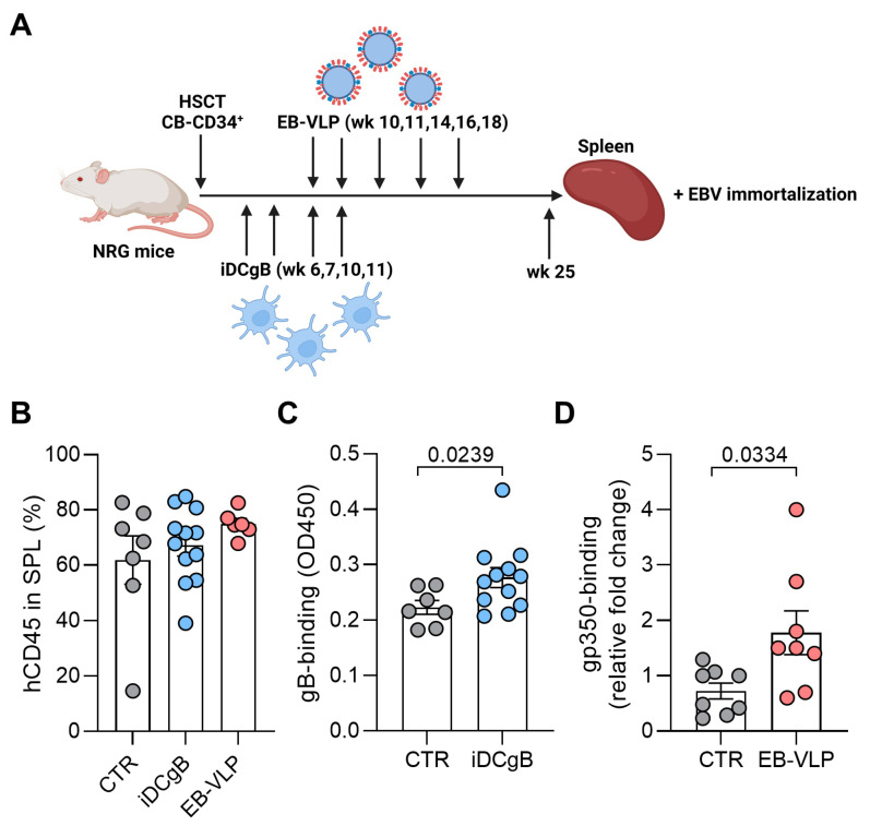
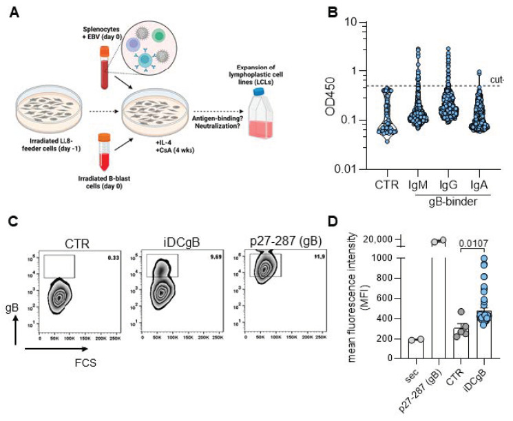
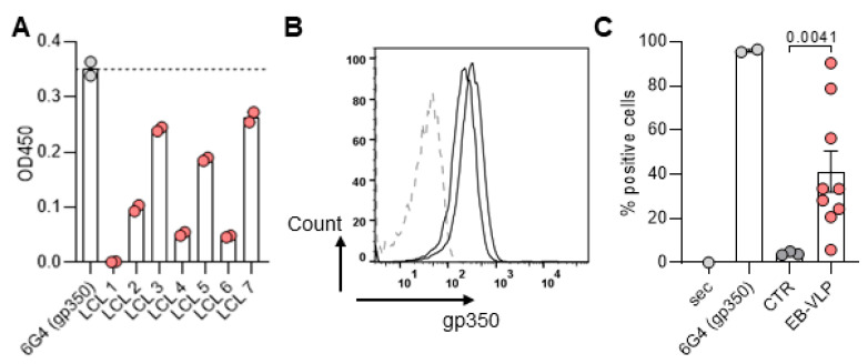
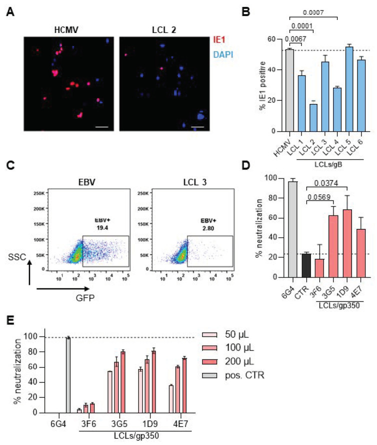
Antiviral neutralizing antibodies (nAbs) are commonly derived from B cells developed in immunized or infected animals and humans. Fully human antibodies are preferred for clinical use as they are potentially less immunogenic. However, the function of B cells varies depending on their homing pattern and an additional hurdle for antibody discovery in humans is the source of human tissues with an immunological microenvironment. Here, we show an efficient method to pharm human antibodies using immortalized B cells recovered from Nod.Rag.Gamma (NRG) mice reconstituting the human immune system (HIS). Humanized HIS mice were immunized either with autologous engineered dendritic cells expressing the human cytomegalovirus gB envelope protein (HCMV-gB) or with Epstein-Barr virus-like particles (EB-VLP). Human B cells recovered from spleen of HIS mice were efficiently immortalized with EBV in vitro. We show that these immortalized B cells secreted human IgGs with neutralization capacities against prototypic HCMV-gB and EBV-gp350. Taken together, we show that HIS mice can be successfully used for the generation and pharming fully human IgGs. This technology can be further explored to generate antibodies against emerging infections for diagnostic or therapeutic purposes.

Keywords: EBV; HCMV; antibodies; dendritic cells; herpesvirus; humanized mice; immortalization; virus-like particles.


Conflict of interest statement

The laboratory of R.S. receives research funding from The Jackson Laboratory for development of humanized mice with improved immunologic functions. The other authors declare no conflict of interest.

Figures

Figure 1
Figure 1
Fully humanized mice immunized with either iDCgB or EB-VLPs produce human IgGs reactive against gB and gp350, respectively. (A) Scheme of immunization of humanized mice and analyses. NRG mice were transplanted with CB-derived CD34+ stem cells. Afterwards, the animals were immunized either four times with iDCgB (autologous to the CB source) or five times with EB-VLPs. 25 weeks after HCT, the mice were sacrificed. Blood and spleen samples were harvested. (B) Reconstitution of humanized mice (CTR = grey; iDCgB = blue and EB-VLP = red) depicted in percentage (%) of human lymphocytes (CD45) in blood after 25 wks. Each dot represents one experimental mouse. (C) Detection of HCMV-gB specific IgG iDC (OD450) in plasma of humanized mice measured by ELISA. CTR not immunized are shown in grey and iDCgB immunized in blue. Each dot represents one mouse. (D) EB-VLPs immunized mice showed gp350-specific human IgG antibodies. The sera from immunized and control mice were used for an ELISA to detect gp350-specific human IgG antibodies. ELISA was performed with undiluted mice plasma obtained from CTR mice (grey) or from mice immunized with EB-VLPs (red). Standard deviation of the mean is indicated and students t-test with Welsh`s correction was used for statistical analysis as indicated.
Figure 2
Figure 2
EBV immortalized B cell lines produce HCMV-gB-specific antibodies. (A) Schematic representation of the in vitro production of humanized mice-derived LCLs. (B) Screening example of lines derived from iDCgB immunized mice by detection of Igs in the cell supernatants binding to HCMV-gB by ELISA. Antibody subtype (IgM, IgG and IgA) is indicated and each dot represents results obtained for one cell line. Cut-off is indicated and was at OD 0.5 and based on IgG background signal obtained from LCL supernatants generated from non-immunized humanized mice (CTR). (C) Gating example of a flow cytometry based screening of B cell lines derived from iDCgB-immunized mice to detect gB-specific IgG. Supernatant was incubated with 293T cells expressing HCMV-gB on the cell surface and a specific binding was detected with a secondary antibody detecting human IgG. As positive control we used a gB specific primary antibody (p27-287). (D) Mean fluorescence intensity (MFI) quantification of all lines screened by flow cytometry. Sec: Secondary antibody used for detection; p27-287 (gB): Control primary antibody and secondary antibody used for detection; CTR: Supernatants from LCLs derived from non-immunized mice and stained with primary and secondary Abs; iDCgB: Supernatants from LCLs derived from iDCgB immunized mice and stained with primary and secondary Abs. Standard deviation of the mean is indicated and students t-test with Welsh`s correction was used for statistical analysis as indicated.
Figure 3
Figure 3
EBV immortalized B cells expressed EBV gp350-specific antibodies. (A) Supernatants LCLs generated from splenocytes from EB-VLPs immunized mice were tested by ELISA against recombinant gp350 protein and a few secreted gp350-specific human IgGs. 6G4 is a monoclonal antibody against gp350. (B) The supernatants of two representative lines were tested by flow cytometry for the presence of antibodies binding to PCI-1 cells stably expressing gp350 on the surface. The cells were stained with LCL supernatant (2 representative examples, black lines). Parental PCI-1 cells were used as negative control (dashed line). (C) Quantification of B cell in percentage of positive cells (%). Sec: Secondary antibody used for detection; 6G4 monoclonal antibody against gp350: Control primary antibody and secondary antibody used for detection; CTR: Supernatants from LCLs derived from non-immunized mice and stained with primary and secondary Abs; EBV-VLP: Supernatants from LCLs derived from VLP immunized mice and stained with primary and secondary Abs. Standard deviation of the mean is indicated and students t-test with Welsh`s correction was used for statistical analysis as indicated.
Figure 4
Figure 4
Supernatants of immortalized B cell lines neutralize HCMV and EBV infections in vitro. (A) The HCMV in vitro neutralization assay was performed with MRC-5 cells infected with the HCMV strain TB40-GLuc. DAPI (blue) was used to quantify the total cells and the fraction of cells expressing HCMV-IE1 (magenta) was used to quantify infection. Microscope magnification is 20× and scale bar represents 50 µm. (B) Percentage of IE1-positive cells within all cells when supernatants of different LCLs targeted against gB were added to the assay. (C) The EBV in vitro neutralization assay was performed with Raji cells infected with the EBV expressing GFP (MOI 0.2). 3 days after infection, the infected cells were quantified by flow cytometry through GFP expression (left panel). (D) Quantified values of EBV neutralization performed with supernatants obtained from different LCLs LCLs targeted against gp350. The 6G4 gp350-specific rat monoclonal antibody was used as positive control (grey). Control LCLs are shown in black. A gp350 negative LCL was used as ref. control. The percentage of inhibition was calculated normalizing to EBV infection. (E) The same experiment was done titrating the amount of supernatants used for neutralization. For all experiments n = 2 was performed and standard deviation of the mean is depicted in the graph. Statistics were performed using One-way-ANOVA and p-values are shown.

Similar articles

References

    1. Gupta A., Gonzalez-Rojas Y., Juarez E., Crespo Casal M., Moya J., Falci D.R., Sarkis E., Solis J., Zheng H., Scott N., et al. Early Treatment for COVID-19 with SARS-CoV-2 Neutralizing Antibody Sotrovimab. N. Engl. J. Med. 2021;385:1941–1950. doi: 10.1056/NEJMoa2107934. - DOI - PubMed
    1. Schampera M.S., Schweinzer K., Abele H., Kagan K.O., Klein R., Rettig I., Jahn G., Hamprecht K. Comparison of cytomegalovirus (CMV)-specific neutralization capacity of hyperimmunoglobulin (HIG) versus standard intravenous immunoglobulin (IVIG) preparations: Impact of CMV IgG normalization. J. Clin. Virol. 2017;90:40–45. doi: 10.1016/j.jcv.2017.03.005. - DOI - PubMed
    1. Niedobitek G., Meru N., Delecluse H.J. Epstein-Barr virus infection and human malignancies. Int. J. Exp. Pathol. 2001;82:149–170. - PMC - PubMed
    1. Zuhair M., Smit G.S.A., Wallis G., Jabbar F., Smith C., Devleesschauwer B., Griffiths P. Estimation of the worldwide seroprevalence of cytomegalovirus: A systematic review and meta-analysis. Rev. Med. Virol. 2019;29:e2034. doi: 10.1002/rmv.2034. - DOI - PubMed
    1. Young L.S., Yap L.F., Murray P.G. Epstein-Barr virus: More than 50 years old and still providing surprises. Nat. Rev. Cancer. 2016;16:789–802. doi: 10.1038/nrc.2016.92. - DOI - PubMed

Publication types

LinkOut - more resources