. 2023 Dec 21;13(1):25.
doi: 10.3390/cells13010025.

Establishment and Characterization of Cell Lines from Canine Metastatic Osteosarcoma

Affiliations

Establishment and Characterization of Cell Lines from Canine Metastatic Osteosarcoma

Ya-Ting Yang et al. Cells. .

Abstract

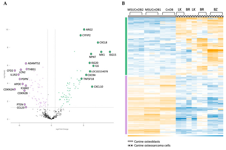
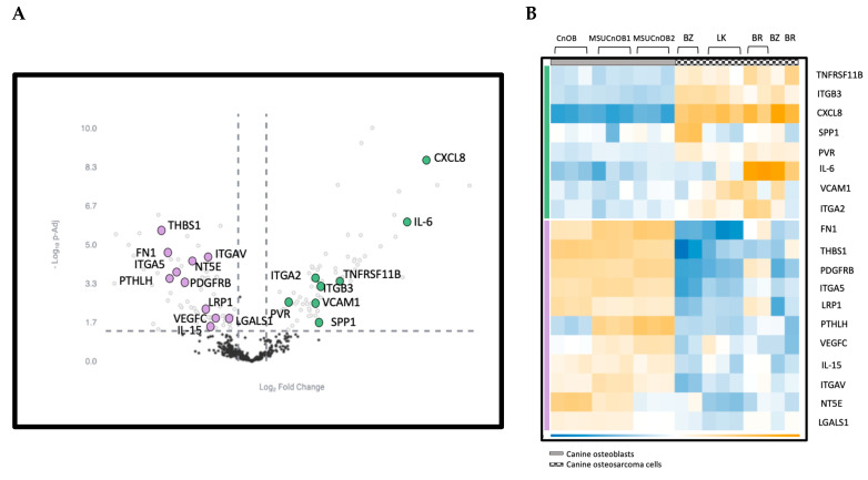
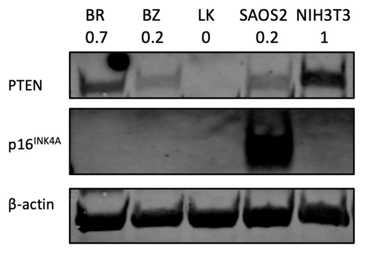
Despite the advancements in treatments for other cancers, the outcomes for osteosarcoma (OSA) patients have not improved in the past forty years, especially in metastatic patients. Moreover, the major cause of death in OSA patients is due to metastatic lesions. In the current study, we report on the establishment of three cell lines derived from metastatic canine OSA patients and their transcriptome as compared to normal canine osteoblasts. All the OSA cell lines displayed significant upregulation of genes in the epithelial mesenchymal transition (EMT) pathway, and upregulation of key cytokines such as CXCL8, CXCL10 and IL6. The two most upregulated genes are MX1 and ISG15. Interestingly, ISG15 has recently been identified as a potential therapeutic target for OSA. In addition, there is notable downregulation of cell cycle control genes, including CDKN2A, CDKN2B and THBS1. At the protein level, p16INK4A, coded by CDKN2A, was undetectable in all the canine OSA cell lines, while expression of the tumor suppressor PTEN was variable, with one cell line showing complete absence and others showing low levels of expression. In addition, the cells express a variety of actionable genes, including KIT, ERBB2, VEGF and immune checkpoint genes. These findings, similar to those reported in human OSA, point to some genes that can be used for prognosis, targeted therapies and novel drug development for both canine and human OSA patients.

Keywords: canine IO panel; cell lines; cytokines; metastasis; osteosarcoma; transcriptome.


Conflict of interest statement

The authors declare no conflict of interest.

Figures

Figure 1
Figure 1
Images of OSA cell lines during culture. Phase contrast images showing growth morphology at 10× magnification. (A) BR cells (passage 47); (B) BZ cells (passage 45); (C) LK cells (passage 35); (D) SAOS2; calibration bar —100 μm.
Figure 2
Figure 2
The canine BR, BZ and LK cell lines were confirmed as osteosarcoma by IHC staining. DAPI signal reflects the staining of nuclei in blue, Alexa Fluor® 488 of osteocalcin in green and eF615® of vimentin in red. Images were acquired at 60× magnification with calibration bar —25 μm. The human OSA cell line SAOS2 was used as a positive control. Osteocalcin is a secreted protein and is being produced continuously, sometimes appearing as cytoplasmic, sometimes nuclear and sometimes covering the cell, especially when the cells are small.
Figure 3
Figure 3
The wound healing assay shows the migration ability variation among OSA cell lines. (A) The represented pictures of all cells show that BR cells migrated and filled the wound area within 12 h, while other wounds remained. (B) Graphical representation of wound healing assay. Each bar represents the average from three measurements.
Figure 4
Figure 4
Volcano scatter plot (A) and associated heatmap (B) of genes in cancer pathways analyzed by nCounter® Canine IO panel. On the volcano plot, dotted vertical lines indicate a 1.5-fold change in normalized gene expression and the dotted horizontal line indicates a p-adjusted value cut-off of 0.05. The overexpressed genes are labeled in green and underexpressed genes are in purple. The 12 most upregulated and the 12 most downregulated genes are labelled for clarity in Figure 4A.
Figure 5
Figure 5
(A) Volcano scatter plot of genes in cancer pathways analyzed by nCounter® Canine IO panel. Dotted vertical lines indicate a 1.5-fold change in normalized gene expression and the dotted horizontal line indicates a p-adjusted value cut-off of 0.05. The upregulated genes are labeled in green and downregulated genes are in purple. (B) Heatmap of genes in the EMT pathway showing differential expression. Genes in the EMT pathway are highlighted in both panels.
Figure 6
Figure 6
PTEN and p16INK4A protein analysis by Western blot. NIH3T3 is a mouse fibroblast cell line used as a positive control for PTEN expression study. Human OSA cell line SAOS2 is used as a positive control for p16INK4A. The number under each cell line designation indicates the PTEN protein band intensity normalized to β-actin and compared to NIH3T3.

Similar articles

Cited by

References

    1. Mirabello L., Troisi R.J., Savage S.A. Osteosarcoma incidence and survival rates from 1973 to 2004: Data from the Surveillance, Epidemiology, and End Results Program. Cancer. 2009;115:1531–1543. doi: 10.1002/cncr.24121. - DOI - PMC - PubMed
    1. Withrow S.J., MacEwen E.G., editors. Small Animal Clinical Oncology. 3rd ed. W. B. Saunders; Philadelphia, PA, USA: 2001.
    1. Rowell J.L., McCarthy D.O., Alvarez C.E. Dog models of naturally occurring cancer. Trends Mol. Med. 2011;17:380–388. doi: 10.1016/j.molmed.2011.02.004. - DOI - PMC - PubMed
    1. Anfinsen K.P., Grotmol T., Bruland O.S., Jonasdottir T.J. Breed-specific incidence rates of canine primary bone tumors—A population based survey of dogs in Norway. Can. J. Vet. Res. 2011;75:209–215. - PMC - PubMed
    1. Yang Y.T., Yuzbasiyan-Gurkan V. Sorafenib and Doxorubicin Show Synergistic Effects in Human and Canine Osteosarcoma Cell Lines. Int. J. Mol. Sci. 2022;23:9345. doi: 10.3390/ijms23169345. - DOI - PMC - PubMed

Publication types

Substances