. 2023 Dec 25;13(1):43.
doi: 10.3390/cells13010043.

Understanding Bartonella-Associated Infective Endocarditis: Examining Heart Valve and Vegetation Appearance and the Role of Neutrophilic Leukocytes

Affiliations

Understanding Bartonella-Associated Infective Endocarditis: Examining Heart Valve and Vegetation Appearance and the Role of Neutrophilic Leukocytes

Kristians Meidrops et al. Cells. .

Abstract

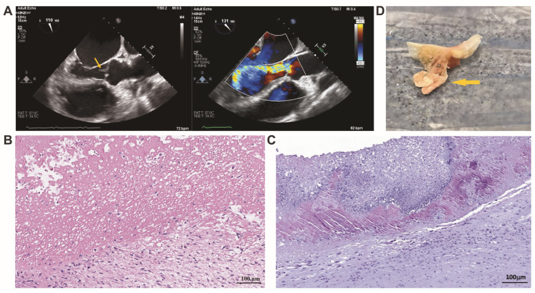
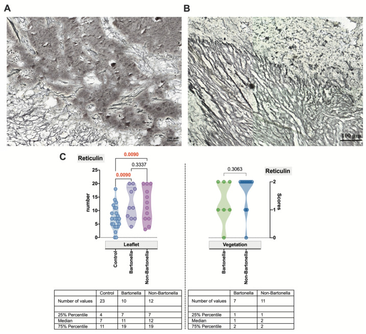
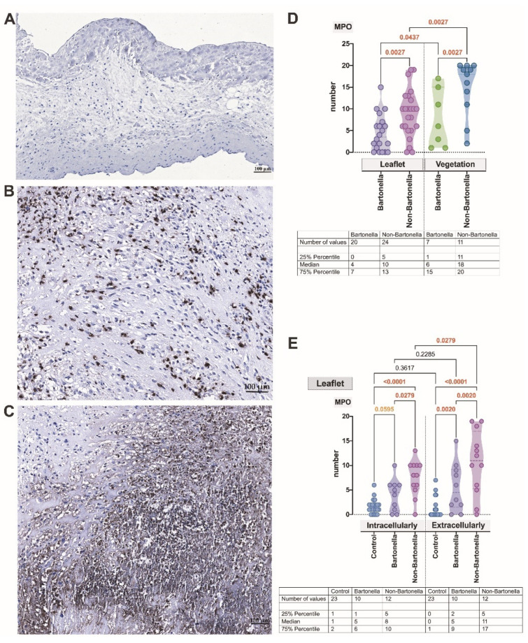
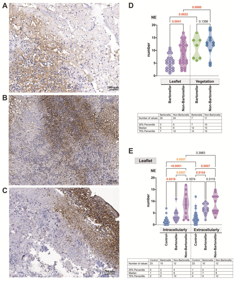
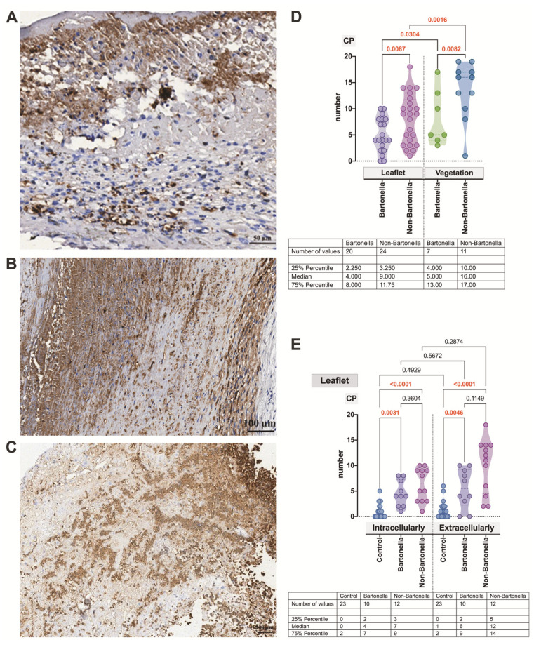
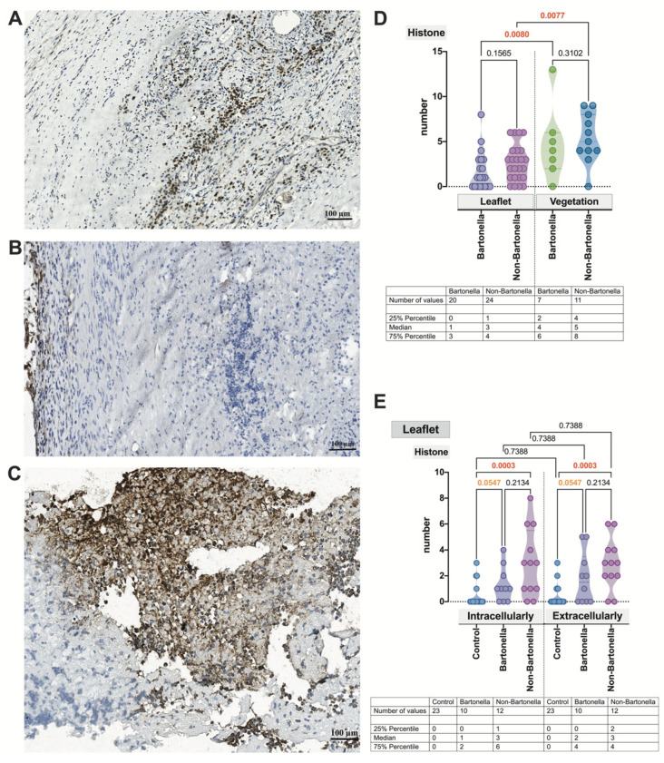
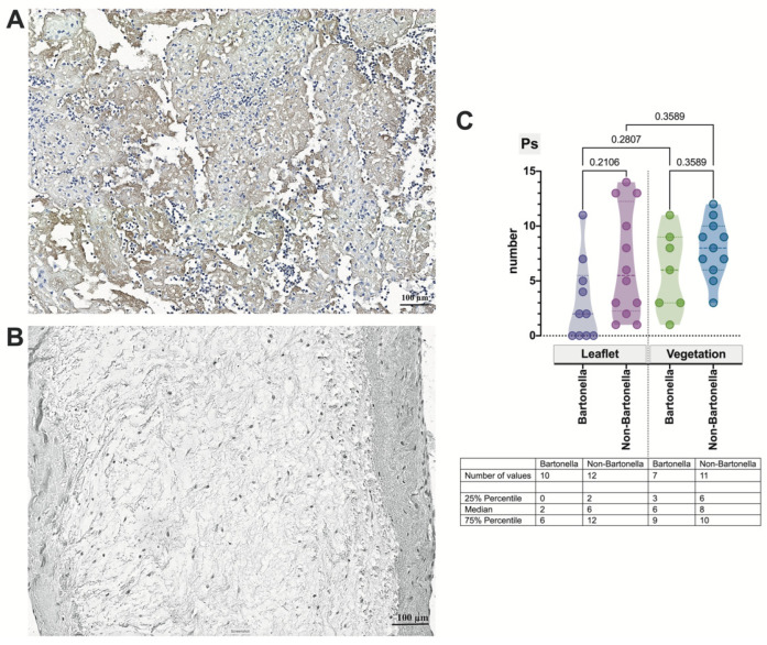
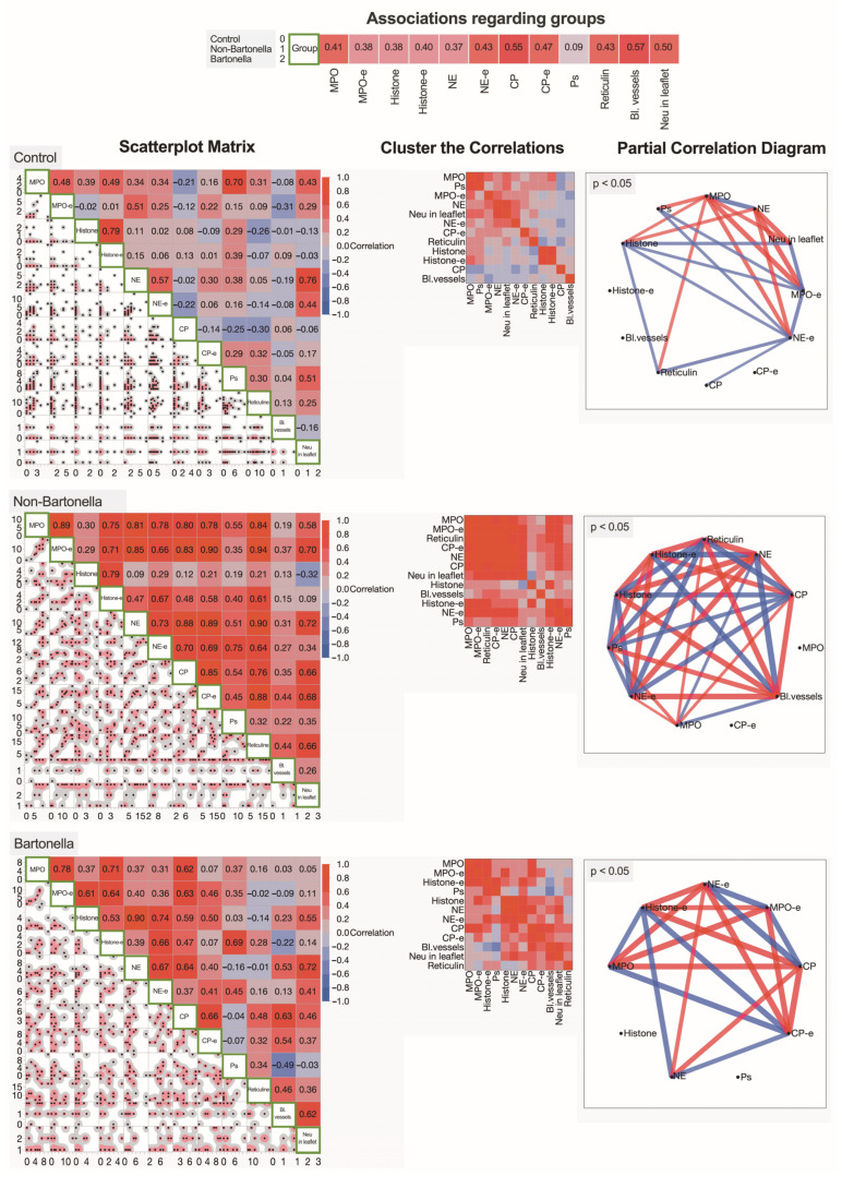
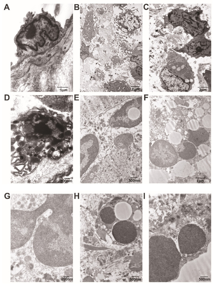
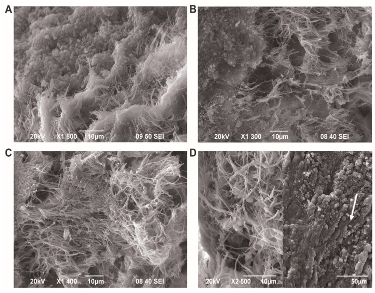
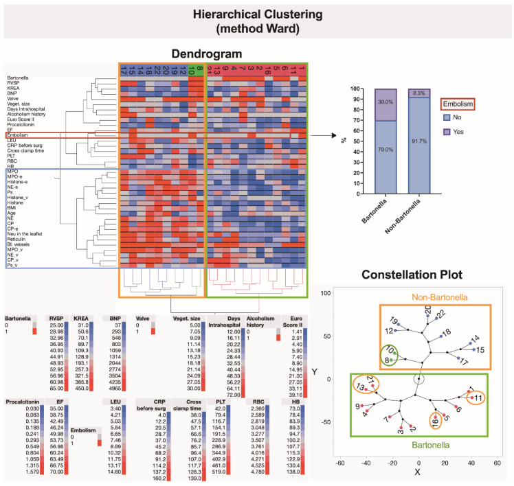
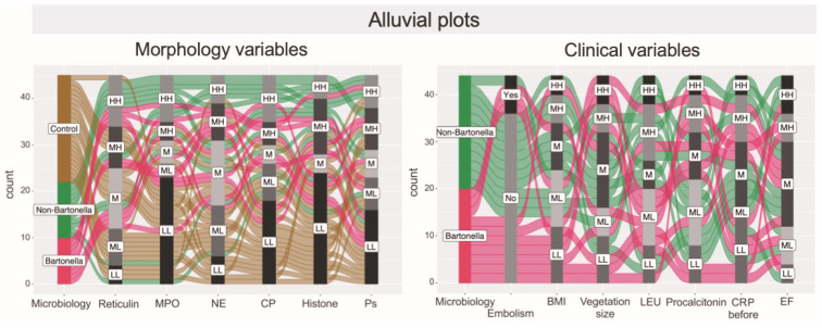
The endocardium and cardiac valves undergo severe impact during infective endocarditis (IE), and the formation of vegetation places IE patients at a heightened risk of embolic complications and mortality. The relevant literature indicates that 50% of IE cases exhibit structurally normal cardiac valves, with no preceding history of heart valve disease. Gram-positive cocci emerge as the predominant causative microorganisms in IE, while Gram-negative Bartonella spp., persisting in the endothelium, follow pathogenic pathways distinct from those of typical IE-causing agents. Employing clinical as well as advanced microbiological and molecular assays facilitated the identification of causative pathogens, and various morphological methods were applied to evaluate heart valve damage, shedding light on the role of neutrophilic leukocytes in host defense. In this research, the immunohistochemical analysis of neutrophilic leukocyte activation markers such as myeloperoxidase, neutrophil elastase, calprotectin, and histone H3, was performed. A distinct difference in the expression patterns of these markers was observed when comparing Bartonella spp.-caused and non-Bartonella spp.-caused IE. The markers exhibited significantly higher expression in non-Bartonella spp.-caused IE compared to Bartonella spp.-caused IE, and they were more prevalent in vegetation than in the valvular leaflets. Notably, the expression of these markers in all IE cases significantly differed from that in control samples. Furthermore, we advocated the use of 16S rRNA Next-Generation Sequencing on excised heart valves as an effective diagnostic tool for IE, particularly in cases where blood cultures yielded negative results. The compelling results achieved in this study regarding the enigmatic nature of Bartonella spp. IE's pathophysiology contribute significantly to our understanding of the peculiarities of inflammation and immune responses.

Keywords: Bartonella spp.; cardiac valves; infective endocarditis; neutrophil extracellular traps; neutrophils; vegetation.


Conflict of interest statement

The authors declare no conflicts of interest. The funders had no role in the design of the study; in the collection, analyses, or interpretation of data; in the writing of the manuscript; or in the decision to publish the results.

Figures

Figure 1
Figure 1
Transoesophageal echocardiography scan of Bartonella spp.-induced aortic valve endocarditis (A). The image on the left, marked with an arrow, depicts vegetation attached to the aortic valve leaflet, while the image on the right, using color Doppler, demonstrates severe aortic valve regurgitation. A representative image depicts the histopathological features of valvular vegetation visually attached to the leaflet in Bartonella spp.-caused IE. The cut-off region of vegetation reveals an amorphous material with a sponge-like appearance, which is invaded by immune cells. Fibroblasts and mononuclear cells are visualized in the leaflet fragment seen in this figure (B). Valvular vegetation in non-Bartonella spp. -caused IE with a bottom-side predominance of neutrophilic leukocytes, macrophages, and some bacteria interspersed by fibrin (C). H&E staining. Scale bar: 100 μm. Macroscopic appearance of excised aortic valve cusp with vegetation. The arrow indicates vegetation attached to the aortic valve leaflet (D).
Figure 2
Figure 2
Representative image demonstrating clumps of bacteria found in vegetation in Bartonella spp. IE (A) and non-Bartonella spp. IE (B) samples. Silver stain demonstrates black coloration in clusters of Bartonella bacilli. A visible metallic tone produced by the silver solution highlights reticular fibers within the valve leaflet. Scale bar: 100 µm. Histochemically confirmed and scored data are plotted for controls, Bartonella spp., and non-Bartonella spp. within the valve leaflet (C: the plot on the left) and for Bartonella spp. and non-Bartonella spp. within vegetation (C: the plot on the right). Each dot represents a single data point. Abbreviations: Ctrl—control; Bartonella—Bartonella spp.-caused IE; non-Bartonella—non-Bartonella spp.-caused IE.
Figure 3
Figure 3
A representative illustration of immunohistochemistry reaction: negative control (A). Micrographs displaying the characteristic appearance of MPO expression observed intra- and extracellularly in the leaflet and valvular vegetation in Bartonella spp.-caused and non-Bartonella spp.-caused IE (B,C). The MPO statistics assessment (D,E). Abbreviations: MPO—myeloperoxidase; Bartonella—Bartonella spp.-caused IE; non-Bartonella—non-Bartonella spp.-caused IE. MPO immunohistochemistry. Scale bar: 100 μm.
Figure 4
Figure 4
Micrographs displaying the characteristic appearance of NE expression observed intra- but mostly extracellularly in valvular vegetation in Bartonella spp.-caused IE (A) and leaflet and valvular vegetation in non-Bartonella spp.-caused IE (B,C). Vegetations are invaded by immune cells. The leaflets exhibit features of neovascularization. The NE statistics assessment (D,E). Abbreviations: NE—neutrophil elastase; Bartonella—Bartonella spp.-caused IE; non-Bartonella—non-Bartonella spp.-caused IE. NE immunohistochemistry. Scale bar: 100 μm.
Figure 5
Figure 5
Micrographs displaying the characteristic appearance of CP expression observed intra- and extracellularly in valvular vegetation in Bartonella spp.-caused IE (A) and the leaflet and valvular vegetation in non-Bartonella spp.-caused IE (B,C). The leaflets exhibit features of neovascularization. The CP statistics assessment (D,E). Abbreviations: CP—calprotectin; Bartonella—Bartonella spp.-caused IE; non-Bartonella—non-Bartonella spp.-caused IE. CP immunohistochemistry. Scale bar: 100 μm.
Figure 6
Figure 6
Micrographs displaying the characteristic appearance of histone H3 expression observed in the valvular leaflet in Bartonella spp.-caused IE (A) and the leaflet and valvular vegetation in non-Bartonella spp.-caused IE (B,C). The leaflets exhibit features of neovascularization. The histone H3 statistics assessment (D,E). Abbreviations: Histone—histone H3; Bartonella—Bartonella spp.-caused IE; non-Bartonella—non-Bartonella spp.-caused IE. Histone H3 immunohistochemistry. Scale bar: 100 μm.
Figure 7
Figure 7
A representative illustration of Ps immunohistochemistry: vegetation attached to the damaged valvular leaflet in non-Bartonella spp.-caused IE (A) and the absence of reaction in the negative control (B). The micrograph demonstrates positive staining, likely reflecting the presence of this transmembrane protein in the blood platelets of vegetation (B,C). The region of interest is heavily colonized by neutrophilic leukocytes. The Ps statistics assessment (C). Abbreviations: Ps—P-selectin; Bartonella—Bartonella spp.-caused IE; non-Bartonella—non-Bartonella spp.-caused IE. Scale bar: 100 μm.
Figure 8
Figure 8
Associations between immunohistochemically obtained expressions of neutrophilic leukocyte activation markers. A scatterplot matrix, along with the corresponding Spearman’s rank correlation coefficients, correlation clusters, and partial correlation diagrams (on the right), is presented for the control group (upper), non-Bartonella spp. IE group (middle), and Bartonella spp. IE group (lower). Abbreviations: MPO—myeloperoxidase, MPO-e—myeloperoxidase expression extracellularly, Histone—histone H3 expression, Histone-e—histone H3 expression extracellularly, NE—neutrophil elastase expression, NE-e—neutrophil elastase expression extracellularly, Ps—P-selectin, Histone_v—histone H3 expression in vegetation, NE—neutrophil elastase, CP—calprotectin, CP-e—calprotectin expression extracellularly, Neu in leaflet—neutrophilic leukocyte count inside valvular leaflet, Bl.vessels—blood vessels inside valvular leaflet.
Figure 9
Figure 9
Representative images obtained using transmission electron microscopy. (A) The endothelium of the valvular leaflet in a Bartonella spp.-caused IE patient is roundly shaped, with few irregular and short microvilli, a wavy nuclear envelope, and marginated chromatin; cytoplasm reveals occasional organelles. (B) A low-power view of the damaged valvular leaflet in a non-Bartonella spp.-caused IE patient. Its content demonstrates various immune cells, including macrophages with well-developed pseudopodia and lipid-laden, are intermixed with collagen microfibrils. (C) In a region nearby, valvular cells reveal a very pronounced and deeply convoluted nuclear envelope; immune cells reveal the extensive expansion of the rough endoplasmic reticulum with content of varying density. (D) An irregularly shaped macrophage with a ruptured plasma membrane reveals a dilated perinuclear space, damaged mitochondria, and some cytoplasmic granules. (E) A neutrophil with a ruptured plasma membrane, wavy-outlined external nuclear membrane, and nuclear inclusion reveals a low-density hyaloplasm containing various granules. Clumps of neutrophil heterochromatin are positioned extracellularly. (F) The free apoptotic nuclei of a neutrophil are surrounded by extracellularly positioned clumps of heterochromatin, cellular debris, and lipid droplets. (G) A high-power view of the neutrophil reveals a nucleus with an irregularly dilated perinuclear space and a ruptured membrane. It is surrounded by a cytoplasm containing granules and autophagic vacuoles. (H) A fragment of the apoptotic neutrophil is enclosed by fibrin, collagen, and cellular debris. (I) A fragment of the neutrophil reveals an irregularly dilated perinuclear space with thread-like material, a ruptured membrane, and various granules positioned both intra- and extracellularly. Scale bars: 1 μm, 2 μm, 2 μm, 500 nm, 500 nm, 1 μm, 200 nm, 500 nm, and 500 nm.
Figure 10
Figure 10
Representative scanning electron micrographs depicting the ultrastructure of the aortic valve endothelial cells and vegetation. (A) Low-power magnification showing the top and depth of vegetation. (B) Low-power depth view of vegetation with fibrin and a few platelets. (C,D) Vegetation side depth shows the sandwiched architecture of vegetation with a network of fibrin bundles and some bacteria trapped. When platelets, bacteria, and leukocytes form vegetation, the surface of tightly aligned endothelial cells (indicated by the white arrow) becomes barely visible. Scale bar: 10 μm; D panel on the right: 50 μm.
Figure 11
Figure 11
Hierarchical clustering shows the similarities of data points related to the studied system. The clades and leaves of a tree include data on the immunohistochemical evaluations of neutrophilic leukocyte activation markers, clinical parameters, complications, including and further specifying embolism, and several laboratory analyses. Variables are scaled in colors from blue to red, representing concentrations ranging from the lowest to the highest. Abbreviations: RVSP—right ventricle systolic pressure; KREA—serum creatinine level, BNP—B-type natriuretic peptide, Veget. size—vegetation size, Euro Score II—EuroScore II risk, EF—ejection fraction of the left ventricle, LEU—leukocyte count, CRP before surg.—C-reactive protein level before surgery, Cross clamp time—aortic cross clamp time, PLT—platelet level, RBC—red blood cell level, HB—hemoglobin level, MPO—myeloperoxidase, MPO-e—myeloperoxidase expression extracellularly, Histone-e—histone H3 expression extracellularly, NE-e—neutrophil elastase expression extracellularly, Ps—P-selectin, Histone_v—histone H3 expression in vegetation, BMI—body mass index, NE—neutrophil elastase, CP—calprotectin, CP-e—calprotectin expression extracellularly, Neu in the leaflet—neutrophilic leukocyte count inside valvular leaflet, Bl.vessels—blood vessels inside valvular leaflet, MPO_v—myeloperoxidase expression in vegetation, NE_v—neutrophil elastase expression in vegetation, CP_v—calprotectin expression in vegetation, Ps_v—P-selectin expression in vegetation.
Figure 12
Figure 12
The alluvial diagrams represent flows among nodes. In the left diagram, individual assessments are presented as rows, while immunohistochemically obtained expressions of neutrophilic leukocyte activation markers are presented as columns. Statistically, the count for each marker is stratified into five different levels. In the groups studied, the width of each line depicted and the flow that stems from it are determined by the proportional fraction of the category total. The left plot clearly shows that in non-Bartonella spp.-caused IE, all immunohistochemical expressions indicative of neutrophilic leukocyte activation are mostly high and medium high. Simultaneously, the right plot shows that the levels of clinical parameters assessed in this study vary among the two groups: BMI, leukocyte count, procalcitonin, and CRO reached the highest level in non-Bartonella spp.-caused IE patients, while the largest vegetations were observed in Bartonella spp.-caused IE patients. Abbreviations: High, High (HH); Medium, High (MH); Medium (M); Medium, Low (ML); Low, Low (LL); MPO—myeloperoxidase; NE—neutrophil elastase; CP—calprotectin; Histone—histone H3; Ps—P-selectin; BMI—body mass index; LEU—leukocyte count; CRP before—C-reactive protein level before surgery; EF—ejection fraction of the left ventricle.

Similar articles

Cited by

References

    1. Talha K.M., Baddour L.M., Thornhill M.H., Arshad V., Tariq W., Tleyjeh I.M., Scott C.G., Hyun M.H., Bailey K.R., Anavekar N.S., et al. Escalating incidence of infective endocarditis in Europe in the 21st century. Open Hear. 2021;8:e001846. doi: 10.1136/openhrt-2021-001846. - DOI - PMC - PubMed
    1. Lisi M., Flamigni F., Russo M., Cameli M., Mandoli G.E., Pastore M.C., Mele D., Campo G., Henein M.Y., Rubolli A. Incidence and mortality of infective endocarditis in the last decade: A single center study. J. Cardiovasc. Med. 2023;24:105–112. doi: 10.2459/JCM.0000000000001410. - DOI - PubMed
    1. Rajani R., Klein J.L. Infective endocarditis: A contemporary update. Clin. Med. 2020;20:31–35. doi: 10.7861/clinmed.cme.20.1.1. - DOI - PMC - PubMed
    1. Sexton D.J., Spelman D. Current best practices and guidelines. Assessment and management of complications in infective endocarditis. Cardiol. Clin. 2003;21:273–282. doi: 10.1016/S0733-8651(03)00031-6. - DOI - PubMed
    1. Chirillo F. New approach to managing infective endocarditis. Trends Cardiovasc. Med. 2021;31:277–286. doi: 10.1016/j.tcm.2020.04.008. - DOI - PubMed

Publication types

Substances