. 2023 Dec 27;13(1):61.
doi: 10.3390/cells13010061.

The Warburg Trap: A Novel Therapeutic Approach for Targeting Osteosarcoma

Affiliations

The Warburg Trap: A Novel Therapeutic Approach for Targeting Osteosarcoma

Joerg Fellenberg et al. Cells. .

Abstract

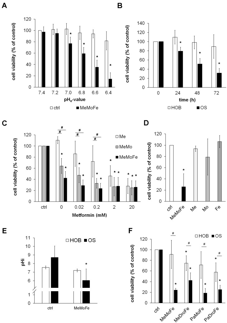
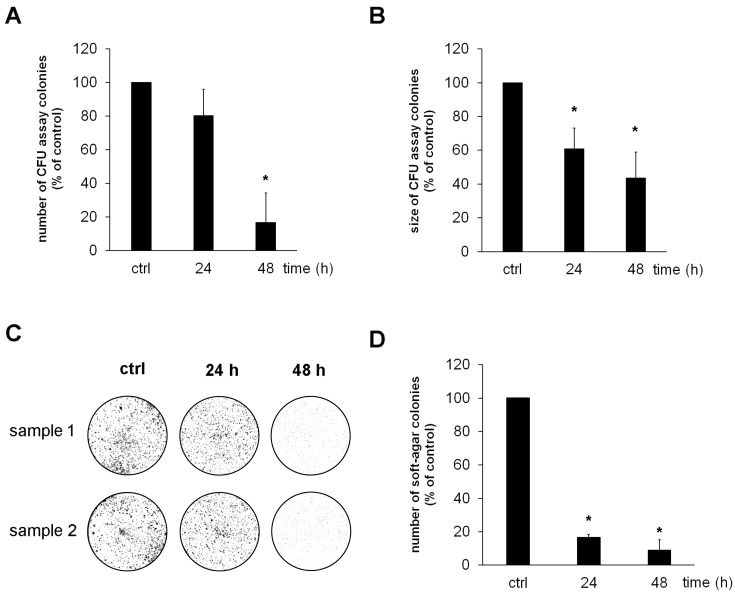
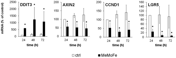
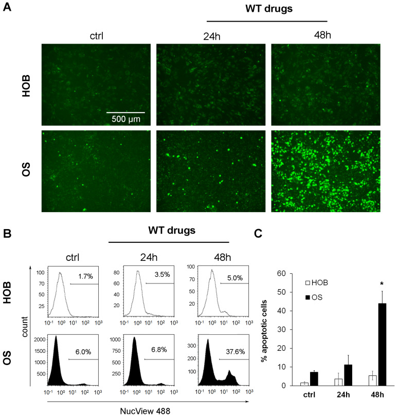
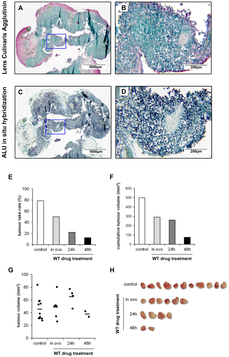
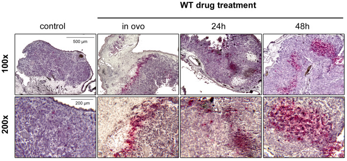
Although urgently needed, no significant improvements in osteosarcoma (OS) therapy have been achieved within the last decades. Here, we present a new therapeutic approach based on drug combinations consisting of mitochondrial complex I (MCI) inhibitors and ionophores that induce cancer cell-specific cell death based on a modulation of cellular energy metabolism and intracellular pH (pHi) named the Warburg Trap (WT). The effects of several drug combinations on intracellular pH, cell viability, colony-forming capacity and expression of WNT-target genes were analysed using OS cell lines and primary human osteoblasts (HOB). Tumour take rates and tumour volumes were analysed in vivo using a chicken chorioallantoic membrane assay (CAM). Several WT drug combinations induced the intracellular acidification and apoptotic cell death in OS cells, whereas HOBs tolerated the treatment. A significant inhibition of the colony-forming ability of OS cells and downregulation of WNT-target genes suggest that cancer stem cells (CSCs) are also targeted by the WT approach. In vivo, we observed a significant reduction in the tumour take rates in response to WT drug treatment. Our data suggest that the Warburg Trap is a promising approach for the development of a novel and effective OS therapy to replace or supplement the current OS chemotherapy.

Keywords: Warburg Trap; apoptosis; cancer stem cells; chemotherapy; osteosarcoma.


Conflict of interest statement

The authors declare no conflicts of interest.

Figures

Figure 1
Figure 1
Cytotoxic effects of Warburg-Trap drugs on cancer cells. (A) Six OS cell lines (n = 6) were treated with a combination of Metformin (1 mM), Monensin (5 nM) and Fenofibrate (10 µM) (MeMoFe) in culture media with pH values ranging from 6.4 to 7.4. Cell viability was quantified after 72 h by WST-1 assay and is expressed as percent of untreated cells cultured at pH 7.4 (* p < 0.01 compared to untreated cells). (B) OS and HOB cell lines (n = 6 each) were treated with a combination of Metformin (1 mM), Monensin (5 nM) and Fenofibrate (10 µM) in an acidic culture medium (pH 6.5). Cell viability was quantified at the indicated time points by WST-1 assay and is expressed as percent of untreated control cells (* p < 0.01 compared to untreated control cells). (C) OS cell lines (n = 6) were treated with Metformin (Me) in the indicated concentrations with or without the addition of Monensin (5 nM) (MeMo) or a combination of Monensin (5 nM) and Fenofibrate (10 µM) (MeMoFe). Cell viability was quantified after 72 h by WST-1 assay and is expressed as percent of untreated control cells (* p < 0.01 compared to untreated control cells, # p < 0.01 compared to cells treated with metformin alone). (D) OS cell lines (n = 6) were treated with a combination of Metformin (1 mM), Monensin (5 nM) and Fenofibrate (10 µM) (MeMoFe) as well as all three drugs alone at the same concentrations. Cell viability was quantified after 72 h by WST-1 assay and is expressed as percent of untreated control cells (* p < 0.01 compared to untreated control cells). (E) WT drugs induce intracellular acidification in OS cells. OS and HOB cell lines (n = 6 each) were cultured for 24 h in an acidic medium (pH 6.5) with or without the addition of Metformin (1 mM), Monensin (5 nM) and Fenofibrate (10 µM) (MeMoFe) before pHi was quantified (* p < 0.01 compared to untreated control cells). (F) Viability of OS and HOB cells after treatment with different WT drug combinations. OS and HOB cells (n = 6 each) were treated with different combinations of Metformin (1 mM), Monensin (5 nM), Fenofibrate (10 µM), Drotaverine (10µM), and Papaverine (10 µM) in acidic culture medium (pH 6.5). Cell viability was quantified after 72 h by WST-1 assay and is expressed as percent of untreated control cells. (* p < 0.05 compared to untreated cells, # p < 0.05 OS compared to HOB cells).
Figure 2
Figure 2
Warburg Trap drugs significantly reduce the colony-forming capacity of OS cells. OS cell lines (n = 6) were seeded at low density in culture plates and treated with the WT drugs (Metformin (1 mM), Monensin (5 nM), and Fenofibrate (10 µM)) in an acidic culture medium (pH 6.5). After 24 h and 48 h of treatment, the culture medium was replaced by a normal cell culture medium with a physiological pH value of 7.4. Colonies that formed during a further seven days of culture were stained with haematoxylin and photographed before (A) the number of colonies and (B) the size of the colonies were quantified using the software ImageJ version 1.48v (* p < 0.01 compared to untreated controls). (C) Representative photographs of stained colonies obtained with cell line 143B analysed in duplicates. (D) Analysis of colony numbers that formed after pre-treatment of osteosarcoma cell lines (n = 6) with WT drugs and subsequent cultivation for 14 days in soft agar (* p < 0.01 compared to untreated controls).
Figure 3
Figure 3
DDIT3-mediated downregulation of WNT target genes by WT drugs. Osteosarcoma cell lines (n = 3) were treated with WT drugs (Metformin (1 mM), Monensin (5 nM), and Fenofibrate (10 µM)) (MeMoFe) for the indicated time before the expression of DDIT3 and the WNT target genes AXIN2, CCND1 and LGR5 were analysed by real-time quantitative RT-PCR analysis. The expression of the reference gene RPS13 (ribosomal protein S13) was used for normalisation. Data are presented as percent of untreated 24 h controls (* p < 0.05 compared to untreated controls).
Figure 4
Figure 4
WT drugs induce apoptosis in OS cells but not in HOBs. OS and HOB cell lines (n = 3 each) were treated with the WT drugs Metformin (1 mM), Monensin (5 nM) and Fenofibrate (10 µM) for 24 and 48 h in an acidic culture medium (pH 6.5). Control cells were cultured in an acidic medium without the addition of WT drugs. After treatment, apoptotic cells were stained with NucView 488. (A) Representative fluorescence images of NucView 488-positive HOB and OS cells (green) at a magnification of 100×. (B) Representative flow cytometry analyses of apoptotic HOB and OS cells. (C) Quantification of apoptotic HOB and OS cells (n = 3 each) (* p < 0.01 compared to untreated controls.
Figure 5
Figure 5
WT drugs inhibit tumour formation in vivo. Characterisation of the resected tumour xenografts was made using (A,B) Lens Culinaris Agglutinin staining, showing the unstained tumour tissue surrounded by the red-stained CAM and (C,D) hybridisation of the xenograft with a probe specific for the human ALU DNA sequence (black) detecting the human 143B OS cells. The blue frames define the inserts shown in a higher magnification on the right. All samples were counterstained with Methyl Green. For WT drug treatment, 143B cells were either pre-treated with the drugs Metformin (1 mM), Monensin (5 nM) and Fenofibrate (10 µM) (MeMoFe) for 24 h and 48 h or the drugs were applied in ovo together with the tumour cells (1 × 10⁶ cells per egg) directly onto the CAM of fertilised chicken eggs (n ≥ 20 per experimental group). After seven days, tumours that have formed were resected and (E) the tumour take rate, (F) the cumulative tumour volumes and (G) the individual tumour volumes were calculated. (H) Images of the resected tumours. Data from a representative experiment are shown.
Figure 6
Figure 6
WT drugs induce apoptosis in vivo. Tumours that formed after pre-treatment of OS cells (HOS 143B) with the drug combination Metformin (1 mM), Monensin (5 nM) and Fenofibrate (10 µM) for 24 h and 48 h or treated directly in ovo were stained for cleaved caspase-3 (red), counterstained with haematoxilin (violet) and photographed at 100× and 200× magnification. Representative photographs are shown.

Similar articles

References

    1. Mirabello L., Troisi R.J., Savage S.A. Osteosarcoma incidence and survival rates from 1973 to 2004: Data from the Surveillance, Epidemiology, and End Results Program. Cancer. 2009;115:1531–1543. doi: 10.1002/cncr.24121. - DOI - PMC - PubMed
    1. Isakoff M.S., Bielack S.S., Meltzer P., Gorlick R. Osteosarcoma: Current Treatment and a Collaborative Pathway to Success. J. Clin. Oncol. 2015;33:3029–3035. doi: 10.1200/JCO.2014.59.4895. - DOI - PMC - PubMed
    1. Mirabello L., Troisi R.J., Savage S.A. International osteosarcoma incidence patterns in children and adolescents, middle ages and elderly persons. Int. J. Cancer. 2009;125:229–234. doi: 10.1002/ijc.24320. - DOI - PMC - PubMed
    1. Gelderblom H., Jinks R.C., Sydes M., Bramwell V.H., van Glabbeke M., Grimer R.J., Hogendoorn P.C., McTiernan A., Lewis I.J., Nooij M.A., et al. Survival after recurrent osteosarcoma: Data from 3 European Osteosarcoma Intergroup (EOI) randomized controlled trials. Eur. J. Cancer. 2011;47:895–902. doi: 10.1016/j.ejca.2010.11.036. - DOI - PubMed
    1. Ta H.T., Dass C.R., Choong P.F., Dunstan D.E. Osteosarcoma treatment: State of the art. Cancer Metastasis Rev. 2009;28:247–263. doi: 10.1007/s10555-009-9186-7. - DOI - PubMed