Review
. 2023 Dec 29;13(1):79.
doi: 10.3390/cells13010079.

Epigenetic Regulation of Neuroinflammation in Alzheimer's Disease

Affiliations
Review

Epigenetic Regulation of Neuroinflammation in Alzheimer's Disease

Yajing Ma et al. Cells. .

Abstract

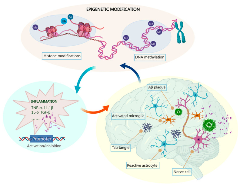
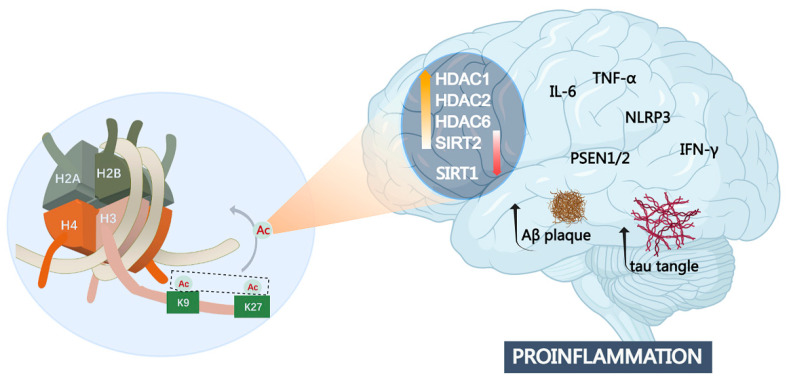
Alzheimer's disease (AD) is a chronic and progressive neurodegenerative disease and clinically manifests with cognitive decline and behavioral disabilities. Over the past years, mounting studies have demonstrated that the inflammatory response plays a key role in the onset and development of AD, and neuroinflammation has been proposed as the third major pathological driving factor of AD, ranking after the two well-known core pathologies, amyloid β (Aβ) deposits and neurofibrillary tangles (NFTs). Epigenetic mechanisms, referring to heritable changes in gene expression independent of DNA sequence alterations, are crucial regulators of neuroinflammation which have emerged as potential therapeutic targets for AD. Upon regulation of transcriptional repression or activation, epigenetic modification profiles are closely involved in inflammatory gene expression and signaling pathways of neuronal differentiation and cognitive function in central nervous system disorders. In this review, we summarize the current knowledge about epigenetic control mechanisms with a focus on DNA and histone modifications involved in the regulation of inflammatory genes and signaling pathways in AD, and the inhibitors under clinical assessment are also discussed.

Keywords: Alzheimer’s disease; DNA methylation; epigenetics; histone modification; inflammation; microglia.


Conflict of interest statement

The authors declare no conflicts of interest, financial or otherwise.

Figures

Figure 1
Figure 1
Epigenetic mechanisms via DNA methylation and histone modifications in neuroinflammation in AD. Inflammatory responses mediated by activated microglia play an essential role in initiation and progression of AD. Aberrant epigenetic modifications of gene promoters of cytokines such as TNF-α, IL-1β, and IL-6 promote the inflammatory response of microglia and astrocyte and provoke the formation of pathological Aβ deposits and neurofibrillary tangles, resulting in the development and aggravation of AD. Abbreviations: Me: methyl; Ac: acetyl; Ub: ubiquitin.
Figure 2
Figure 2
DNA de/methylation status is closely related to the expression of inflammatory cytokines in activated microglia. DNA hypermethylation is associated with gene silencing, while DNA hypomethylation correlates with gene activation. Abbreviations: pin 1: peptidyl-prolyl cis/trans isomerase; CLDN 5: claudin-5; TET: ten-eleven-translocation protein.
Figure 3
Figure 3
Histone methylation at specific sites causes gene transcriptional activation or repression. Dysregulated histone methylation has a vital role in microglial cell polarization to affect the proinflammatory cytokine production to cause neurotoxicity. Abbreviations: AR, androgen receptor; ER: estrogen receptor; JMJD3: Jumonji domain-containing protein-3.
Figure 4
Figure 4
Histone hypoacetylation at both H3K9 and H3K27 sites involved in AD pathogenesis. Aberrant expression of multiple histone deacetylases such as HDAC1, HDAC2, HDAC6, SIRT1, and SIFT2 is closely related to inflammatory response, as well as Aβ deposits and tau pathology. Abbreviations: NLRP3: NOD-like receptor thermal protein domain-associated protein 3; PSEN 1: Presenilin 1.
Figure 5
Figure 5
Schematic of cascade enzymatic reaction of ubiquitination. First, E1 enzymes activate ubiquitin in an ATP-dependent manner to form a thioester bond between the C-terminal glycine of ubiquitin and the cysteine site of E1. The activated ubiquitin subsequently forms a new thioester bond with the cysteine site of E2-conjugating enzyme. The resultant Ub-E2 conjugate cooperates with the E3 ligase to transfer the ubiquitin on the substrate to complete the ubiquitin labeling of the target protein, which is then degraded by the 26S proteasome. On the other hand, ubiquitin-specific protease (USP)-mediated deubiquitination enables the dissociation of ubiquitin from the substrate.
Figure 6
Figure 6
Chemical structures of HDACs and LSD1 inhibitors in clinical trials.

References

    1. Long J.M., Holtzman D.M. Alzheimer Disease: An Update on Pathobiology and Treatment Strategies. Cell. 2019;179:312–339. doi: 10.1016/j.cell.2019.09.001. - DOI - PMC - PubMed
    1. Ju Y., Tam K.Y. Pathological mechanisms and therapeutic strategies for Alzheimer’s disease. Neural Regen. Res. 2022;17:543–549. doi: 10.4103/1673-5374.320970. - DOI - PMC - PubMed
    1. Leng F., Edison P. Neuroinflammation and microglial activation in Alzheimer disease: Where do we go from here? Nat. Rev. Neurol. 2021;17:157–172. doi: 10.1038/s41582-020-00435-y. - DOI - PubMed
    1. Cornejo F., von Bernhardi R. Role of scavenger receptors in glia-mediated neuroinflammatory response associated with Alzheimer’s disease. Mediat. Inflamm. 2013;2013:895651. doi: 10.1155/2013/895651. - DOI - PMC - PubMed
    1. Kinney J.W., Bemiller S.M., Murtishaw A.S., Leisgang A.M., Salazar A.M., Lamb B.T. Inflammation as a central mechanism in Alzheimer’s disease. Alzheimer’s Dement. 2018;4:575–590. doi: 10.1016/j.trci.2018.06.014. - DOI - PMC - PubMed

Publication types

Substances