. 2024 Jan 4;13(1):102.
doi: 10.3390/cells13010102.

An Approach to Intersectionally Target Mature Enteroendocrine Cells in the Small Intestine of Mice

Affiliations

An Approach to Intersectionally Target Mature Enteroendocrine Cells in the Small Intestine of Mice

Christian Vossen et al. Cells. .

Abstract

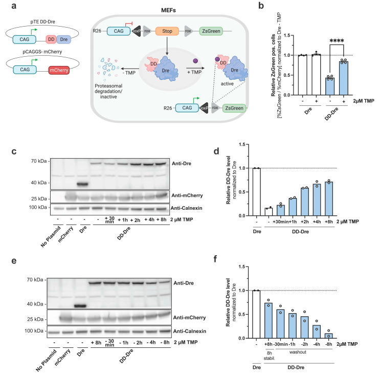
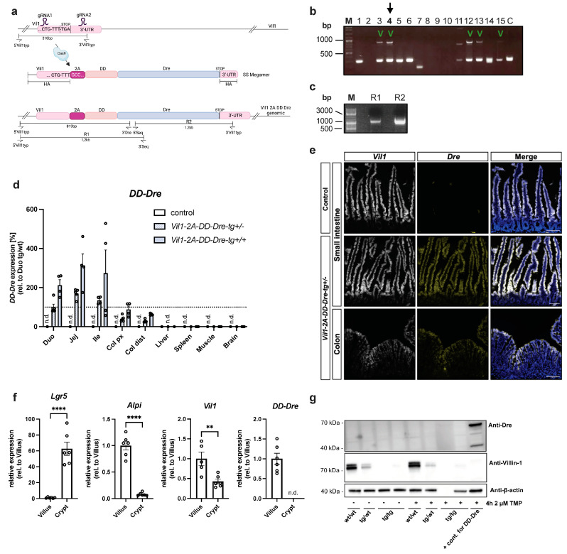
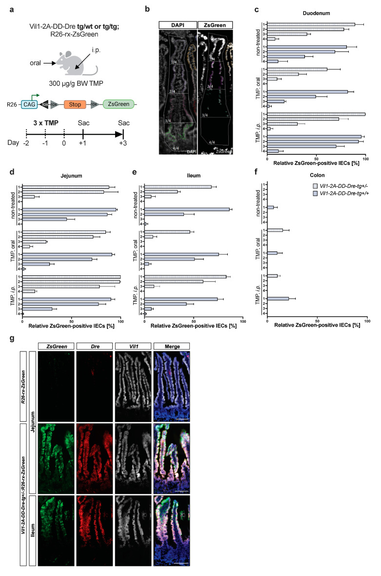
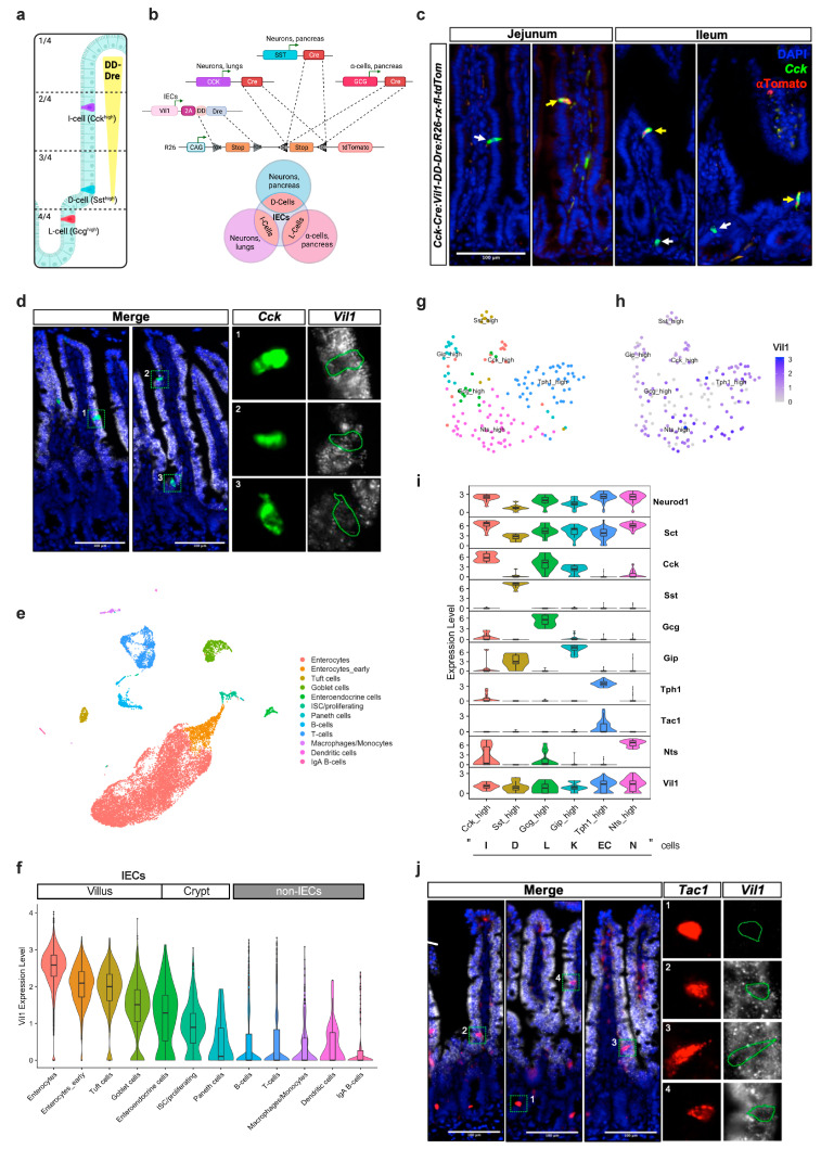
Enteroendocrine cells (EECs) constitute only a small proportion of Villin-1 (Vil1)-expressing intestinal epithelial cells (IECs) of the gastrointestinal tract; yet, in sum, they build the largest endocrine organ of the body, with each of them storing and releasing a distinct set of peptides for the control of feeding behavior, glucose metabolism, and gastrointestinal motility. Like all IEC types, EECs are continuously renewed from intestinal stem cells in the crypt base and terminally differentiate into mature subtypes while moving up the crypt-villus axis. Interestingly, EECs adjust their hormonal secretion according to their migration state as EECs receive altering differentiation signals along the crypt-villus axis and thus undergo functional readaptation. Cell-specific targeting of mature EEC subtypes by specific promoters is challenging because the expression of EEC-derived peptides and their precursors is not limited to EECs but are also found in other organs, such as the brain (e.g., Cck and Sst) as well as in the pancreas (e.g., Sst and Gcg). Here, we describe an intersectional genetic approach that enables cell type-specific targeting of functionally distinct EEC subtypes by combining a newly generated Dre-recombinase expressing mouse line (Vil1-2A-DD-Dre) with multiple existing Cre-recombinase mice and mouse strains with rox and loxP sites flanked stop cassettes for transgene expression. We found that transgene expression in triple-transgenic mice is highly specific in I but not D and L cells in the terminal villi of the small intestine. The targeting of EECs only in terminal villi is due to the integration of a defective 2A separating peptide that, combined with low EEC intrinsic Vil1 expression, restricts our Vil1-2A-DD-Dre mouse line and the intersectional genetic approach described here only applicable for the investigation of mature EEC subpopulations.

Keywords: Cck-expressing I cell; Cre/loxP; Dre/rox; EEC; Gcg-expressing L cell; Sst-expressing D cell; enteroendocrine cells of small intestine.


Conflict of interest statement

The authors declare no conflict of interest.

Figures

Figure 1
Figure 1
Generation of a TMP-inducible Dre system. (a) pTE-DD-Dre and pCAGGS-mCherry plasmids were transiently transfected into mouse embryonic fibroblasts carrying an R26-rx-ZsGreen reporter construct (MEF). Without trimethoprim (TMP), destabilizing domain (DD) targets DD-Dre for proteasomal degradation, whereas TMP treatment stabilizes DD-Dre, enabling excision of the rox-flanked STOP cassette of R26-rx-ZsGreen resulting in green fluorescence. (b) Quantification of ZsGreen+ cells normalized to mCherry+ cells (transfection control) of non-induced or TMP-treated MEFs transfected with the indicated plasmids. ZsGreen+/mCherry+ cells were quantified by FACS 48 h after transfection and normalized to pTE-Dre transfected cells w/o TMP. Gating strategy is shown in Figure S1a,b. Statistical analysis was performed using Student’s t-test. Representative results of three independent experiments are shown (n = 4). (c) Western blot analysis using anti-Dre, anti-mCherry, and anti-Calnexin (loading control) antibodies of MEFs transiently transfected w/o plasmid or with pCAGGS-mCherry control or with pTE-Dre or pTE-DD-Dre treated with 1µM TMP for the indicated time spans to verify DD-Dre inducibility. Representative fluorescence microscopy images of co-transfected mCherry are shown in Figure S1c. (n = 2). (d) Quantification of DD-Dre protein levels relative to Calnexin loading control in c) normalized to Dre protein level. (e) Western blot analysis using anti-Dre, anti-mCherry, and anti-Calnexin (loading control) antibodies of MEFs transiently transfected w/o plasmid or with pCAGGS-mcherry control with pTE-Dre or pTE-DD-Dre treated with 1 µM TMP for 8 h and consecutive TMP wash-out for indicated time points to verify DD-Dre instability. Representative fluorescence microscopy images of co-transfected mCherry are shown in Figure S1d. (n = 2). (f) Quantification of DD-Dre protein relative to Calnexin loading control in (e) normalized to Dre protein level. Dashed lines represent respective normalization to one. Data are represented as mean ± SEM. **** p ≤ 0.0001.
Figure 2
Figure 2
Generation of Vil1-2A-DD-Dre transgenic mice. (a) CRISPR/Cas9 strategy to insert ssMegamer encoding 2A-DD-Dre into the stop codon of the endogenous Vil1 gene. Vil1-2A-DD-Dre mice were generated by oocyte injection of C57BL/6N origin. gRNA1 and gRNA2 flank the stop codon of the Vil1 gene, where, when co-injected with Cas9 protein, genomic DNA is cleaved. A ssMegamer repair template was co-injected that carried homology arms to the Vil1 gene as well as an in-frame fusion of 2A-DD-Dre, which, upon correct integration, resulted in Vil1-2A-DD-Dre genomic insertion. Genotyping was performed using external primers 5′Vil1typ with 3′Vil1typ and 3′Dre to result in a 310 bp wt band and an 819 bp insertion band. (b) Genotyping PCR of founder mice. The 5 animals that tested positive for 2A-DD-Dre insertion are marked. (c) Founder 4 was used to amplify 1.2 kb R1 and R2 PCR products (from (a) that were verified for correct integration by sequencing). (d) Female and male Vil1-2A-DD-Dre-tg+/− mice were intercrossed to obtain control, Vil1-2A-DD-Dre-tg+/−, and Vil1-2A-DD-Dre-tg+/+ mice. DD-Dre expression in the duodenum, jejunum, ileum, proximal, and distal colon, as well as in liver, spleen, and muscle, and the brain was examined by qPCR derived from indicated mice (each circle one mouse). (e) Representative images of RNAScope ISH against Vil1 and Dre mRNA on small intestinal and colon sections of indicated mice. Scale bar: 100 µm. (f) qPCR-based expression analysis of DD-Dre using mRNA derived from villus- or crypt-isolated IECs (each circle one mouse). Statistical analysis was performed using Student’s t-test. (g) Western blot analysis using anti-Dre, anti-VIL1, and anti-b-Actin (loading control) antibodies of lysates from IECs isolated from indicated mice that were non-induced or treated ex vivo with TMP for 4 h. Data are represented as mean ± SEM. ** p ≤ 0.01, **** p ≤ 0.0001.
Figure 3
Figure 3
Efficient TMP-independent DD-Dre-mediated recombination of rox-flanked stop cassette in the small intestine of Vil1-2A-DD-Dre mice. (a) Vil1-2A-DD-Dre-tg+/−; or tg+/+ were intercrossed with a R26-rx-ZsGreen reporter strain and either treated with 300 µg/g bodyweight TMP orally or by i.p. injection on 3 consecutive days. Controls did not receive TMP. Mice were sacrificed 1–3 days after the last TMP administration to isolate intestinal samples and quantify DD-Dre activity. (b) Representative image of intestinal ZsGreen fluorescence counterstained with DAPI. The crypt–villus axes were divided into 4 quadrants from apical to basal to quantify percentage of ZsGreen+ positive cells in (c) duodenum, (d) jejunum, (e) ileum, and (f) colon of indicated mice; light blue heterozygous, dark blue homozygous Vil1-2A-DD-Dre-tg mice. (g) Representative images of multiplex RNAScope ISH of jejunal and ileal sections of indicated mice using probes to detect ZsGreen, Dre, and Vil1 mRNA. Scale bars: 100 µm.
Figure 4
Figure 4
An intersectional approach to target mature EEC populations. (a) Scheme of intestinal distribution of Cck expressing I cells, Sst expressing D cells, and Gcg expressing L cells along the crypt–villus quadrants. I cells can be found either near the crypt–villus junction or higher up the villus axis. (b) Intersectional strategy using Vil1-2A-DD-Dre; R26-rx-fl-tdTomato with combinations of Cck-Cre, Sst-Cre, and Gcg-Cre mice to target specifically mature I, D, and L cells, respectively. Note that DD-Dre is expressed only in mature IECs, whereas Cck-Cre is expressed in I cells, neurons, and lungs; Sst-Cre is expressed in D cells, neurons, and pancreas; Gcg-Cre is expressed in L cells and pancreatic alpha cells. (c) Anti-tdTomato IHC combined with RNAScope ISH against Cck counterstained with DAPI of jejunum and ileum of indicated mice. Yellow arrows indicate Cck/tdTom double-positive cells. White arrows indicate Cck-only positive cells. (d) Multiplex RNAscope ISH of Vil1 and Cck counterstained with DAPI revealed lower Vil1 expression in I cells than in neighboring enterocytes. Scale bars: 100 µm. (e) UMAP graph of single-cell transcriptomics of alive small intestinal cells of 12-week-old mice (3 mice were pooled). (f) Violin plot for Vil1 expression across the indicated cell clusters. (g) Supervised re-clustering of EEC types according to their hormonal expression. (h) Vil1 expression in EEC subtypes. (i) Violin plots showing expression levels of EEC subtype markers and Vil1 in the defined EEC populations from (g). (j) Multiplex RNAscope ISH of Vil1 and Tac1 counterstained with DAPI confirmed Vil1 expression.

Similar articles

References

    1. Barker N., van Es J.H., Kuipers J., Kujala P., van den Born M., Cozijnsen M., Haegebarth A., Korving J., Begthel H., Peters P.J., et al. Identification of Stem Cells in Small Intestine and Colon by Marker Gene Lgr5. Nature. 2007;449:1003–1007. doi: 10.1038/nature06196. - DOI - PubMed
    1. Darwich A.S., Aslam U., Ashcroft D.M., Rostami-Hodjegan A. Meta-Analysis of the Turnover of Intestinal Epithelia in Preclinical Animal Species and Humans. Drug Metab. Dispos. 2014;42:2016–2022. doi: 10.1124/dmd.114.058404. - DOI - PubMed
    1. Gehart H., Van Es J.H., Hamer K., Beumer J., Kretzschmar K., Dekkers J.F., Rios A., Clevers H. Identification of Enteroendocrine Regulators by Real-Time Single-Cell Differentiation Mapping. Cell. 2019;176:1158–1173.e16. doi: 10.1016/j.cell.2018.12.029. - DOI - PubMed
    1. Latorre R., Sternini C., De Giorgio R., Greenwood-Van Meerveld B. Enteroendocrine Cells: A Review of Their Role in Brain–Gut Communication. Neurogastroenterol. Motil. 2016;28:620–630. doi: 10.1111/nmo.12754. - DOI - PMC - PubMed
    1. Beumer J., Gehart H., Clevers H. Enteroendocrine Dynamics—New Tools Reveal Hormonal Plasticity in the Gut. Endocr. Rev. 2020;41:bnaa018. doi: 10.1210/endrev/bnaa018. - DOI - PMC - PubMed

Publication types