. 2024 Jan 4;13(1):105.
doi: 10.3390/cells13010105.

Plasma Membrane Blebbing Is Controlled by Subcellular Distribution of Vimentin Intermediate Filaments

Affiliations

Plasma Membrane Blebbing Is Controlled by Subcellular Distribution of Vimentin Intermediate Filaments

Aleksandra S Chikina et al. Cells. .

Abstract

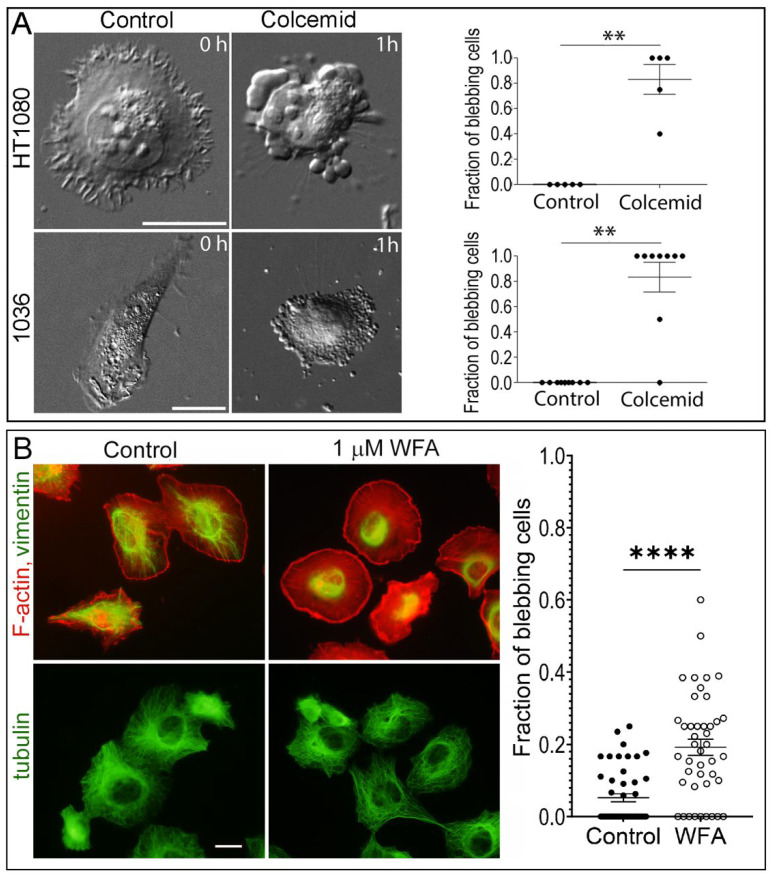
The formation of specific cellular protrusions, plasma membrane blebs, underlies the amoeboid mode of cell motility, which is characteristic for free-living amoebae and leukocytes, and can also be adopted by stem and tumor cells to bypass unfavorable migration conditions and thus facilitate their long-distance migration. Not all cells are equally prone to bleb formation. We have previously shown that membrane blebbing can be experimentally induced in a subset of HT1080 fibrosarcoma cells, whereas other cells in the same culture under the same conditions retain non-blebbing mesenchymal morphology. Here we show that this heterogeneity is associated with the distribution of vimentin intermediate filaments (VIFs). Using different approaches to alter the VIF organization, we show that blebbing activity is biased toward cell edges lacking abundant VIFs, whereas the VIF-rich regions of the cell periphery exhibit low blebbing activity. This pattern is observed both in interphase fibroblasts, with and without experimentally induced blebbing, and during mitosis-associated blebbing. Moreover, the downregulation of vimentin expression or displacement of VIFs away from the cell periphery promotes blebbing even in cells resistant to bleb-inducing treatments. Thus, we reveal a new important function of VIFs in cell physiology that involves the regulation of non-apoptotic blebbing essential for amoeboid cell migration and mitosis.

Keywords: blebbing; cell cortex; mesenchymal-to-amoeboid transition; vimentin intermediate filaments.


Conflict of interest statement

The authors declare no conflict of interest.

Figures

Figure 1
Figure 1
Correlative phase contrast microscopy and PREM of cells treated with 200 µM CK-666 for 1 h and then allowed to restore lamellipodia after drug washout. (AC) Example of a cell that switched to blebbing under CK-666 treatment. (DF) Example of a cell that did not switch to blebbing under CK-666 treatment. (A,D) Phase contrast images taken before CK-666 application (left), under 1 h CK-666 treatment (middle) and 1 h after CK-666 washout (right). Blebs and lamellipodia are marked by arrows and arrowheads, respectively. (B,E) Correlative PREM images with NMII immunogold staining after detergent extraction and gelsolin treatment of the cells shown in (A) and (D), respectively. (C,F) Enlarged boxed regions from (B) and (E), respectively. IFs are pseudocolored in blue; unlabeled long filaments represent microtubules. Boxed regions in main panels are enlarged in insets, where microtubules are pseudocolored in red and IFs in blue. Bars, 10 µm (A,B,D,E), 500 nm (C,F), and 50 nm ((C,F) insets).
Figure 2
Figure 2
Vimentin enrichment at the cell periphery negatively correlates with cell edge blebbing. (A,B) Correlative DIC (inserts) and anti-vimentin immunofluorescence microscopy (main panels) of HT1080 cells treated with 200 μM CK-666. (A) In the non-blebbing cell, the VIF network is distributed throughout the cell up to the cell periphery. (B) In the blebbing cell, the VIF network is sparse at the cell periphery and concentrates in the perinuclear region. (C) Center-to-periphery distribution of the mean immunofluorescence intensity of vimentin in blebbing (red, n = 24 cells) and non-blebbing (blue, n = 19 cells) HT1080 cells treated with CK-666 and in untreated normal 1036 fibroblasts (green, n = 7 cells); mean ± SEM; *, p < 0.05; unpaired t-test with correction for multiple comparisons using Holm–Sidak method comparing data for blebbing and non-blebbing HT1080 cells for each zone. (D,E) Correlative DIC microscopy (left panels) and SIM of untreated blebbing (D) and non-blebbing (E) HT1080 cells stained with vimentin antibody and phalloidin for F-actin. Cell contours are outlined in yellow. Scale bars: 15 µm (A,B) and 10 µm (D,E).
Figure 3
Figure 3
Subcellular distribution of VIFs correlates with and predicts blebbing behavior in HT1080 cells. (A,B) EGFP-vimHT1080 cells treated with 200 µM CK-666 shown by DIC (left) and vimentin immunofluorescence microscopy (middle), and as a merged image (right). (A) Cell exhibiting non-polarized blebbing around the cell perimeter (arrowheads) has a relatively symmetric perinuclear distribution of VIFs with low levels of VIFs at the cell periphery. (B) A cell exhibiting polarized blebbing at one side of the cell (arrowhead) has an asymmetric distribution of VIFs enriched at the non-blebbing edge (arrow) and depleted at the blebbing edge (arrowhead). (CE) Correlative DIC microscopy ((C), frames from time-lapse video), and SIM (D,E) of the HT1080 cell with asymmetric blebbing induced by treatment with 200 µM CK-666 for 1 h. Blebs form at the cell edge devoid of a dense VIF network ((E), green outline), but not at the cell edge enriched in VIFs ((E), yellow outline). Vimentin is visualized by immunostaining; actin is stained by phalloidin. (FJ) Pre-existing VIF distribution predicts blebbing activity after application of CK-666. (F,H) Distribution of VIFs either throughout the cell (F) or in the perinuclear region (H) in EGFP-vimHT1080 cells before CK-666 application. (G,I) Frames from time-lapse DIC sequences of the cells shown in F and H, respectively, after CK-666 application. The cell with widely distributed VIFs (F) retracts but does not switch to blebbing for up to ~25 min after addition of 200 µM CK-666 (G). The cell with pre-existing perinuclear enrichment of VIFs (H) switches to blebbing within 30 sec after addition of 200 µM CK-666 (I). Time in min:sec. Scale bar 20 µm. (J) Percentage of area occupied by VIFs in cells exhibiting or not exhibiting blebbing after treatment with 200 µM CK-666 for 1 h. Box plot shows the quartiles (box), the 5th and 95th percentiles (whiskers), and outliers (dots); ****, p < 0.0001; unpaired t-test.
Figure 4
Figure 4
Subcellular distribution of VIFs correlates with blebbing behavior during mitosis. Mitotic EGFP-vimentin-expressing HT1080 cells shown by DIC (left column) and fluorescence microscopy (middle column), and by DIC/EGFP overlay (right column). (A) A cell at an early cytokinesis stage. VIFs (green) are enriched at the cleavage furrow and the nuclei, while blebbing occurs at the polar regions depleted of VIFs. (B,C) A mitotic cell shown during late cytokinesis (B) and after abscission (C). Time in min:sec. The daughter cell with broadly distributed VIFs (arrow) has lamellipodia, whereas the other daughter cell with perinuclear accumulation of VIFs (arrowhead) exhibits blebbing at late cytokinesis (B), but stops blebbing as soon as its VIF network is redistributed toward the periphery after abscission (C). Scale bars, 20 µm.
Figure 5
Figure 5
Pharmacological displacement of VIFs from cell periphery promotes blebbing. (A) Treatment of HT1080 (top) and 1036 (bottom) cells with 0.2 µM colcemid induces blebbing even without CK-666 treatment. Live-cell imaging by DIC microscopy before (left) and 1 h after colcemid treatment. Graphs show fractions of blebbing cells in individual experiments with and without colcemid treatment. Each data point represents an independent experiment (n = 71 HT1080 cells and 49 1036 cells across all experiments with 5–9 cells per experiment); lines show mean ± SEM; **, p < 0.0079, Mann–Whitney test. (B) Treatment of HT1080 cells with 1 μM WFA for 3 h. Immunofluorescence staining with antibodies to vimentin or α-tubulin and phalloidin. Scale bars, 20 μm. Graph shows fractions of blebbing cells per field of view in control (matching amount of DMSO) and WFA-treated cells (n = 547 control cells from 49 fields of view and 566 WFA-treated cells from 43 fields of view from 3 independent experiments); lines show mean ± SEM; ****, p < 0.0001, Mann–Whitney test.
Figure 6
Figure 6
Genetic depletion of VIFs promotes blebbing. (AC) Blebbing in HT1080scramble cells stably expressing scrambled shRNA (control) and vimentin-silenced HT1080shVim cells (shVim). (A) Representative Western blot of vimentin in control and HT1080shVim cells (top) and quantification of vimentin knockdown efficiency (bottom, % of control, mean ± SEM; n = 3 lysates/group; p < 0.05, t-test). Actin serves as loading control. (B) Immunofluorescence staining of vimentin (left) and relative immunofluorescence intensities of vimentin (right; mean ± SEM, n = 30 for each culture, ****, p < 0.0001, t-test) in control and HT1080shVim cells. Expression of shVim decreased the abundance of VIFs, which remained only in the perinuclear area. (C) Fractions of blebbing cells in control or HT1080shVim populations after treatment with 200 µM CK-666 or a matching amount of DMSO (mean +/− SEM from 3 independent experiments; n = 262, 312, and 397 for individual replicates). Mean values from each replicate were pooled and analyzed using Holm–Sidak’s multiple comparisons test, *, p < 0.05; **, p < 0.01. (D,E) Vimentin-null MFT-16 mouse fibroblasts exhibit greater fractions of blebbing cells after treatment with 100 µM CK-666, as compared with wild-type MFT-6 and vimentin-reconstituted MFT-16HVim fibroblasts. (D) Examples of mesenchymal, intermediate, and blebbing cell phenotypes. Scale bars, 10 µm. (E) Percentage of cells with different morphologies in the indicated conditions. Data show mean ± SEM for blebbing cells from three experiments; n = 65–203 cells per condition in each experiment. Statistical significance was determined using paired t-test; NS, not significant difference (p > 0.05); *, p < 0.05; **, p < 0.01.

Similar articles

Cited by

References

    1. Charras G.T., Yarrow J.C., Horton M.A., Mahadevan L., Mitchison T.J. Non-equilibration of hydrostatic pressure in blebbing cells. Nature. 2005;435:365–369. doi: 10.1038/nature03550. - DOI - PMC - PubMed
    1. Paluch E.K., Raz E. The role and regulation of blebs in cell migration. Curr. Opin. Cell Biol. 2013;25:582–590. doi: 10.1016/j.ceb.2013.05.005. - DOI - PMC - PubMed
    1. Charras G.T., Hu C.K., Coughlin M., Mitchison T.J. Reassembly of contractile actin cortex in cell blebs. J. Cell Biol. 2006;175:477–490. doi: 10.1083/jcb.200602085. - DOI - PMC - PubMed
    1. Charras G., Paluch E. Blebs lead the way: How to migrate without lamellipodia. Nat. Rev. Mol. Cell Biol. 2008;9:730–736. doi: 10.1038/nrm2453. - DOI - PubMed
    1. Sedzinski J., Biro M., Oswald A., Tinevez J.Y., Salbreux G., Paluch E. Polar actomyosin contractility destabilizes the position of the cytokinetic furrow. Nature. 2011;476:462–466. doi: 10.1038/nature10286. - DOI - PubMed

Publication types

LinkOut - more resources