

Evaluation of Rheumatic Diseases Affecting the Temporomandibular Joint: A Cone Beam Computed Tomography Study and Literature Review
- PMID: 38201313
- PMCID: PMC10795630
- DOI: 10.3390/diagnostics14010004
Evaluation of Rheumatic Diseases Affecting the Temporomandibular Joint: A Cone Beam Computed Tomography Study and Literature Review
Abstract
Introduction: Due to the silent manifestation of temporomandibular joint (TMJ), dentists and rheumatologists may neglect treatment for this joint.
Aims: The aim of this study was to investigate the TMJ components in patients with various rheumatic diseases and to compare them with a control group based on cone beam computed tomography (CBCT) images.
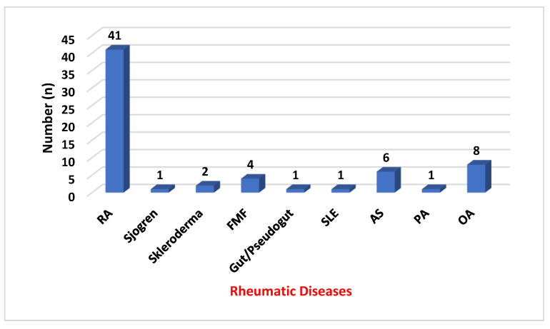
Materials and methods: This study comprised an assessment of the CBCT images of 65 patients (130 temporomandibular joints) with various rheumatic diseases (mostly rheumatoid arthritis) affecting the TMJ. Moreover, 65 patients (130 temporomandibular joints) with a similar age and gender distribution were examined as the control group. Pathologies were classified into a total of 12 types for the presence of any osseous changes in the condylar head or articular fossa or for joint space narrowing. Statistical analysis of all data was performed with SPSS version 18. The conformity of continuous variables to a normal distribution was examined by the Kolmogorov-Smirnov test. The Mann-Whitney U test was used to compare the means of two independent groups. The Pearson Chi-square test, Yates correction and Fisher's exact test were used in the analysis of categorical variables.
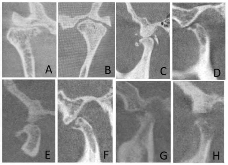
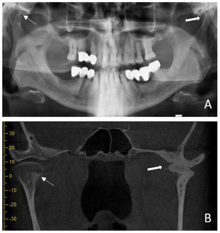
Results: The mean age of the patient and control groups was 50 ± 13 and 48 ± 16, respectively, and no statistically significant difference was found between the patient and control groups in terms of age distribution (p = 0.123). Condylar erosion, condylar flattening, subcondylar sclerosis, osteophytes, subcortical cysts, articular eminence resorption and articular eminence flattening rates were found to be statistically significantly higher in the patient group than in the control group (p < 0.05).
Conclusions: Dentomaxillofacial radiologists should examine the bony components of the TMJ in patients with rheumatic diseases, and a multidisciplinary approach involving a dental specialist and rheumatologist is required.
Keywords: cone beam computed tomography; rheumatic diseases; temporomandibular joint.
Conflict of interest statement
The authors declare no conflict of interest.
Figures
Similar articles
-
Evaluation of the effects of bisphosphonate therapy on the temporomandibular joint using cone beam computed tomography.Oral Radiol. 2025 Jul;41(3):430-437. doi: 10.1007/s11282-025-00816-3. Epub 2025 Mar 12. Oral Radiol. 2025. PMID: 40072733
-
Evaluation of osteoarthritic changes in the temporomandibular joint and their correlations with age: A retrospective CBCT study.Dent Med Probl. 2020 Jan-Mar;57(1):67-72. doi: 10.17219/dmp/112392. Dent Med Probl. 2020. PMID: 31997586
-
Assessment of bony changes in temporomandibular joint in patients using cone beam computed tomography - a cross sectional study.Head Face Med. 2023 Oct 28;19(1):47. doi: 10.1186/s13005-023-00392-z. Head Face Med. 2023. PMID: 37898789 Free PMC article.
-
Cone-Beam Computed Tomography for Temporomandibular Joint Imaging.Cureus. 2022 Nov 14;14(11):e31515. doi: 10.7759/cureus.31515. eCollection 2022 Nov. Cureus. 2022. PMID: 36532912 Free PMC article. Review.
-
The diagnostic accuracy of cone beam computed tomography in detecting temporomandibular joint bony disorders: a systematic review.Pol J Radiol. 2024 Jun 13;89:e292-e301. doi: 10.5114/pjr/187943. eCollection 2024. Pol J Radiol. 2024. PMID: 39040559 Free PMC article. Review.
Cited by
-
Temporomandibular joint arthritis in rheumatic diseases patients: which are the effective rehabilitative approaches for pain relief? A systematic review.BMC Musculoskelet Disord. 2025 Feb 18;26(1):159. doi: 10.1186/s12891-024-08196-1. BMC Musculoskelet Disord. 2025. PMID: 39966784 Free PMC article.
References
-
- Helenius L.M., Tervahartiala P., Helenius I., Al-Sukhun J., Kivisaari L., Suuronen R., Kautiainen H., Hallikainen D., Lindqvist C., Leirisalo-Repo M. Clinical radiographic and MRI findings of the temporomandibular joint in patients with different rheumatic diseases. Int. J. Oral Max. Surg. 2006;35:983–989. doi: 10.1016/j.ijom.2006.08.001. - DOI - PubMed
-
- Cunha S.C.d., Nogueira R.V.B., Duarte Â.P., Vasconcelos B.C.d.E., Almeida R.d.A.C. Analysis of helkimo and craniomandibular indexes for temporomandibular disorder diagnosis on rheumatoid arthritis patients. Rev. Bras. Otorrinolaringol. 2007;73:19–26. doi: 10.1590/S0034-72992007000100004. - DOI - PMC - PubMed
LinkOut - more resources
Full Text Sources
