Machine Learning Algorithm: Texture Analysis in CNO and Application in Distinguishing CNO and Bone Marrow Growth-Related Changes on Whole-Body MRI
- PMID: 38201370
- PMCID: PMC10804385
- DOI: 10.3390/diagnostics14010061
Machine Learning Algorithm: Texture Analysis in CNO and Application in Distinguishing CNO and Bone Marrow Growth-Related Changes on Whole-Body MRI
Abstract
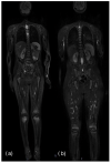
Objective: The purpose of this study is to analyze the texture characteristics of chronic non-bacterial osteomyelitis (CNO) bone lesions, identified as areas of altered signal intensity on short tau inversion recovery (STIR) sequences, and to distinguish them from bone marrow growth-related changes through Machine Learning (ML) and Deep Learning (DL) analysis.
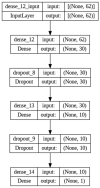
Materials and methods: We included a group of 66 patients with confirmed diagnosis of CNO and a group of 28 patients with suspected extra-skeletal systemic disease. All examinations were performed on a 1.5 T MRI scanner. Using the opensource 3D Slicer software version 4.10.2, the ROIs on CNO lesions and on the red bone marrow were sampled. Texture analysis (TA) was carried out using Pyradiomics. We applied an optimization search grid algorithm on nine classic ML classifiers and a Deep Learning (DL) Neural Network (NN). The model's performance was evaluated using Accuracy (ACC), AUC-ROC curves, F1-score, Positive Predictive Value (PPV), Mean Absolute Error (MAE) and Root-Mean-Square Error (RMSE). Furthermore, we used Shapley additive explanations to gain insight into the behavior of the prediction model.
Results: Most predictive characteristics were selected by Boruta algorithm for each combination of ROI sequences for the characterization and classification of the two types of signal hyperintensity. The overall best classification result was obtained by the NN with ACC = 0.91, AUC = 0.93 with 95% CI 0.91-0.94, F1-score = 0.94 and PPV = 93.8%. Between classic ML methods, ensemble learners showed high model performance; specifically, the best-performing classifier was the Stack (ST) with ACC = 0.85, AUC = 0.81 with 95% CI 0.8-0.84, F1-score = 0.9, PPV = 90%.
Conclusions: Our results show the potential of ML methods in discerning edema-like lesions, in particular by distinguishing CNO lesions from hematopoietic bone marrow changes in a pediatric population. The Neural Network showed the overall best results, while a Stacking classifier, based on Gradient Boosting and Random Forest as principal estimators and Logistic Regressor as final estimator, achieved the best results between the other ML methods.
Keywords: bone marrow; children; chronic non-bacterial osteomyelitis; machine learning; texture analysis; whole-body magnetic resonance imaging.
Conflict of interest statement
The authors declare no conflict of interest.
Figures


References
-
- d’Angelo P., de Horatio L.T., Toma P., Müller L.-S.O., Avenarius D., von Brandis E., Zadig P., Casazza I., Pardeo M., Pires-Marafon D., et al. Chronic nonbacterial osteomyelitis—Clinical and magnetic resonance imaging features. Pediatr. Radiol. 2021;51:282–288. doi: 10.1007/s00247-020-04827-6. - DOI - PMC - PubMed
LinkOut - more resources
Full Text Sources

