

Subtypes of Melanomas Associated with Different Degrees of Actinic Elastosis in Conventional Histology, Irrespective of Age and Body Site, Suggesting Chronic Ultraviolet Light Exposure as Driver for Lentigo Maligna Melanoma and Nodular Melanoma
- PMID: 38201430
- PMCID: PMC10778567
- DOI: 10.3390/cancers16010001
Subtypes of Melanomas Associated with Different Degrees of Actinic Elastosis in Conventional Histology, Irrespective of Age and Body Site, Suggesting Chronic Ultraviolet Light Exposure as Driver for Lentigo Maligna Melanoma and Nodular Melanoma
Abstract
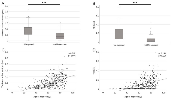
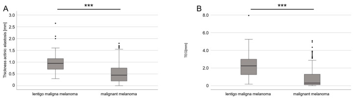
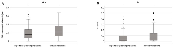
(1) Background: Ultraviolet (UV) radiation and sunburns are associated with an increased incidence of acquired nevi and melanomas. However, the data are controversial as to whether chronic UV exposure or high intermittent UV exposure is the major carcinogenic factor in melanocytic tumors. In this study, we compared the degree of actinic elastosis (AE) as a surrogate for lifetime UV exposure in nevi and different clinical melanoma subtypes (i.e., superficial spreading melanoma (SSM), nodular malignant melanoma (NMM), acral lentiginous melanoma (ALM), and lentigo maligna melanoma (LMM)) with respect to clinical variables (age, sex, and body site). (2) Methods: We defined a semi-quantitative score for the degree of AE ranging from 0 = none to 3 = total loss of elastic fibers (basophilic degeneration) and multiplied it by the perilesional vertical extent (depth), measured histometrically (tumor-associated elastosis grade (TEG)). We matched the TEG of n = 595 melanocytic lesions from 559 patients with their clinical variables. (3) Results: The TEG was correlated with age and UV-exposed body sites. Furthermore, the TEG was significantly higher in LMM than in all other types of melanomas and the TEG in NMM was higher than in SSM, irrespective of patient age and tumor site. (4) Conclusions: High cumulative UV exposure is more strongly associated with LMM and NMM than with other melanoma subtypes.
Keywords: actinic elastosis; carcinogenesis; histopathology; melanoma; nevogenesis.
Conflict of interest statement
D.N. received financial support (speaker’s honoraria, advisory boards, travel expense reimbursements or grants) from: Abbvie, Almirall, Boehringer Ingelheim, Bristol-Myers-Squib, GlaxoSmithKline, Incyte, Janssen-Cilag, Kyowa Kirin, LEO Pharma, Lilly, L’Oreal/Cerave, MSD, Novartis, Pfizer, and UCB Pharma. K.D. received financial support (speaker’s honoraria, advisory boards, travel expense reimbursements or grants) from: Abbvie, Bristol-Myers-Squib, Novartis, and Pierre-Fabre. The remaining authors declare no competing financial interests regarding the content of the manuscript. The funders had no role in the design of the study; in the collection, analyses, or interpretation of data; in the writing of the manuscript; or in the decision to publish the results.
Figures

Similar articles
-
Retrospective Single-Center Case Study of Clinical Variables and the Degree of Actinic Elastosis Associated with Rare Skin Cancers.Biology (Basel). 2024 Jul 16;13(7):529. doi: 10.3390/biology13070529. Biology (Basel). 2024. PMID: 39056721 Free PMC article.
-
Degree of Actinic Elastosis Is a Surrogate of Exposure to Chronic Ultraviolet Radiation and Correlates More Strongly with Cutaneous Squamous Cell Carcinoma than Basal Cell Carcinoma.Life (Basel). 2023 Mar 17;13(3):811. doi: 10.3390/life13030811. Life (Basel). 2023. PMID: 36983966 Free PMC article.
-
Sun exposure and host phenotype as predictors of cutaneous melanoma associated with neval remnants or dermal elastosis.Int J Cancer. 2006 Aug 1;119(3):636-42. doi: 10.1002/ijc.21907. Int J Cancer. 2006. PMID: 16572428
-
Role of In Vivo Reflectance Confocal Microscopy in the Analysis of Melanocytic Lesions.Acta Dermatovenerol Croat. 2018 Apr;26(1):64-67. Acta Dermatovenerol Croat. 2018. PMID: 29782304 Review.
-
Histopathologic classification and prognostic factors of melanoma: a 2021 update.Ital J Dermatol Venerol. 2021 Jun;156(3):300-321. doi: 10.23736/S2784-8671.21.06958-3. Epub 2021 May 13. Ital J Dermatol Venerol. 2021. PMID: 33982546 Review.
Cited by
-
Impact of Patient's Age and Physician's Professional Background on the Number Needed to Treat in Malignant Melanoma Detection.Cancers (Basel). 2024 Nov 29;16(23):4014. doi: 10.3390/cancers16234014. Cancers (Basel). 2024. PMID: 39682200 Free PMC article.
-
The influence of viscosity of hydrogels on the spreading and migration of cells in 3D bioprinted skin cancer models.Front Cell Dev Biol. 2024 May 21;12:1391259. doi: 10.3389/fcell.2024.1391259. eCollection 2024. Front Cell Dev Biol. 2024. PMID: 38835508 Free PMC article. Review.
-
Retrospective Single-Center Case Study of Clinical Variables and the Degree of Actinic Elastosis Associated with Rare Skin Cancers.Biology (Basel). 2024 Jul 16;13(7):529. doi: 10.3390/biology13070529. Biology (Basel). 2024. PMID: 39056721 Free PMC article.
-
Survival status of women with cervical cancer in Sub-Saharan Africa: a systematic review and meta-analysis, 2024.Front Oncol. 2025 Jan 7;14:1491840. doi: 10.3389/fonc.2024.1491840. eCollection 2024. Front Oncol. 2025. PMID: 39839767 Free PMC article.
References
-
- International Agency for Research on Cancer . In: WHO Classification of Skin Tumours. 4th ed. Elder D.E., editor. International Agency for Research on Cancer; Lyon, France: 2018.
LinkOut - more resources
Full Text Sources
