

Intragenic EGFR::EGFR.E1E8 Fusion (EGFRvIII) in 4331 Solid Tumors
- PMID: 38201434
- PMCID: PMC10778229
- DOI: 10.3390/cancers16010006
Intragenic EGFR::EGFR.E1E8 Fusion (EGFRvIII) in 4331 Solid Tumors
Abstract
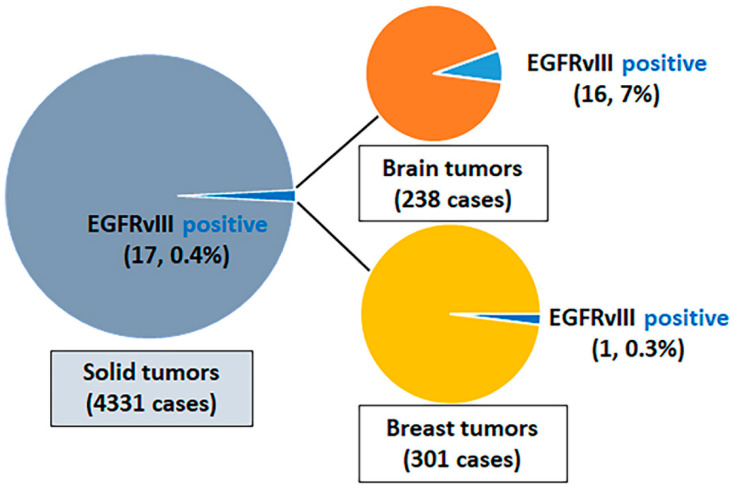
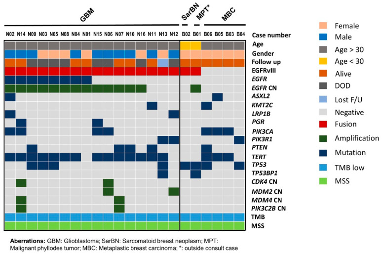
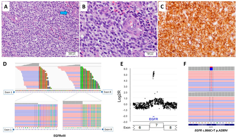
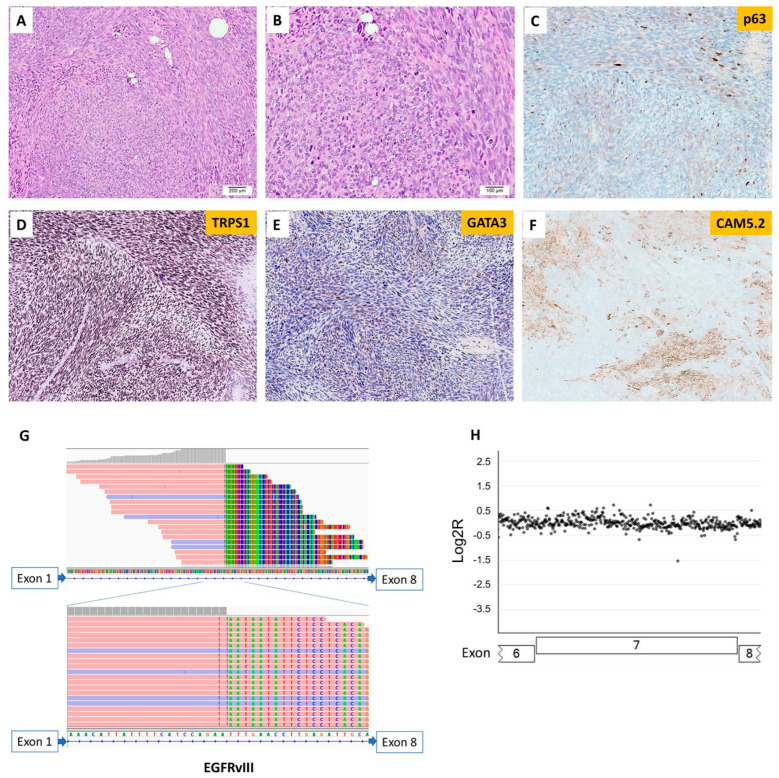
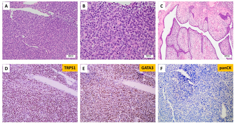
Epidermal growth factor receptor variant III (EGFRvIII, the deletion of exons 2-7) is a recurrent intragenic EGFR::EGFR.E1E8 fusion that occurs in high-grade gliomas. The presence of EGFRvIII in other solid tumors has not been well characterized. We retrospectively reviewed advanced malignant solid tumor cases tested by a custom hybrid capture 610-gene next-generation sequencing platform from 2021 to 2022. EGFRvIII was identified in 17 of 4331 (0.4%) cases, including 16 of 238 (7%) brain tumors and 1/301 (0.3%) breast tumors. EGFRvIII-positive brain tumors were all glioblastoma IDH-wildtype, most with concurrent TERT promoter mutation (14 of 16), EGFR amplification (13 of 16), and EGFR mutation (8 of 16). The only EGFRvIII-positive breast lesion was a sarcomatoid neoplasm in a young female patient. A separate breast case tested outside our institution with reported EGFRvIII was noted in a young female patient with a malignant phyllodes tumor with stromal overgrowth. Microscopically, both EGFRvIII-positive breast tumors showed high-grade sarcomatoid morphology with brisk mitotic activity. In summary, EGFRvIII is rare, occurring primarily in glioblastoma and rarely in breast sarcomatoid neoplasm, with no instances identified in other tumor types in our series. This select group of patients may benefit from chemotherapy and/or targeted anti-EGFR therapy.
Keywords: EGFR::EGFR.E1E8 fusion; EGFRvIII; breast sarcomatoid neoplasm; glioblastoma; malignant phyllodes tumor.
Conflict of interest statement
The authors declare no conflict of interest.
Figures
Similar articles
-
Epidermal Growth Factor Receptor Variant III (EGFRvIII) Positivity in EGFR-Amplified Glioblastomas: Prognostic Role and Comparison between Primary and Recurrent Tumors.Clin Cancer Res. 2017 Nov 15;23(22):6846-6855. doi: 10.1158/1078-0432.CCR-17-0890. Epub 2017 Aug 29. Clin Cancer Res. 2017. PMID: 28855349
-
Simultaneous detection of EGFR amplification and EGFRvIII variant using digital PCR-based method in glioblastoma.Acta Neuropathol Commun. 2020 Apr 17;8(1):52. doi: 10.1186/s40478-020-00917-6. Acta Neuropathol Commun. 2020. PMID: 32303258 Free PMC article. Clinical Trial.
-
Molecular characterization of EGFR and EGFRvIII signaling networks in human glioblastoma tumor xenografts.Mol Cell Proteomics. 2012 Dec;11(12):1724-40. doi: 10.1074/mcp.M112.019984. Epub 2012 Sep 10. Mol Cell Proteomics. 2012. PMID: 22964225 Free PMC article.
-
The EGFR variant III mutant as a target for immunotherapy of glioblastoma multiforme.Eur J Pharmacol. 2017 Sep 5;810:70-82. doi: 10.1016/j.ejphar.2017.05.064. Epub 2017 Jun 2. Eur J Pharmacol. 2017. PMID: 28583430 Review.
-
The epidermal growth factor receptor variant III (EGFRvIII): where wild things are altered.FEBS J. 2013 Nov;280(21):5350-70. doi: 10.1111/febs.12393. Epub 2013 Jul 8. FEBS J. 2013. PMID: 23777544 Review.
Cited by
-
EGFR transcript variants: Potential diagnostic biomarker for glioblastoma, IDH-wildtype?J Neuropathol Exp Neurol. 2025 Apr 25:nlaf041. doi: 10.1093/jnen/nlaf041. Online ahead of print. J Neuropathol Exp Neurol. 2025. PMID: 40277394 No abstract available.
-
Robotic vs laparoscopic approaches of pancreatic resection: a systematic review and meta-analysis.J Robot Surg. 2025 Jun 16;19(1):295. doi: 10.1007/s11701-025-02446-7. J Robot Surg. 2025. PMID: 40522553 Review.
-
Precision Neuro-Oncology in Glioblastoma: AI-Guided CRISPR Editing and Real-Time Multi-Omics for Genomic Brain Surgery.Int J Mol Sci. 2025 Jul 30;26(15):7364. doi: 10.3390/ijms26157364. Int J Mol Sci. 2025. PMID: 40806492 Free PMC article. Review.
-
Alternative Splicing in Tumorigenesis and Cancer Therapy.Biomolecules. 2025 May 29;15(6):789. doi: 10.3390/biom15060789. Biomolecules. 2025. PMID: 40563429 Free PMC article. Review.
References
LinkOut - more resources
Full Text Sources
Research Materials
Miscellaneous
