. 2023 Dec 20;16(1):34.
doi: 10.3390/cancers16010034.

Sunitinib Treatment of VHL C162F Cells Slows Down Proliferation and Healing Ability via Downregulation of ZHX2 and Confers a Mesenchymal Phenotype

Affiliations

Sunitinib Treatment of VHL C162F Cells Slows Down Proliferation and Healing Ability via Downregulation of ZHX2 and Confers a Mesenchymal Phenotype

Stéphanie Buart et al. Cancers (Basel). .

Abstract

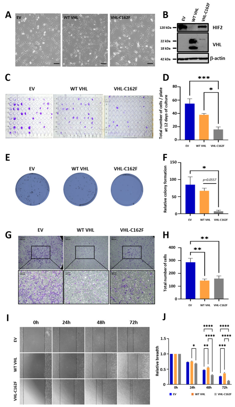
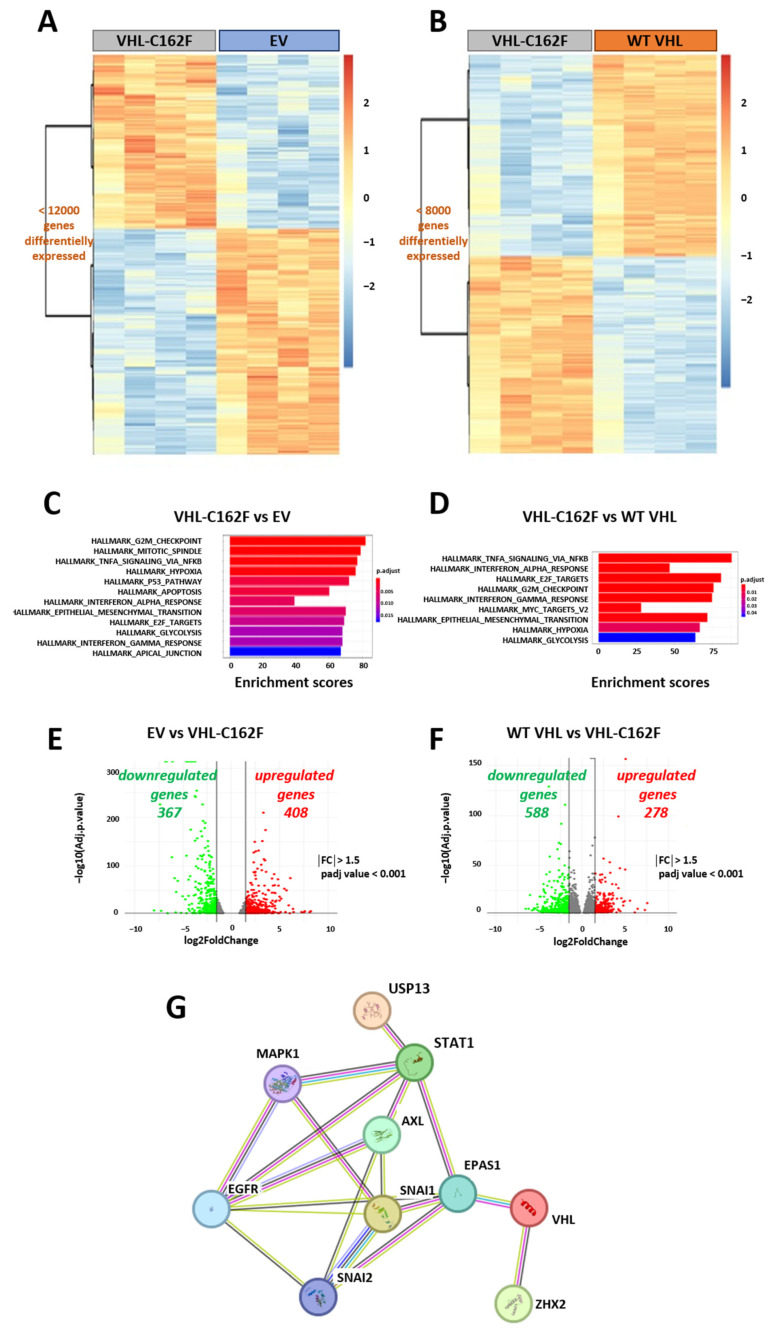
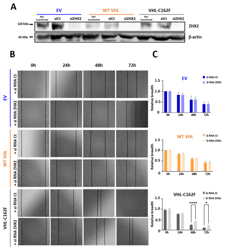
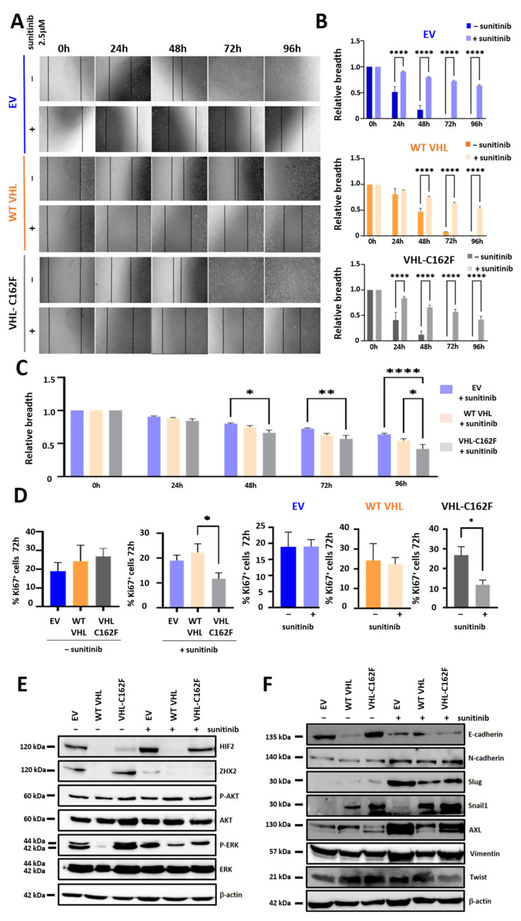
von Hippel-Lindau (VHL) disease, due to mutations of the tumor suppressor VHL gene, is a rare hereditary syndrome with a high risk of developing clear cell renal cell carcinoma (ccRCC). We asked whether the VHL-C162F mutation interferes with proliferation, migration, healing and forming colony ability by using wild-type VHL (WT VHL) and VHL-C162F reconstituted cells. We then analyzed the in vitro impact of the sunitinib treatment on VHL-C162F cells. We showed that VHL-C162F mutations have no impact on cell morphology, colony formation and migration ability but confer a significant higher healing ability than in WT VHL cells. RNA sequencing analysis revealed that VHL-C162F mutation upregulates genes involved in hypoxia and epithelial mesenchymal transition (EMT) pathways by comparison with VHL WT cells. We next showed a decrease in healing ability in VHL-C162F cells depleting on ZHX2, an oncogenic driver of ccRCC, highlighting the potential involvement of ZHX2 in aggressiveness of the VHL-C162F cells. Moreover, we found that sunitinib treatment inhibits ZHX2 expression and induces a reduced proliferation correlating with downregulation of P-ERK. Sunitinib treatment also conferred a more mesenchymal profile to VHL-C162F cells with significant downregulation of E-cadherin and upregulation of N-cadherin, Slug and AXL. Sunitinib therapy may therefore promote disease progression in VHL-C162F patients.

Keywords: VHL-C162F mutation; ZHX2; ccRCCs; epithelial mesenchymal transition; hypoxia-related genes; sunitinib.


Conflict of interest statement

The authors declare no conflict of interest. The funders had no role in the design of the study, in the collection, analyses, or interpretation of data, in the writing of the manuscript, or in the decision to publish the results.

Figures

Figure 1
Figure 1
Morphology, colony formation, migration potential and healing ability of C162F VHL mutated cells. (A): Phase contrast microscopy images showing the similar morphology of the three cells lines, EV, WT VHL, and VHL-C162F. Scale bar, 100 µm. (B): Western blot analysis of HIF-2α and VHL proteins from EV, WT VHL and VHL-C162F cells. β-Actin is used as a loading control. (C): Single cell culture by sorting for the three cell lines. Photographs correspond to one representative 96-well microplate. (D): The count of the colony corresponds to the total number of EV, WT VHL and C162F cells in one 96-well microplates (n = 6). * p ≤ 0.05 and *** p ≤ 0.001 using One Way ANOVA analysis and Bonferroni test. (E): Clonogenic assay using EV, WT VHL, and VHL-C162F cell lines. Photographs correspond to one representative well of a 6-well microplate. (F): The count of the colony is the mean of nine wells (n = 3). * p ≤ 0.05 using One Way ANOVA analysis and Tukey test. (G): The migration ability of EV, WT VHL and VHL-C162F was determined by transwell cell migration assays. Respectively, scale bars are 500 μm and 200 μm for photographs up and down. (H): The total number of migrated cells is shown in the bar graphs (n = 3). ** p ≤ 0.01 using One Way ANOVA analysis and Tukey test. (I): Representative scratch wound images showing the healing ability in EV, WT VHL and VHL-C162F cells at 0, 24, 48 and 72 h. (J): Graphs represent normalized relative breadth measured at 24, 48 and 72 h for the 3 cells lines (n = 12). * p ≤ 0.05, ** p ≤ 0.01, *** p ≤ 0.001 and **** p ≤ 0.0001 using One Way ANOVA analysis and Bonferroni test. The uncropped bolts are shown in Supplementary Materials Figure S1.
Figure 2
Figure 2
RNA-Seq analysis revealed discrete transcriptional differences between WT VHL and VHL-C162F cells reflecting an increase in cell plasticity in mutated cells. (A): Heatmap from RNA-Seq analysis comparing EV and VHL-C162F cells in triplicate. (B): Heatmap from RNA-Seq analysis comparing WT VHL and VHL-C162F cells in triplicate. (C): Differentially enriched gene sets nominated from GSEA analysis in VHL-C162F vs. EV cells classified by enrichment scores. A positive score indicates enrichment. (D): Differentially enriched gene sets nominated from GSEA analysis in VHL-C162F vs. WT VHL cells classified by enrichment scores. A positive score indicates enrichment. (E): Volcano plot of genes differentially expressed (Log10 fold change) in VHL-C162F vs. EV cells and (F): in VHL-C162F cells vs. WT VHL cells. For VHL-C162F vs. EV, a total of 367 downregulated genes and 408 upregulated genes were identified with a fold change >1.5, FDR < 0.001. For VHL-C162F vs. WT VHL, a total of 588 downregulated genes and 278 upregulated genes were identified with a fold change >1.5, padj value < 0.001. (G): Protein–Protein Interaction Networks showing Functional Enrichment Analysis by STRING. VHL interacts with some proteins as EPAS1 (HIF2α), STAT1, USP13, ZHX2, EGFR, AXL, SNAI1, SNAI2 (Slug) and MAPK1.
Figure 3
Figure 3
Loss of ZHX2 significantly reduced the healing ability of VHL-C162F mutated cells. (A): Western blot analysis of ZHX2 and β actin in cells from wound healing assay. Protein extracts were from EV, WT VHL and VHL-C162F cells not transfected and transfected with siRNA control or siRNA ZHX2. β-Actin is used as a loading control. (B): Representative scratch wound images showing the healing ability in EV, WT VHL and VHL-C162F cells transfected with siRNA control or with a mix of siRNA ZHX2. Photographs were taken at 0, 24, 48 and 72 h. (C): Graphs represent normalized relative breadth measured at 24, 48 and 72 h for the 3 cells lines transfected with siRNA control or ZHX2 siRNAS (n = 3). * p ≤ 0.05 and **** p ≤ 0.0001 using One Way ANOVA analysis and Bonferroni test. The uncropped bolts are shown in Supplementary Materials Figure S2.
Figure 4
Figure 4
Sunitinib significantly reduced the healing ability of VHL-C162F mutated cells by inhibiting the expression of ZHX2 and inducing a more mesenchymal profile with upregulation of N-cadherin, Slug and AXL. (A): Representative scratch wound images showing the reduced healing ability in EV, WT VHL and VHL-C162F cells treated with sunitinib at 2.5 μM. Photographs were taken at 0, 24, 48, 72 and 96 h. (B): The healing ability is compared for each cell line not treated versus treated with sunitinib 2.5 μM at 0, 24, 48, 72 and 96 h (n = 10). **** p ≤ 0.0001 using One Way ANOVA analysis and Bonferroni test. (C): The healing ability is compared for the three cell lines treated with sunitinib 2.5 μM (n = 10). * p ≤ 0.05, ** p ≤ 0.01 and **** p ≤ 0.0001 using One Way ANOVA analysis and Bonferroni test. (D): Intracellular expression of Ki67 in the three cell lines not treated and treated to sunitinib 2.5 μM at 72 h. Graphs represent percentages of Ki67 cells in the three cell lines not treated and treated with sunitinib 2.5 μM at 72 h (n = 5). * p ≤ 0.05 using One Way ANOVA analysis and Bonferroni test. Graphs represent percentages of Ki67 in EV or WT VHL or C162F cells not treated and treated with sunitinib 2.5 μM at 72 h (n = 5). * p ≤ 0.05 using Mann-Whitney U test. (E): Western blot analysis of HIF2, ZHX2, P-AKT, AKT, P-ERK, ERK and β actin in cells lines not treated and treated with sunitinib 2.5 μM. β-Actin is used as a loading control. Photographs correspond to one representative experiment. Quantifications with ImageJ are in Figure S1. (F): Western blot analysis of E-cadherin, N-cadherin, Slug, Snail1, AXL, Vimentin, Twist and β actin in cells lines not treated and treated with sunitinib 2.5 μM. β-Actin is used as a loading control. Photographs correspond to one representative experiment. Quantifications with ImageJ are in Figure S3. The uncropped bolts are shown in Supplementary Materials Figures S4–S9.

Similar articles

Cited by

References

    1. Sharma A., Mukewar S., Vege S.S. Clinical Profile of Pancreatic Cystic Lesions in von Hippel-Lindau Disease: A Series of 48 Patients Seen at a Tertiary Institution. Pancreas. 2017;46:948–952. doi: 10.1097/MPA.0000000000000875. - DOI - PubMed
    1. Rathmell W.K., Chen S. VHL inactivation in renal cell carcinoma: Implications for diagnosis, prognosis and treatment. Expert Rev. Anticancer. Ther. 2008;8:63–73. doi: 10.1586/14737140.8.1.63. - DOI - PMC - PubMed
    1. Maher E.R., Neumann H.P.H., Richard S. von Hippel–Lindau disease: A clinical and scientific review. Eur. J. Hum. Genet. 2011;19:617–623. doi: 10.1038/ejhg.2010.175. - DOI - PMC - PubMed
    1. Kim E., Zschiedrich S. Renal Cell Carcinoma in von Hippel-Lindau Disease-From Tumor Genetics to Novel Therapeutic Strategies. Front. Pediatr. 2018;6:16. doi: 10.3389/fped.2018.00016. - DOI - PMC - PubMed
    1. Tabata M., Sato Y., Kogure Y., McClure M.B., Oshikawa-Kumade Y., Saito Y., Shingaki S., Ito Y., Yuasa M., Koya J., et al. Inter- and intra-tumor heterogeneity of genetic and immune profiles in inherited renal cell carcinoma. Cell Rep. 2023;42:112736. doi: 10.1016/j.celrep.2023.112736. - DOI - PubMed