Review
. 2023 Dec 20;16(1):36.
doi: 10.3390/cancers16010036.

Natural Anticancer Peptides from Marine Animal Species: Evidence from In Vitro Cell Model Systems

Affiliations
Review

Natural Anticancer Peptides from Marine Animal Species: Evidence from In Vitro Cell Model Systems

Mariangela Librizzi et al. Cancers (Basel). .

Abstract

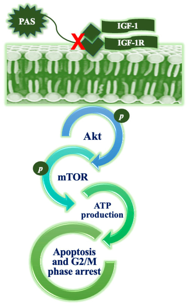
Anticancer peptides are short and structurally heterogeneous aminoacidic chains, which display selective cytotoxicity mostly against tumor cells, but not healthy cells, based on their different cell surface properties. Their anti-tumoral activity is carried out through interference with intracellular homeostasis, such as plasmalemma integrity, cell cycle control, enzymatic activities and mitochondrial functions, ultimately acting as angiogenesis-, drug resistance- and metastasis-inhibiting agents, immune stimulators, differentiation inducers and necrosis or extrinsic/intrinsic apoptosis promoters. The marine environment features an ever-growing level of biodiversity, and seas and oceans are poorly exploited mines in terms of natural products of biomedical interest. Adaptation processes to extreme and competitive environmental conditions led marine species to produce unique metabolites as a chemical strategy to allow inter-individual signalization and ensure survival against predators, infectious agents or UV radiation. These natural metabolites have found broad use in various applications in healthcare management, due to their anticancer, anti-angiogenic, anti-inflammatory and regeneration abilities. The aim of this review is to pick selected studies that report on the isolation of marine animal-derived peptides and the identification of their anticancer activity in in vitro cultures of cancer cells, and list them with respect to the taxonomical hierarchy of the source organism.

Keywords: Annelida; Arthropoda; Chordata; Cnidaria; Echinodermata; Mollusca; Porifera; anticancer peptides; marine drugs; tumor cells.


Conflict of interest statement

The authors declare no conflict of interest.

Figures

Figure 1
Figure 1
Specimen of C. basilana. Credit: RECOLNAT (ANR-11-INBS-0004)—Marie Hennion—(CC-BY 4.0). http://mediaphoto.mnhn.fr/media/1457603880888AD4dw03lT3Rng1tE (accessed on 6 March 2023).
Figure 2
Figure 2
Structures of microcionamide A (1), C (2) and D (3) [24].
Figure 3
Figure 3
Specimen of G. corticostylifera. Author: Eduardo Hajdu (CC BY-NC-SA 4.0). https://www.marinespecies.org/porifera/porifera.php?p=image&tid=191313&pic=148142 (accessed on 6 March 2023).
Figure 4
Figure 4
Structures of geodiamolides A and B (1, X = I and Br, respectively), and geodiamolides H and I (2, X = I and Br, respectively) [26].
Figure 5
Figure 5
Specimen of C. lamellata, an example of Cymbastela sp. (CC.BY.4.0). https://en.wikipedia.org/wiki/Cymbastela_lamellata#/media/File:Cymbastela_tricalyciformis_(Bergquist,_1970)_(AM_MA36086-1).jpg (accessed on 16 January 2023).
Figure 6
Figure 6
Structure of hemiasterlins. Hemiasterlin: R1 and R2 = Me; hemiasterlin A: R1 = H, R2 = Me; hemiasterlin B: R1 and R2 = H. Redrawn from [29].
Figure 7
Figure 7
Specimen of A. elegantissima. Author: Neil McDaniel (CC BY-NC-SA 4.0). https://www.marinespecies.org/aphia.php?p=image&tid=283347&pic=117079 (accessed on 14 March 2023).
Figure 8
Figure 8
Specimen of A. anjunae. Author: Gaurav Patil (CC BY-NC). https://www.inaturalist.org/photos/61697779 (accessed on 20 September 2023).
Figure 9
Figure 9
Specimen of M. meretrix. Author: Joop Trausel and Frans Slieker (CC BY-NC-SA 4.0). https://www.marinespecies.org/aphia.php?p=image&tid=224891&pic=67475 (accessed on 16 January 2023).
Figure 10
Figure 10
Specimen of R. philippinarum. Author: Joop Trausel and Frans Slieker (CC BY-NC-SA 4.0). https://www.marinespecies.org/photogallery.php?album=700&pic=106682 (accessed on 14 March 2023).
Figure 11
Figure 11
Specimen of A. broughtonii. Author: Joop Trausel and Frans Slieker (CC BY-NC-SA 4.0). https://www.marinespecies.org/aphia.php?p=image&tid=504357&pic=50786 (accessed on 6 March 2023).
Figure 12
Figure 12
Specimen of A. kagoshimensis. Author: Joop Trausel and Frans Slieker (CC BY-NC-SA 4.0). https://www.marinespecies.org/photogallery.php?album=700&pic=50806 (accessed on 20 September 2023).
Figure 13
Figure 13
Scheme depicting the proposed mechanism of action of PAS, which induces cell cycle arrest and apoptosis downstream to the suppression of IGF-1R/Akt/mTOR signaling and ATP production. Redrawn from [52].
Figure 14
Figure 14
Specimen of T. granosa. Author: Joop Trausel and Frans Slieker (CC BY-NC-SA 4.0). https://www.marinespecies.org/photogallery.php?album=700&pic=51141 (accessed on 16 January 2023).
Figure 15
Figure 15
Specimen of D. auricularia. Author: Philippe Bourjon (CC BY-NC-SA 3.0). https://commons.wikimedia.org/wiki/File:Dolabella_auricularia.jpg (accessed on 16 January 2023).
Figure 16
Figure 16
Specimen of B. exarata. Author: Kirsten Van Laethem (CC BY-NC-SA). https://www.marinespecies.org/aphia.php?p=image&tid=533828&pic=126086 (accessed on 6 March 2023).
Figure 17
Figure 17
Specimen of S. esculenta. Author: Harum Koh (CC BY-SA 2.0). https://upload.wikimedia.org/wikipedia/commons/b/b1/Japan_squid%2C_Sepia_esculenta_%2815601195858%29.jpg (accessed on 16 January 2023).
Figure 18
Figure 18
Specimen of P. aibuhitensis. Author: Hong Zhou (CC BY-NC SA 3.0). https://eol.org/media/30500878 (accessed on 6 March 2023).
Figure 19
Figure 19
Specimens of L. vannamei. Author: Fotokannan (CC BY-SA 4.0). https://upload.wikimedia.org/wikipedia/commons/8/85/Pacific_white_shrimp.jpg?uselang=it (accessed on 20 September 2023).
Figure 20
Figure 20
Specimen of P. monodon. Author: CSIRO (CC-BY-3.0). https://upload.wikimedia.org/wikipedia/commons/2/2e/CSIRO_ScienceImage_2992_The_Giant_Tiger_Prawn.jpg (accessed on 14 March 2023).
Figure 21
Figure 21
Specimen of S. paramamosain. Author: Wibowo Djatmiko (CC BY-SA 4.0). https://en.wikipedia.org/wiki/Scylla_paramamosain#/media/File:Scyl_param_180225-5311834_mrd.JPG (accessed on 6 March 2023).
Figure 22
Figure 22
An example of the horseshoe crab, Tachypleus tridentatus. Author: Didier Descouens (CC BY-SA 4.0). https://upload.wikimedia.org/wikipedia/commons/f/f4/Limules.jpg?uselang=it (accessed on 14 March 2023).
Figure 23
Figure 23
Specimen of C. savignyi. Author: Robin Gwen Agarwal (CC BY-NC). https://inaturalist.nz/photos/1083563 (accessed on 20 September 2023).
Figure 24
Figure 24
Specimen of R. porosa. (CC-BY-SA-2.5). https://en.wikipedia.org/wiki/Ocellate_spot_skate#/media/File:Okamejei_kenojei2.jpg (accessed on 14 March 2023).
Figure 25
Figure 25
Specimen of E. coioides (CC-BY-SA-4.0). https://commons.wikimedia.org/wiki/File:Epinephelus_coioides_%28Orange_spotted_grouper%29_in_the_Philippines.jpg (accessed on 20 September 2023).
Figure 26
Figure 26
Specimen of O. niloticus. Author: Germano Roberto Schüür (CC-BY-SA-4.0). https://commons.wikimedia.org/w/index.php?curid=40488546 (accessed on 20 September 2023).
Figure 27
Figure 27
Proposed mechanism of action of TP3 on brain cancer cells, based upon the inhibition of FAK and RAS signalization, leading to the suppression of metalloproteases MMP2 and MMP9 in the tumor microenvironment [110] (CC-BY-4.0).
Figure 28
Figure 28
Proposed mechanism of action of TP3 on osteosarcoma cells, based upon the elevation of mitochondrial ROS production (↑), impairment of the activities of OXPHOS complexes and induction of caspase-9/3-mediated apoptosis. TP3-promoted apoptosis is also dependent upon the modulation of the expression levels of proteins associated with mitochondrial dynamics, such as OPA1, MFN1/2, FIS1 and DRP1. They lead to enhanced mitochondrial fission and, ultimately, to the destruction of mitochondrial function. The reducing agent mitoTempo may counteract ROS-mediated apoptosis [111].
Figure 29
Figure 29
Specimen of O. mossambicus (CC-BY-SA-3.0). https://commons.wikimedia.org/wiki/File:Oreochromis_mossambicus.jpg (accessed on 6 March 2023).
Figure 30
Figure 30
Specimen of P. marmoratus. Author: Philippe Bourjon (CC-BY-SA-4.0). https://commons.wikimedia.org/wiki/File:Pardachirus_marmoratus_%28Soleidae%29.jpg (accessed on 20 September 2023).
Figure 31
Figure 31
Proposed mechanism of action of pardaxin in fibrosarcoma cells. After cellular uptake, pardaxin selectively targets the endoplasmic reticulum, leading to Ca++ release and induction of c-FOS expression. Concurrently, an ROS-mediated stress response and MAPK signaling (e.g., ERK and JNK) contributes to mitochondrial dysfunction, activation of c-JUN/c-FOS complex and its downstream promoting effect on apoptosis [117].
Figure 32
Figure 32
Proposed mechanism of action of pardaxin in ovarian cancer cells. After cellular uptake, pardaxin induces ROS overproduction (↑) in mitochondria, reinforced also by the attenuation of OXPHOS enzymatic complexes (↓), an imbalance in MMP, the up-regulation of t-Bid and Bax and activation of caspase-9 and -3 cascade, leading to the mitochondrial pathway of apoptosis. Mitochondrial fragmentation also occurs in parallel with autophagosome formation, thereby suggesting the activation of mitophagy [118] (CC-BY-4.0).
Figure 33
Figure 33
Specimen of P. americanus. Author: Smithsonian Environmental Research Center (CC-BY-2.0). https://commons.wikimedia.org/wiki/File:Pseudopleuronectes_americanus_%28S0892%29_%2812658628105%29.jpg (accessed on 16 January 2023).
Figure 34
Figure 34
Proposed mechanism of action of NRC-03 on oral squamous cancer cells. The peptide targets the mitochondria and nucleus, causing mitochondria swelling, membrane blebbing and DNA fragmentation. ROS are produced in mitochondria via respiratory complex I (↑) in response to the increased oxygen consumption rate, and they activate MAPK/ERK and NF-κB signalization. NRC-03 also up-regulates cyclophilin D, thus stimulating mitochondrial pore opening and loss of transmembrane potential (↓ Δψm) that leads to the switching-on of apoptosis [121] (CC-BY-4.0).

Similar articles

Cited by

References

    1. Ghaly G., Tallima H., Dabbish E., Badr ElDin N., Abd El-Rahman M.K., Ibrahim M.A.A., Shoeib T. Anti-Cancer Peptides: Status and Future Prospects. Molecules. 2023;28:1148. doi: 10.3390/molecules28031148. - DOI - PMC - PubMed
    1. Xie M., Liu D., Yang Y. Anti-cancer peptides: Classification, mechanism of action, reconstruction and modification. Open Biol. 2020;10:200004. doi: 10.1098/rsob.200004. - DOI - PMC - PubMed
    1. Norouzi P., Mirmohammadi M., Houshdar Tehrani M.H. Anticancer peptides mechanisms, simple and complex. Chem. Biol. Interact. 2022;368:110194. doi: 10.1016/j.cbi.2022.110194. - DOI - PubMed
    1. Chinnadurai R.K., Khan N., Meghwanshi G.K., Ponne S., Althobiti M., Kumar R. Current research status of anti-cancer peptides: Mechanism of action, production, and clinical applications. Biomed. Pharmacother. 2023;164:114996. doi: 10.1016/j.biopha.2023.114996. - DOI - PubMed
    1. Rogers A.D., Appeltans W., Assis J., Balance L.T., Cury P., Duarte C., Favoretto F., Hynes L.A., Kumagai J.A., Lovelock C.E., et al. Discovering marine biodiversity in the 21st century. Adv. Mar. Biol. 2022;93:23–115. - PubMed

LinkOut - more resources