. 2023 Dec 20;16(1):43.
doi: 10.3390/cancers16010043.

Congenital Tumors-Magnetic Resonance Imaging Findings with Focus on Rare Tumors

Affiliations

Congenital Tumors-Magnetic Resonance Imaging Findings with Focus on Rare Tumors

Piotr Kwasniewicz et al. Cancers (Basel). .

Abstract

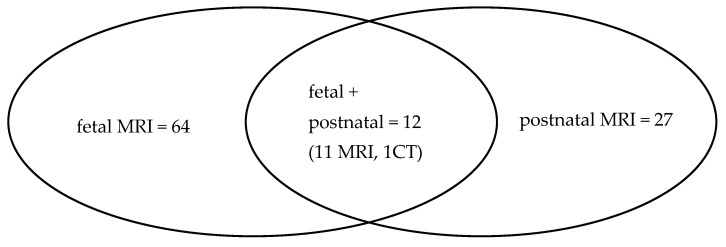
Congenital tumors are rare and, owing to this rarity, there is limited information on many of them. A total of 839 fetal and postnatal MRI studies performed in the first 3 months of life were retrospectively reviewed. They were performed with the use of 1.5 T scanners. Seventy-six tumors were diagnosed based on fetal MRI between 20 and 37 gestational weeks, and 27 were found after birth, from 1 day of age to 3 months of life. Teratomas were the most common tumors in our dataset, mainly in the sacrococcygeal region (SCT), followed by cardiac rhabdomyomas and subependymal giant cell astrocytomas (SEGA) associated with TSC, and neuroblastomas. The group of less common tumors consisted of infantile fibrosarcomas, malignant rhabdoid tumors, mesoblastic nephromas and Wilms tumor, craniopharyngiomas, brain stem gliomas, desmoplastic infantile astrocytoma, choroid plexus carcinoma, glioblastoma, hemangiopericytoma, rhabdomyosarcoma, melanoma, mesenchymal hamartomas of the chest wall and the liver, and juvenile xanthogranuloma, with special consideration of blue rubber bleb nevus syndrome. MRI plays a significant role in further and better characterization of congenital tumors, leading to a correct diagnosis in many cases, which is crucial for pregnancy and neonatal management and psychological preparation of the parents. No diagnosis is impossible and can be absolutely excluded.

Keywords: congenital tumor; fetus; infant; magnetic resonance imaging (MRI).


Conflict of interest statement

The authors declare no conflicts of interest.

Figures

Figure 1
Figure 1
Selection of the study group. * brain malformations such as callosal agenesis, heterotopias, schizencephaly, holoprosencephaly, rhombencephalosynapsis, hydrocephalus, etc.; skeletal deformations; cardiac malformations; renal agenesis; hernias. ** congenital cysts, i.e., duplication cysts, ovarian cysts, adrenal cysts; CCAM; cystic lymphatic malformations; etc.
Figure 2
Figure 2
Fetal and postnatal diagnoses in the presented material.
Figure 3
Figure 3
Different locations of teratomas (arrows) on fetal MRI (SSFSE/T2W and FIESTA images). (a) Intracranial; (b) facial; (c) anterior mediastinal; (d) abdominal; (e) sacrococcygeal (SCT type II).
Figure 4
Figure 4
Tuberous sclerosis complex (TSC) on fetal MRI. Cardiac rhabdomyoma: (a) sagittal FIESTA; (b) axial SSFSE/T2. Subependymal giant cell astrocytoma (SEGA): (c) T1WI; (d) SSFSE/T2WI.
Figure 5
Figure 5
Infantile fibrosarcoma (arrows) in a fetus at a GA of 29 weeks. Huge heterogenous solid and cystic back mass infiltrating vertebral arches. (a) Axial DWI; (b) sagittal SSFSE/T2WI; (c) axial SSFSE/T2WI; (d) axial FIESTA.
Figure 6
Figure 6
Malignant rhabdoid tumor (MRT) of the brachial plexus (arrows) with infiltration of the right clavicle, right lung, and muscles in a newborn girl. Inhomogeneous mass with low ADC values. (a) Sagittal T2WI; (b) coronal STIR; (c) axial DWI; (d) corresponding ADC map.
Figure 7
Figure 7
Wilms tumor arising from the left kidney in a newborn—encapsulated, heterogenous with necrosis, mass effect, contrast enhancement and restricted diffusion in solid parts. (a) Axial CE-T1WI; (b) axial T2WI; (c) DWI; (d) corresponding ADC map; (e) coronal FIESTA; (f) sagittal T2WI.
Figure 8
Figure 8
Diffuse brain stem glioma. Fetal MRI (arrows): (a) sagittal T2WI; (b) axial FIESTA. (c,d) US images of a brain stem mass reaching through the mastoid fontanelle after birth (asteriks).
Figure 9
Figure 9
Extra-pleural hemangiopericytoma/solitary fibrous tumor in a fetus (arrows). A mass of mixed signal intensity deriving from the face, with peripheral deposits of hemosiderin. (a) Sagittal SSFSE/T2WI; (b) axial GRE EPI; (c) axial FIESTA. Postnatal MRI: (d) sagittal T2WI; (e) CE-T1WI+Gd showing inhomogeneous contrast enhancement.
Figure 10
Figure 10
Rhabdomyosarcoma (arrows) in a newborn with a soft tissue mass in a forearm, adjacent to the ulna and radius, causing periosteal reaction, with restricted diffusion and inhomogeneous, moderate contrast enhancement. (a) US transverse section; (b,c) axial T2WI; (d) coronal CE-T1WI; (e) coronal STIR.
Figure 11
Figure 11
Malignant melanoma in a neonate (arrows). Solid, firm, occipital soft tissue lesion with strong contrast enhancement, without infiltration of the skull bones. (a) axial T2WI; (b) axial CE-T1WI; (c) axial GRE/T2*WI; (d,e) US sections.
Figure 12
Figure 12
Mesenchymal hamartomas of the chest wall. Bilateral lesions of the ribs in a fetus. (a) Coronal SSFSE/T2WI; (b) axial GRE EPI. Follow-up MRI after birth. (c) Axial T2WI; (d) axial T1WI; (e) DWI; (f) US transverse section; (g) coronal T2WI.
Figure 13
Figure 13
Juvenile xanthogranuloma. MRI of a neonate with irregular, solid, enhancing masses that are difficult to separate from internal organs. Increased volume of fluid in the abdominal cavity. (a,e,f) T2WI+fatsat: axial, coronal, and sagittal, respectively; (b) DWI; (c) corresponding ADC map; (d) axial CE-T1WI+fatsat.
Figure 14
Figure 14
Another case of BRBNS in a fetus. (a) Coronal SSFSE/T2WI. Postnatal MRI of the intra-abdominal mass with peripheral contrast enhancement and hemorrhagic foci (bright spots on T1-weighted images); (b) coronal T1WI; (c) coronal CE-T1WI+fatsat; (arrow points at another small lesion in the left liver lobe).

References

    1. Woodward P.J., Sohaey R., Kennedy A., Koeller K.K. A comprehensive review of fetal tumors with pathologic correlation. Radiographics. 2005;25:215–242. doi: 10.1148/rg.251045156. - DOI - PubMed
    1. Isaacs H. Perinatal (congenital and neonatal) neoplasms: A report of 110 cases. Pediatr. Pathol. 1985;3:165–210. doi: 10.3109/15513818509078782. - DOI - PubMed
    1. Moore S.W., Satg’e D., Sasco A.J., Zimmermann A., Plaschkes J. The epidemiology of neonatal tumours. Report of an international working group. Pediatr. Surg. Int. 2003;19:509–519. doi: 10.1007/s00383-003-1048-8. - DOI - PubMed
    1. Alamo L., Beck-Popovic M., Gudinchet F., Meuli R. Congenital tumors: Imaging when life just begins. Insights Imaging. 2011;2:297–308. doi: 10.1007/s13244-011-0073-8. - DOI - PMC - PubMed
    1. Bekiesinska-Figatowska M., Jurkiewicz E., Duczkowski M., Duczkowska A., Romaniuk-Doroszewska A., Brągoszewska H., Ceran A. Congenital CNS tumors diagnosed on prenatal MRI. Neuroradiol. J. 2011;24:477–481. doi: 10.1177/197140091102400402. - DOI - PubMed

LinkOut - more resources