. 2023 Dec 24;16(1):106.
doi: 10.3390/cancers16010106.

Improving HCC Prognostic Models after Liver Resection by AI-Extracted Tissue Fiber Framework Analytics

Affiliations

Improving HCC Prognostic Models after Liver Resection by AI-Extracted Tissue Fiber Framework Analytics

Rokas Stulpinas et al. Cancers (Basel). .

Abstract

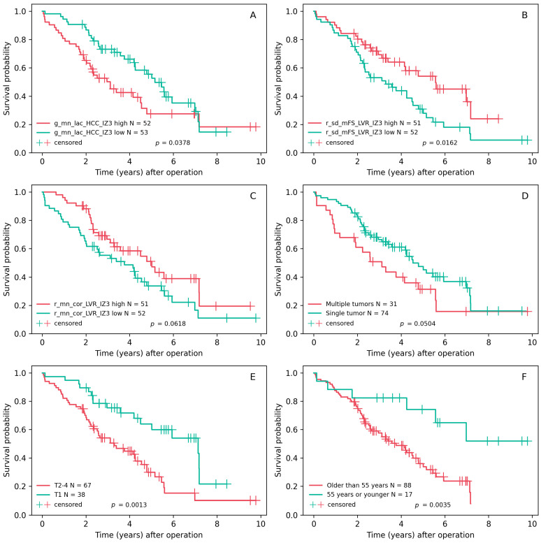
Despite advances in diagnostic and treatment technologies, predicting outcomes of patients with hepatocellular carcinoma (HCC) remains a challenge. Prognostic models are further obscured by the variable impact of the tumor properties and the remaining liver parenchyma, often affected by cirrhosis or non-alcoholic fatty liver disease that tend to precede HCC. This study investigated the prognostic value of reticulin and collagen microarchitecture in liver resection samples. We analyzed 105 scanned tissue sections that were stained using a Gordon and Sweet's silver impregnation protocol combined with Picric Acid-Sirius Red. A convolutional neural network was utilized to segment the red-staining collagen and black linear reticulin strands, generating a detailed map of the fiber structure within the HCC and adjacent liver tissue. Subsequent hexagonal grid subsampling coupled with automated epithelial edge detection and computational fiber morphometry provided the foundation for region-specific tissue analysis. Two penalized Cox regression models using LASSO achieved a concordance index (C-index) greater than 0.7. These models incorporated variables such as patient age, tumor multifocality, and fiber-derived features from the epithelial edge in both the tumor and liver compartments. The prognostic value at the tumor edge was derived from the reticulin structure, while collagen characteristics were significant at the epithelial edge of peritumoral liver. The prognostic performance of these models was superior to models solely reliant on conventional clinicopathologic parameters, highlighting the utility of AI-extracted microarchitectural features for the management of HCC.

Keywords: CNN; artificial intelligence; digital pathology; hepatocellular carcinoma; hexagonal grid; liver; overall survival; prognostic modelling.


Conflict of interest statement

The authors declare no conflicts of interest.

Figures

Figure 1
Figure 1
Study design: (A) surgical liver resection due to HCC at the Vilnius University hospital Santaros Klinikos (Vilnius, Lithuania); (B) specimens tested at the National Center of Pathology (Vilnius, Lithuania); (C) samples stained using a modified Gordon and Sweet’s silver impregnation protocol, combined with Picric Acid–Sirius Red; (D) slides scanned at 20× magnification (0.5 μm per pixel) using an Aperio® AT2 DX scanner; (E) tissue segmentation using HALO® AI on the manual annotations; (F) epithelial edge detection and ranking of the hexagonal grid tiles according to tissue class proportions; (G) segmentation of reticulin and collagen fibers using a pretrained convolutional neural network, producing an image of red and green fibers against the black background for viewing and analysis; (H) calculating pixel-level, fiber-level, and image-level features describing the microarchitecture of the fibers within each hexagon; (I) data from individual hexagons are aggregated across predetermined tissue regions to provide case-level features for prognostic modeling.
Figure 2
Figure 2
Staining and segmentation: (AD) hematoxylin–eosin (H&E) staining is inferior to GSPS in the frame of this study due to its limited capacity to highlight the fibrous structures, such as the tumor capsule and septation of cirrhotic liver parenchyma; (EH) a modified Gordon and Sweet’s silver impregnation protocol combined with Picric Acid–Sirius Red (GSPS). The nodularity of the cirrhotic liver and the encapsulation of the tumor are highlighted by red-stained bands of collagen (F); note the regular black linear reticulin network of the liver (G) in contrast to the loss of reticulin in HCC (H); (IL) the CNN-generated pixel-precise mask of red (collagen) and green (reticulin) fibers set against a contrasting black background, optimized for viewing and morphometric feature extraction.
Figure 3
Figure 3
Hexagonal tiling, ranking, and zones of interest: (A) HALO® AI (Indica Labs, Albuquerque, NM, USA) classifier result on the manually placed HCC annotation; (B) ranking of the hexagonal tiles according to their shortest distance from the tumor edge (rank 0 hexagon); (C) regions of interest within the HCC annotation; (D) classifier result for the manually annotated non-malignant liver parenchyma; (E) ranking of the hexagons on the sides of non-malignant epithelial edge; (F) regions of interest within the peritumoral liver annotation.
Figure 4
Figure 4
(AF) Kaplan–Meier overall survival plots and the cutoff values for the components of models with good discriminative ability (C-index > 0.7); the best univariate predictor is also added.
Figure 5
Figure 5
The heatmap of factor loadings (sorted by the loadings in Factor 1). Positive loadings in dark green boxes, negative loadings in red. Color intensity indicates magnitude, with dark green for high positive loadings and light green for low positive loadings.

Similar articles

References

    1. Toh M.R., Wong E.Y.T., Wong S.H., Ng A.W.T., Loo L.H., Chow P.K.H., Ngeow J. Global Epidemiology and Genetics of Hepatocellular Carcinoma. Gastroenterology. 2023;164:766–782. doi: 10.1053/j.gastro.2023.01.033. - DOI - PubMed
    1. Eswaran S.L., Reau N.S. Hepatocellular Carcinoma: 5 Things to Know. [(accessed on 15 September 2023)]. Available online: https://www.medscape.com/viewarticle/925146?form=fpf.
    1. Chrysavgis L., Giannakodimos I., Diamantopoulou P., Cholongitas E. Non-alcoholic fatty liver disease and hepatocellular carcinoma: Clinical challenges of an intriguing link. World J. Gastroenterol. 2022;28:310–331. doi: 10.3748/wjg.v28.i3.310. - DOI - PMC - PubMed
    1. Vogel A., Meyer T., Sapisochin G., Salem R., Saborowski A. Hepatocellular carcinoma. Lancet. 2022;400:1345–1362. doi: 10.1016/S0140-6736(22)01200-4. - DOI - PubMed
    1. Younossi Z.M., Wong G., Anstee Q.M., Henry L. The Global Burden of Liver Disease. Clin. Gastroenterol. Hepatol. 2023;21:1978–1991. doi: 10.1016/j.cgh.2023.04.015. - DOI - PubMed

LinkOut - more resources