Review
. 2023 Dec 29;16(1):169.
doi: 10.3390/cancers16010169.

Reconstruction of the Anterior Skull Base Using the Nasoseptal Flap: A Review

Affiliations
Review

Reconstruction of the Anterior Skull Base Using the Nasoseptal Flap: A Review

Michael T Werner et al. Cancers (Basel). .

Abstract

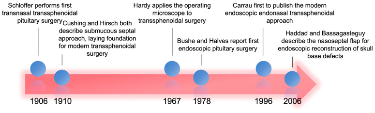
The nasoseptal flap is a workhorse reconstructive option for anterior skull base defects during endonasal surgery. This paper highlights the versatility of the nasoseptal flap. After providing a brief historical perspective, this review will focus on the relevant primary literature published in the last ten years. We will touch upon new applications of the flap, how the flap has been modified to expand its reach and robustness, and some of the current limitations. We will conclude by discussing what the future holds for improving upon the design and use of the nasoseptal flap in anterior skull base reconstruction.

Keywords: anterior skull base reconstruction; expanded endoscopic endonasal surgery; nasoseptal flap; pedicled flaps; transnasal transsphenoidal pituitary surgery.


Conflict of interest statement

The authors declare no conflicts of interest.

Figures

Figure 1
Figure 1
Timeline covering 100 years of milestones leading to modern endoscopic endonasal skull base surgery and use of NSF [17,18].
Figure 2
Figure 2
Schematic view of the nasal septum with surgical landmarks, predicted posterior septal artery vascular pedicle distribution, and nasoseptal flap incisions demarcated. The incisions can be carried anteriorly or laterally as depicted by the dashed arrows. IT: inferior turbinate; MT: middle turbinate; ST: superior turbinate; SO: sphenoid ostium; Ch: choana.
Figure 3
Figure 3
(A) Sagittal view of CT sinus without contrast through frontal sinus outflow tract demonstrating the general reach of the nasoseptal flap (blue). The posterior wall of the frontal sinus and the lower third of the clivus are typically too distal for the conventional nasoseptal flap (red). (B) Coronal view demonstrating lateral reach covering the ethmoidal roof and medial bony orbit (blue) with distal limits at the lateral orbital wall (red). Image from Radiopaedia.org. Fr: Frontal sinus; Et: Ethmoid air cells; Sp: Sphenoid sinus; MT: middle turbinate; IT: inferior turbinate; MS: maxillary sinus; NS: nasal septum.
Figure 4
Figure 4
Alterations to the original nasoseptal flap design to increase coverage. (A) The inferior incision is lateralized to the nasal wall and third incision is made along the maxillary crest which augments the final surface area of the flap. (B) The superior incision is carried laterally superior to the sphenoid ostium and brought anteriorly along the inferior turbinate and then swung circumferentially across the nasal roof to meet with the inferior incision on the anterior septum. (C) The mucosa around the choana is released and then a superior incision is brought across the nasal floor to the head of the inferior turbinate to incorporate an inferior turbinate flap. IT: inferior turbinate; NF: nasal floor; NS: nasal septum; Ch: choana; PSA: posterior septal artery; SO: sphenoid os.
Figure 5
Figure 5
Depiction of the reverse flap to limit exposed septal cartilage and minimize crusting. After raising the conventional nasoseptal flap, a posterior bony septectomy is made, and the contralateral septal mucosa is incised and draped over the ipsilateral cartilage. IT: inferior turbinate; MT: middle turbinate; ST: superior turbinate; Ch: choana; SO: sphenoid os.

Similar articles

Cited by

References

    1. Patel M.R., Stadler M.E., Snyderman C.H., Carrau R.L., Kassam A.B., Germanwala A.V., Gardner P., Zanation A.M. How to Choose? Endoscopic Skull Base Reconstructive Options and Limitations. Skull Base. 2010;20:397–403. doi: 10.1055/s-0030-1253573. - DOI - PMC - PubMed
    1. Zwagerman N.T., Wang E.W., Shin S.S., Chang Y., Fernandez-miranda J.C., Snyderman C.H., Gardner P.A. Does Lumbar Drainage Reduce Postoperative Cerebrospinal Fluid Leak after Endoscopic Endonasal Skull Base Surgery? A Prospective, Randomized Controlled Trial. J. Neurosurg. 2019;131:1172–1178. doi: 10.3171/2018.4.JNS172447. - DOI - PubMed
    1. Bernal-sprekelsen M., Rioja E., Enseñat J., Enriquez K., Viscovich L., Agredo-lemos F.E., Alobid I. Management of Anterior Skull Base Defect Depending on Its Size and Location. BioMed Res. Int. 2014;2014:346873. doi: 10.1155/2014/346873. - DOI - PMC - PubMed
    1. Banks C.A., Palmer J.N., Chiu A.G., Malley B.W.O., Woodworth B.A., Kennedy D.W. Endoscopic Closure of CSF Rhinorrhea: 193 Cases over 21 Years. Otolaryngol. Head Neck Surg. 2009;140:826–833. doi: 10.1016/j.otohns.2008.12.060. - DOI - PubMed
    1. Ivan M.E., Bryan Iorgulescu J., El-Sayed I., McDermott M.W., Parsa A.T., Pletcher S.D., Jahangiri A., Wagner J., Aghi M.K. Risk Factors for Postoperative Cerebrospinal Fluid Leak and Meningitis after Expanded Endoscopic Endonasal Surgery. J. Clin. Neurosci. 2015;22:48–54. doi: 10.1016/j.jocn.2014.08.009. - DOI - PubMed

LinkOut - more resources