

The Use of Low-Dose Chest Computed Tomography for the Diagnosis and Monitoring of Pulmonary Infections in Patients with Hematologic Malignancies
- PMID: 38201613
- PMCID: PMC10778314
- DOI: 10.3390/cancers16010186
The Use of Low-Dose Chest Computed Tomography for the Diagnosis and Monitoring of Pulmonary Infections in Patients with Hematologic Malignancies
Abstract
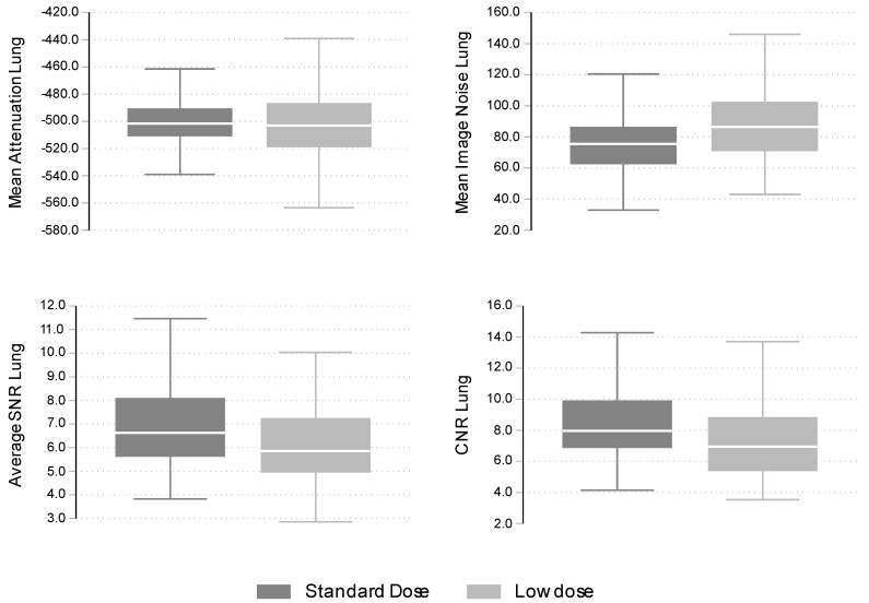
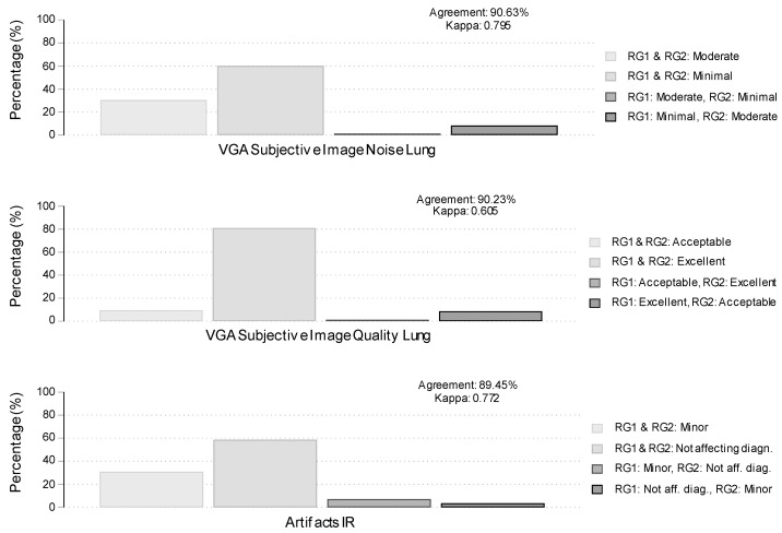
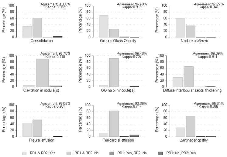
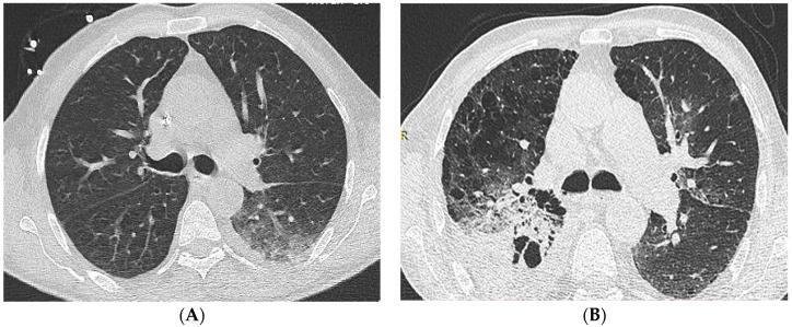
The study aimed to assess the image quality and diagnostic performance of low-dose Chest Computed Tomography (LDCCT) in detecting pulmonary infections in patients with hematologic malignancies. A total of 164 neutropenic patients underwent 256 consecutive CT examinations, comparing 149 LDCCT and 107 Standard-Dose Chest CT (SDCCT) between May 2015 and June 2019. LDCCT demonstrated a 47% reduction in radiation dose while maintaining acceptable image noise and quality compared to SDCCT. However, LDCCT exhibited lower sensitivity in detecting consolidation (27.5%) and ground glass opacity (64.4%) compared to SDCCT (45.8% and 82.2%, respectively) with all the respective p-values from unadjusted and adjusted for sex, age, and BMI analyses being lower than 0.006 and the corresponding Odds Ratios of detection ranging from 0.30 to 0.34. Similar trends were observed for nodules ≥3 mm and ground glass halo in nodules but were not affected by sex, age and BMI. No significant differences were found for cavitation in nodules, diffuse interlobular septal thickening, pleural effusion, pericardial effusion, and lymphadenopathy. In conclusion, LDCCT achieved substantial dose reduction with satisfactory image quality but showed limitations in detecting specific radiologic findings associated with pulmonary infections in neutropenic patients compared to SDCCT.
Keywords: CT noise reduction algorithms; diagnostic performance; hematologic malignancies; low-dose chest CT; lung abnormalities; neutropenic patients; radiation dose; standard-dose chest CT.
Conflict of interest statement
The authors declare no conflicts of interest.
Figures
Similar articles
-
Evaluation of pulmonary metastases in children by non-contrast chest computed tomography.J Med Assoc Thai. 2013 Mar;96(3):334-9. J Med Assoc Thai. 2013. PMID: 23539938
-
Ultra-Low Dose CT Chest in Acute COVID-19 Pneumonia: A Pilot Study from India.Diagnostics (Basel). 2023 Jan 18;13(3):351. doi: 10.3390/diagnostics13030351. Diagnostics (Basel). 2023. PMID: 36766456 Free PMC article.
-
Adaptive iterative dose reduction using 3D processing for reduced- and low-dose pulmonary CT: comparison with standard-dose CT for image noise reduction and radiological findings.AJR Am J Roentgenol. 2012 Oct;199(4):W477-85. doi: 10.2214/AJR.11.8275. AJR Am J Roentgenol. 2012. PMID: 22997397
-
Chest CT findings in patients with coronavirus disease 2019 (COVID-19): a comprehensive review.Diagn Interv Radiol. 2021 Sep;27(5):621-632. doi: 10.5152/dir.2020.20212. Diagn Interv Radiol. 2021. PMID: 33135665 Free PMC article. Review.
-
Chest CT Imaging Signature of Coronavirus Disease 2019 Infection: In Pursuit of the Scientific Evidence.Chest. 2020 Nov;158(5):1885-1895. doi: 10.1016/j.chest.2020.06.025. Epub 2020 Jun 25. Chest. 2020. PMID: 32592709 Free PMC article.
References
-
- Heussel C.P., Kauczor H.U., Heussel G.E., Fischer B., Begrich M., Mildenberger P., Thelen M. Pneumonia in febrile neutropenic patients and in bone marrow and blood stem-cell transplant recipients: Use of high-resolution computed tomography. J. Clin. Oncol. 1999;17:796–805. doi: 10.1200/JCO.1999.17.3.796. - DOI - PubMed
LinkOut - more resources
Full Text Sources
