Rare Pediatric Cerebellar High-Grade Gliomas Mimic Medulloblastomas Histologically and Transcriptomically and Show p53 Mutations
- PMID: 38201659
- PMCID: PMC10778382
- DOI: 10.3390/cancers16010232
Rare Pediatric Cerebellar High-Grade Gliomas Mimic Medulloblastomas Histologically and Transcriptomically and Show p53 Mutations
Abstract
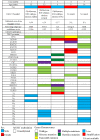
Pediatric high-grade gliomas (HGG) of the cerebellum are rare, and only a few cases have been documented in detail in the literature. A major differential diagnosis for poorly differentiated tumors in the cerebellum in children is medulloblastoma. In this study, we described the histological and molecular features of a series of five pediatric high-grade gliomas of the cerebellum. They actually showed histological and immunohistochemical features that overlapped with those of medulloblastomas and achieved high scores in NanoString-based medulloblastoma diagnostic assay. Methylation profiling demonstrated these tumors were heterogeneous epigenetically, clustering to GBM_MID, DMG_K27, and GBM_RTKIII methylation classes. MYCN amplification was present in one case, and PDGFRA amplification in another two cases. Interestingly, target sequencing showed that all tumors carried TP53 mutations. Our results highlight that pediatric high-grade gliomas of the cerebellum can mimic medulloblastomas at histological and transcriptomic levels. Our report adds to the rare number of cases in the literature of cerebellar HGGs in children. We recommend the use of both methylation array and TP53 screening in the differential diagnoses of poorly differentiated embryonal-like tumors of the cerebellum.
Keywords: MYCN; PDGFRA; children; high-grade gliomas; p53.
Conflict of interest statement
The authors declare that they have no competing interests.
Figures


Similar articles
-
Molecular landscape of pediatric type IDH wildtype, H3 wildtype hemispheric glioblastomas.Lab Invest. 2022 Jul;102(7):731-740. doi: 10.1038/s41374-022-00769-9. Epub 2022 Mar 24. Lab Invest. 2022. PMID: 35332262
-
Cerebellar High-Grade Glioma: A Translationally Oriented Review of the Literature.Cancers (Basel). 2022 Dec 28;15(1):174. doi: 10.3390/cancers15010174. Cancers (Basel). 2022. PMID: 36612169 Free PMC article. Review.
-
Bithalamic gliomas may be molecularly distinct from their unilateral high-grade counterparts.Brain Pathol. 2018 Jan;28(1):112-120. doi: 10.1111/bpa.12484. Epub 2017 Mar 12. Brain Pathol. 2018. PMID: 28032389 Free PMC article.
-
Novel oncogenic PDGFRA mutations in pediatric high-grade gliomas.Cancer Res. 2013 Oct 15;73(20):6219-29. doi: 10.1158/0008-5472.CAN-13-1491. Epub 2013 Aug 22. Cancer Res. 2013. PMID: 23970477 Free PMC article.
-
Cerebellar tumors.Handb Clin Neurol. 2018;155:289-299. doi: 10.1016/B978-0-444-64189-2.00019-6. Handb Clin Neurol. 2018. PMID: 29891066 Review.
Cited by
-
Dissecting the Natural Patterns of Progression and Senescence in Pediatric Low-Grade Glioma: From Cellular Mechanisms to Clinical Implications.Cells. 2024 Jul 19;13(14):1215. doi: 10.3390/cells13141215. Cells. 2024. PMID: 39056798 Free PMC article. Review.
References
-
- Ostrom Q.T., de Blank P.M., Kruchko C., Petersen C.M., Liao P., Finlay J.L., Stearns D.S., Wolff J.E., Wolinsky Y., Letterio J.J., et al. Alex’s Lemonade Stand Foundation Infant and Childhood Primary Brain and Central Nervous System Tumors Diagnosed in the United States in 2007–2011. Neuro Oncol. 2015;16((Suppl. S10)):x1–x36. doi: 10.1093/neuonc/nou327. - DOI - PMC - PubMed
-
- Karremann M., Rausche U., Roth D., Kühn A., Pietsch T., Gielen G.H., Warmuth-Metz M., Kortmann R.D., Straeter R., Gnekow A., et al. Cerebellar location may predict an unfavourable prognosis in paediatric high-grade glioma. Br. J. Cancer. 2013;109:844–851. doi: 10.1038/bjc.2013.404. - DOI - PMC - PubMed
Grants and funding
LinkOut - more resources
Full Text Sources
Research Materials
Miscellaneous

