. 2024 Jan 4;16(1):169.
doi: 10.3390/nu16010169.

Phogrin Regulates High-Fat Diet-Induced Compensatory Pancreatic β-Cell Growth by Switching Binding Partners

Affiliations

Phogrin Regulates High-Fat Diet-Induced Compensatory Pancreatic β-Cell Growth by Switching Binding Partners

Chisato Kubota et al. Nutrients. .

Abstract

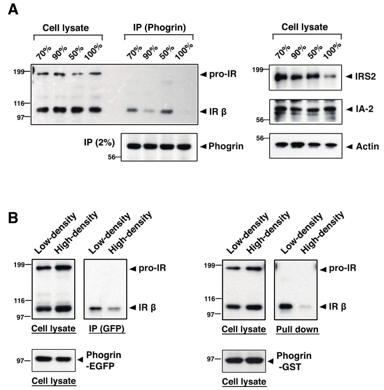
The receptor protein tyrosine phosphatase phogrin primarily localizes to hormone secretory granules in neuroendocrine cells. Concurrent with glucose-stimulated insulin secretion, phogrin translocates to pancreatic β-cell plasma membranes, where it interacts with insulin receptors (IRs) to stabilize insulin receptor substrate 2 (IRS2) that, in turn, contributes to glucose-responsive β-cell growth. Pancreatic β-cell development was not altered in β-cell-specific, phogrin-deficient mice, but the thymidine incorporation rate decreased in phogrin-deficient islets with a moderate reduction in IRS2 protein expression. In this study, we analyzed the β-cell response to high-fat diet stress and found that the compensatory expansion in β-cell mass was significantly suppressed in phogrin-deficient mice. Phogrin-IR interactions occurred only in high-fat diet murine islets and proliferating β-cell lines, whereas they were inhibited by the intercellular binding of surface phogrin under confluent cell culture conditions. Thus, phogrin could regulate glucose-stimulated compensatory β-cell growth by changing its binding partner from another β-cell phogrin to IR in the same β-cells.

Keywords: high-fat diet; insulin receptor substrate; insulin signaling; islet antigen; pancreatic β-cell mass; secretory granules.


Conflict of interest statement

The authors declare no conflicts of interest.

Figures

Figure 1
Figure 1
Pancreatic β-cell mass and islet size distribution in phogrin-deficient mice: (A) Immunohistochemical analyses of pancreas tissue from control (Ctrl: Cre+/−_Phogrin+/+) or knockout (βKO: Cre+/−_Phogrinfl/fl) 7-week-old male mice using anti-insulin antibody. Areas of β-cells relative to pancreas area were quantified and summed, and data are presented as means ± standard errors of the mean (SEM; n = 4). (B) Using the data (A), the number of pancreatic islets of each size (β-cell area, mm2) was shown as a percentage of the total number. (C) The same analyses on 12- or 20–22-week-old mice are shown as in (B) (n = 4).
Figure 2
Figure 2
Reduced compensatory β-cell expansion in HFD-fed, phogrin-knockout (βKO) mice: (A) Body weight in Ctrl or βKO mice on a normal diet (NFD) or a high-fat. high-sucrose diet (HFD). Data are mean ± standard errors of the mean (SEM) from 7–31 male mice per group. (B) Glucose tolerance in 22-week-old Ctrl or βKO mice after 17 weeks on NFD or HFD. Data are means ± SEM from 13–25 male mice per group. (C) Immunohistochemical analyses of pancreas tissue from NFD- or HFD-fed Ctrl or βKO mice after 20 weeks using anti-insulin antibody. Each image is one of five composed sections for each mouse. Areas of β-cells relative to pancreas area were quantified and data are presented as means ± SEM (n = 4, * p < 0.05). (D) β-cell replication measured using a BrdU incorporation assay on NFD- or HFD-fed Ctrl or βKO mice after 3 days. Images are various sized islets from βKO mice fed a NFD (mouse No. 215) or HFD (No. 216). Results are shown as a ratio of BrdU-positive cells (green) to insulin-positive cells (red). Data are presented as means ± SEM (n = 4).
Figure 3
Figure 3
Phogrin interacts with insulin receptors (IRs) in islets from high-fat, high-sucrose diet (HFD)-fed mice. Islets isolated from wild-type mice fed a normal diet (NFD) or HFD were lysed, and 0.5 mg of each extract were immunoprecipitated with anti-phogrin rabbit antibody. Each immunoprecipitate and an original lysate aliquot (5 μg) were analyzed via immunoblotting with antibodies against insulin receptor β-subunit (IRβ) (upper panel). Phogrin amounts in each precipitate (5%) and lysate were determined via immunoblotting with anti-phogrin murine antibody (lower panels).
Figure 4
Figure 4
Intracellular binding of phogrin to IR is attenuated in high-density conditions: (A) MIN6 cells cultured at various cell densities (50–100%) for at least 24 h were extracted and an equal amount of each extract (1.5 mg) was immunoprecipitated with anti-phogrin antibody. The insulin receptor (IR) amount in each precipitate was determined via immunoblotting (left upper panel) and the immunoprecipitated phogrin level was also determined (left lower panel). IRS2, IA-2, and β-actin expression levels in each cell extract were analyzed via immunoblotting (right panels). (B) MIN6 cells expressing phogrin–EGFP (left panels) or phogrin–GST (right panels) were cultured at 90% (high) or 50% (low) cell density. Cell extracts were either immunoprecipitated with anti-GFP monoclonal antibody or pulled down with glutathione sepharose. The amount of IR in each precipitate and IR and phogrin expression levels in each lysate were determined via immunoblotting.
Figure 5
Figure 5
Intercellular interaction of phogrin occurs in high-density co-cultures: Cell extracts (2.0 mg protein) from MIN6/phogrin–EGFP and MIN6/phogrin–GST cultured together at high (100%) or low (75%) density for 48 h or extract mixtures (2.0 mg) from cells cultured separately were pulled down using glutathione sepharose. The presence of phogrin–EGFP in each precipitate (lower panel) and expression levels of phogrin–EGFP and phogrin–GST in each lysate (upper panels) were determined via immunoblotting.
Figure 6
Figure 6
A model for phogrin-mediated regulation of β-cell compensative proliferation. Insulin resistance progression leads to β-cell damage or shedding, resulting in a transient β-cell decrease in the islets. Phogrin binds to IR on the plasma membrane and induces autocrine insulin signaling via IRS2 protein; consequently, β-cells proliferate and form a regular islet. Intercellular phogrin binding occurs at high cell density state and irreversible PTP1B inactivation induces IRS2 degradation, hence the proliferative signal stops. ROS, reactive oxygen species.

Similar articles

References

    1. Wasmeier C., Hutton J.C. Molecular cloning of phogrin, a protein-tyrosine phosphatase homologue localized to insulin secretory granule membranes. J. Biol. Chem. 1996;271:18161–18170. doi: 10.1074/jbc.271.30.18161. - DOI - PubMed
    1. Lu J., Li Q., Xie H., Chen Z.J., Borovitskaya A.E., Maclaren N.K., Notkins A.L., Lan M.S. Identification of a second transmembrane protein tyrosine phosphatase, IA-2beta, as an autoantigen in insulin-dependent diabetes mellitus: Precursor of the 37-kDa tryptic fragment. Proc. Natl. Acad. Sci. USA. 1996;93:2307–2311. doi: 10.1073/pnas.93.6.2307. - DOI - PMC - PubMed
    1. Cui L., Yu W., DeAizpurua H.J., Schmidli R.S., Pallen C.J. Cloning and characterization of islet cell antigen-related protein-tyrosine phosphatase (PTP), a novel receptor-like PTP and autoantigen in insulin-dependent diabetes. J. Biol. Chem. 1996;271:24817–24823. doi: 10.1074/jbc.271.40.24817. - DOI - PubMed
    1. Chiang M.K., Flanagan J.G. PTP-NP, a new member of the receptor protein tyrosine phosphatase family, implicated in development of nervous system and pancreatic endocrine cells. Development. 1996;122:2239–2250. doi: 10.1242/dev.122.7.2239. - DOI - PubMed
    1. Solimena M., Dirkx R., Hermel J.M., Pleasic-Williams S., A Shapiro J., Caron L., Rabin D.U. ICA 512, an autoantigen of type I diabetes, is an intrinsic membrane protein of neurosecretory granules. EMBO J. 1996;15:2102–2114. doi: 10.1002/j.1460-2075.1996.tb00564.x. - DOI - PMC - PubMed

LinkOut - more resources