

Prognostic Value of the Area of Lung Involved in Severe and Non-Severe Bronchiolitis: An Observational, Ultrasound-Based Study
- PMID: 38202091
- PMCID: PMC10780043
- DOI: 10.3390/jcm13010084
Prognostic Value of the Area of Lung Involved in Severe and Non-Severe Bronchiolitis: An Observational, Ultrasound-Based Study
Abstract
Background: Point of care lung ultrasound (LUS) has a definite role in viral bronchiolitis when combined with clinical data. Previous data showed a bigger involvement of the superior lung zones in more severe cases. The aim of the present study is to describe whether different lung areas are implicated to different degrees in patients admitted to a Pediatric Intensive Care Unit (PICU) and needing ventilation compared to those with less severe forms.
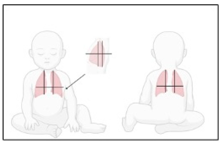
Methods: observational, prospective study. LUS scores of single lung areas and clinical data were collected for all children aged 0-12 months presenting with bronchiolitis to the participating centers and used as covariates for logistic regression having "PICU admission" as outcome. A subsequent analysis was carried out to investigate factors concurring with different lung zones' involvement.
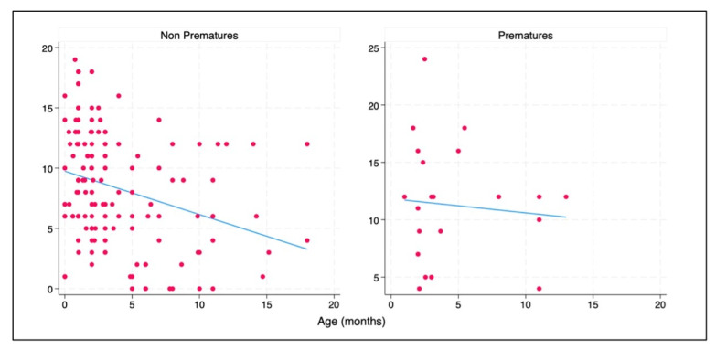
Results: 173 patients were enrolled. Difficulty in feeding, presence of wheezing, SpO2 were all risk factors for PICU admission. Superior lung areas' LUS scores presented higher Odds Ratios for PICU admission and need for ventilation than inferior ones. Age and prematurity concurred in determining their higher LUS scores.
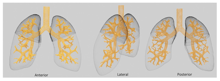
Conclusions: Superior lobes' greater involvement could be favored by the geometrical distribution of relative bronchi, exiting with an acute angle from mainstem bronchi in small children where airway caliber is small and only small volumes of secretions can be occlusive.
Keywords: Continuous Positive Airway Pressure (CPAP); Pediatric Intensive Care Unit (PICU); bronchial geometry; bronchiolitis; lung regional differences; superior lung lobes.
Conflict of interest statement
The authors declare no conflict of interest.
Figures
References
-
- Shi T., McAllister D.A., O’Brien K.L., Simoes E.A.F., Madhi S.A., Gessner B.D., Polack F.P., Balsells E., Acacio S., Aguayo C., et al. Global, regional, and national disease burden estimates of acute lower respiratory infections due to respiratory syncytial virus in young children in 2015: A systematic review and modelling study. Lancet. 2017;390:946–958. doi: 10.1016/S0140-6736(17)30938-8. - DOI - PMC - PubMed
-
- De Luca M., D’Amore C., Romani L., Tripiciano C., Clemente V., Mercadante S., Perrotta D., Nunziata J., Cecchetti C., Rossetti E., et al. Severe viral respiratory infections in the pre-COVID era: A 5-year experience in two pediatric intensive care units in Italy. Influenza Resp. Viruses. 2023;17:e13038. doi: 10.1111/irv.13038. - DOI - PMC - PubMed
-
- Abdel Kader M., Abou Samra M.F., Abdel Aal S.M.S., Shehata N., Khalifa A. The utility of lung ultrasound in evaluation of infants with suspected bronchiolitis. Egypt. J. Radiol. Nucl. Med. 2016;47:1057–1064. doi: 10.1016/j.ejrnm.2016.06.009. - DOI
LinkOut - more resources
Full Text Sources
