

Digital Ulcers and Ventricular Arrhythmias as Red Flags to Predict Replacement Myocardial Fibrosis in Systemic Sclerosis
- PMID: 38202095
- PMCID: PMC10779804
- DOI: 10.3390/jcm13010089
Digital Ulcers and Ventricular Arrhythmias as Red Flags to Predict Replacement Myocardial Fibrosis in Systemic Sclerosis
Abstract
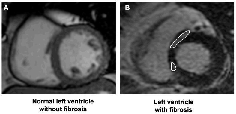
Background: Cardiac involvement in systemic sclerosis (SSc) affects the prognosis of the disease. Echocardiography is the first line imaging tool to detect cardiac involvement, but it is not able to routinely detect myocardial fibrosis. Late gadolinium enhancement (LGE) cardiovascular magnetic resonance (CMR) is the gold standard for replacement myocardial fibrosis assessment, but its availability is currently limited.
Aim: We aimed to assess the clinical and instrumental parameters that would be useful for predicting the presence of LGE-CMR, to achieve a better selection of patients with SSc that could benefit from third-level CMR imaging.
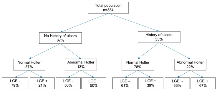
Methods: 344 SSc patients underwent a comprehensive echocardiogram and LGE-CMR on the same day; for 189 patients, a 24 h ECG Holter monitoring was available.
Results: CMR showed non-junctional replacement myocardial fibrosis via LGE in 25.1% patients. A history of digital ulcers (OR 2.188; 95% C.I. 1.069-4.481) and ventricular arrhythmias at ECG Holter monitoring (OR 3.086; 95% C.I. 1.191-7.998) were independent predictors of replacement myocardial fibrosis.
Conclusions: CMR can detect patterns of clinical and subclinical cardiac involvement, which are frequent in SSc. A history of digital ulcers and evidence of ventricular arrhythmias at ECG Holter monitoring are red flags for the presence of replacement myocardial fibrosis in CMR. The association between digital ulcers and myocardial fibrosis suggests that a similar pathological substrate of abnormal vascular function may underlie peripheral vascular and cardiac complications.
Keywords: ECG Holter monitoring; cardiac involvement; cardiac magnetic resonance; digital ulcers; late gadolinium enhancement; myocardial fibrosis; systemic sclerosis.
Conflict of interest statement
L.G. has received consultancy fees from GE Healthcare, Philips Healthcare, EchoNous and Caption Health outside the submitted work. C.B. received consulting fees from Eli-Lilly, Boehringer Ingelheim outside the submitted work. Research grants from Gruppo Italiano Lotta alla Sclerodermia (GILS), European Scleroderma Trials and Research Group (EUSTAR), Foundation for Research in Reumatology (FOREUM), Scleroderma Clinical Trials Consortium (SCTC), Scleroderma Research Foundation and educational grants from AbbVie were received outside the submitted work. C.C. received consulting fees and/or honoraria from SOBI, Novartis, Pfizer, Roche and Jannsen Boehringer Ingelheim outside the submitted work. G.D.L. received honoraria from SOBI, Novartis, Pfizer, MSD and Celgene outside the submitted work. L.D. has consultancy relationships with Abbvie, AstraZeneca, Biogen, Boehringer-Ingelheim, BMS, Eli Lilly, Galapagos, GSK, Janssen, Kiniksa Pharmaceuticals, Novartis, Pfizer and SOBI; research grants from BMS, Celltrion, Kiniksa pharmaceuticals, Pfizer and SOBI; and has served as speaker for Novartis and SOBI outside the submitted work. M.M.-C. received consultancies from Actelion, Janssen, Inventiva, Bayer, Biogen, Boehringer, CSL Behring, Corbus, Galapagos, Mitsubishi, Samsung, Regeneron, Acceleron, MSD, Chemomab, Lilly, Pfizer and Roche outside the submitted work. Other authors: none declared.
Figures
References
-
- Varga J., Trojanowska M., Kuwana M. Pathogenesis of systemic sclerosis: Recent insights of molecular and cellular mechanisms and therapeutic opportunities. J. Scleroderma Relat. Disord. 2017;2:137–152. doi: 10.5301/jsrd.5000249. - DOI
-
- Mavrogeni S.I., Kitas G.D., Dimitroulas T., Sfikakis P.P., Seo P., Gabriel S., Patel A.R., Gargani L., Bombardieri S., Matucci-Cerinic M., et al. Cardiovascular magnetic resonance in rheumatology: Current status and recommendations for use. Int. J. Cardiol. 2016;217:135–148. doi: 10.1016/j.ijcard.2016.04.158. - DOI - PubMed
-
- Bruni C., Buch M.H., Furst D.E., De Luca G., Djokovic A., Dumitru R.B., Giollo A., Polovina M., Steelandt A., Bratis K., et al. Primary systemic sclerosis heart involvement: A systematic literature review and preliminary data-driven, consensus-based WSF/HFA definition. J. Scleroderma Relat. Disord. 2022;7:24–32. doi: 10.1177/23971983211053246. - DOI - PMC - PubMed
LinkOut - more resources
Full Text Sources
