. 2023 Dec 23;13(1):90.
doi: 10.3390/jcm13010090.

Robotic Lateral Pelvic Lymph Node Dissection in Rectal Cancer: A Feasibility Study from a European Centre

Affiliations

Robotic Lateral Pelvic Lymph Node Dissection in Rectal Cancer: A Feasibility Study from a European Centre

Tou Pin Chang et al. J Clin Med. .

Abstract

Introduction: The role of robotic lateral pelvic lymph node dissection (LPLND) for lateral pelvic nodal disease (LPND) in rectal cancer has yet to be investigated in the Western hemisphere. This study aims to investigate the safety and feasibility of robotic LPLND by utilising a well-established totally robotic TME protocol.

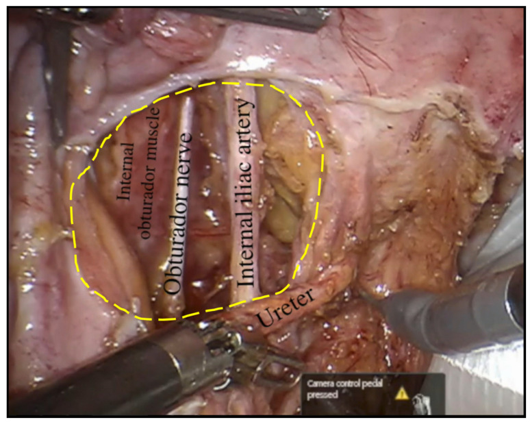
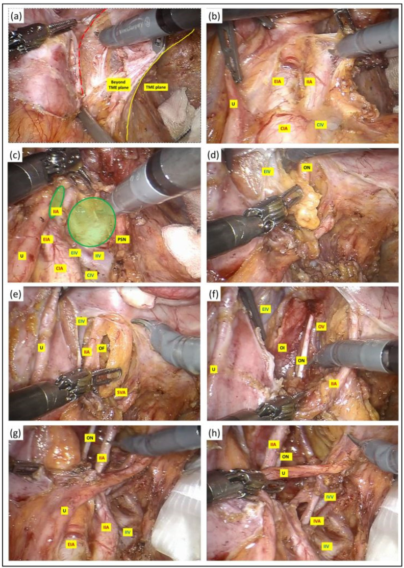
Methods: We conducted a retrospective study on 17 consecutive patients who underwent robotic LPLND for LPND ± TME for rectal cancer between 2015 and 2021. A single docking totally robotic approach from the left hip with full splenic mobilisation was performed using the X/Xi da Vinci platform. All patients underwent a tri-compartmental robotic en bloc excision of LPND with preservation of the obturator nerve and pelvic nerve plexus, leaving a well-skeletonised internal iliac vessel and its branches.

Results: The median operative time was 280 min, which was 40 min longer than our standard robotic TME. The median BMI was 26, and there were no conversions. The median inpatient stay was 7 days with no Clavien-Dindo > 3 complications. One patient (6%) developed local recurrence and metastatic disease within 5 months. The proportion of histologically confirmed LPND was 41%, of which 94% were well to moderately differentiated adenocarcinoma. Median pre-operative lateral pelvic node size was significantly higher in positive nodes (14 mm vs. 8 mm (p = 0.01)). All patients had clear resection margins on histology.

Discussion: Robotic LPLND is safe and feasible with good peri-operative and short-term outcomes, with the ergonomic advantages of a robotic TME docking protocol readily transferrable in LPLND.

Keywords: lateral pelvic lymph node dissection; minimally invasive surgery; robotic colorectal; robotic rectal cancer.


Conflict of interest statement

JSK is a proctor with intuitive surgical. All other authors declare no conflict of interest.

Figures

Figure 1
Figure 1
Flow chart of patient selection for robotic LPLND; CRM: Circumferential resection margin; CRT: chemoradiotherapy/chemotherapy; PET-CT: position emission tomography; rTME: robotic total mesorectal excision; rLPLND: robotic lateral pelvic lymph node dissection.
Figure 2
Figure 2
Robotic port placements (numbered circles) for LPLND ± TME along a 3 cm lateralised diagonal line from an initial imaginary line between the mid-inguinal point (blue circle) and the left costal margin (blue circle) lateralised at 8 cm from the midline. The assistant port (A) is placed at least a fist breadth away from the robotic ports to allow optimal instrument entry to the pelvis between the third and fourth robotic ports (yellow dotted lines). The ‘No port zone’ (NPZ) denotes the area within two fingers breadth from adjacent bony landmarks, which should be devoid of any ports.
Figure 3
Figure 3
The obturator fossa in the left pelvic side wall following completion of robotic LPLND with the internal obturator muscle depicting the lateral boundary of the fossa at its depth and the central compartment, which consists of the left obturator nerve and left internal iliac artery with the left ureter returned to its anatomical position.
Figure 4
Figure 4
(a) A vertical peritoneal incision was made at the medial umbilical fold, and this was extended to the level of common iliac artery bifurcation. The dissection was continued along the external iliac vessels (red dotted line) and the lymph nodes along these vessels were excised in their entirety with their surrounding lymphatic tissue package kept intact. (bd) Once the external iliac vein had been dissected free, the third robotic arm with a double fenestrated forceps was deployed against the ureter for gentle retraction to facilitate entry into the obturator fossa between the internal and external iliac vessels (green circles), clearing the lymphatic package in a craniocaudal fashion. The extent of the dissection anteriorly was at the level where the plane started to fuse with the prevesical tissue and the femoral canal. Further division of nodal tissues at this stage was performed between clips to prevent lymphatic leak. (e) As the dissection approached the depth of the obturator fossa, the obturator vessels and nerve were encountered medial to the external iliac vein and lateral to the superior vesical artery. We paid careful attention not to injure or divide the obturator nerve to prevent post-operative neuropathy. The obturator artery and vein, on the other hand, were divided between clips if clinically indicated. (f) The nodal dissection was continued along the internal iliac artery and its anterior divisions to expose the superior and inferior vesical, obturator and umbilical artery from the anterior branch and along the posterior branch to allow en bloc excision of the pathological lymph node mass with its surrounding lymphatic tissue package kept intact, and this was retrieved in a bag. The lateral boundary of dissection at the depth of the obturator fossa was the obturator internus, whereas medially, the dissection at this depth continued into the TME plane that was created at the earlier stage. (g,h) Following completion of LPLND, the remaining structures in the pelvic side wall consist of well-skeletonised internal iliac vessels and their branches, the obturator nerve and the pelvic nerve plexus preserved in its entirety with no intervening residual fat or nodal tissues. U: ureter; CIA: common iliac artery; EIA: external iliac artery; IIA: internal iliac artery; CIV: common iliac vein; EIV: external iliac vein; PSN: presacral nerve plexus; OF: obturator fossa; IVA: inferior vesicle artery; IVV: inferior vesicle vein; OI: obturator internus; ON: obturator nerve.

Similar articles

Cited by

References

    1. Liu Q., Luo D., Cai S., Li Q., Li X. Circumferential resection margin as a prognostic factor after rectal cancer surgery: A large population-based retrospective study. Cancer Med. 2018;7:3673–3681. doi: 10.1002/cam4.1662. - DOI - PMC - PubMed
    1. Watanabe T., Muro K., Ajioka Y., Hashiguchi Y., Ito Y., Saito Y., Hamaguchi T., Ishida H., Ishiguro M., Ishihara S., et al. Japanese Society for Cancer of the Colon and Rectum (JSCCR) guidelines 2016 for the treatment of colorectal cancer. Int. J. Clin. Oncol. 2018;23:1–34. doi: 10.1007/s10147-017-1101-6. - DOI - PMC - PubMed
    1. Shimoyama M., Yamazaki T., Suda T., Hatakeyama K. Prognostic significance of lateral lymph node micrometastases in lower rectal cancer: An immunohistochemical study with CAM5.2. Dis. Colon. Rectum. 2003;46:333–339. doi: 10.1007/s10350-004-6552-y. - DOI - PubMed
    1. Akiyoshi T., Watanabe T., Miyata S., Kotake K., Muto T., Sugihara K. Results of a Japanese nationwide multi-institutional study on lateral pelvic lymph node metastasis in low rectal cancer: Is it regional or distant disease? Ann. Surg. 2012;255:1129–1134. doi: 10.1097/SLA.0b013e3182565d9d. - DOI - PubMed
    1. Sauer R., Becker H., Hohenberger W., Rödel C., Wittekind C., Fietkau R., Martus P., Tschmelitsch J., Hager E., Hess C.F., et al. Preoperative versus postoperative chemoradiotherapy for rectal cancer. N. Engl. J. Med. 2004;351:1731–1740. doi: 10.1056/NEJMoa040694. - DOI - PubMed