

The Symptomatic Calcification and Ossification of the Ligamentum Flavum in the Spine: Our Experience and Review of the Literature
- PMID: 38202112
- PMCID: PMC10780021
- DOI: 10.3390/jcm13010105
The Symptomatic Calcification and Ossification of the Ligamentum Flavum in the Spine: Our Experience and Review of the Literature
Abstract
Introduction: We report our experience regarding the clinical features and pathological findings of the calcification of the ligamentum flavum (CLF) and ossification of the ligamentum flavum (OLF) in the spine. In addition, we reviewed the previous studies on CLF and OLF to enhance the understanding of these conditions.
Materials and methods: We compared the clinical, radiological, and histopathological features of CLF and OLF.
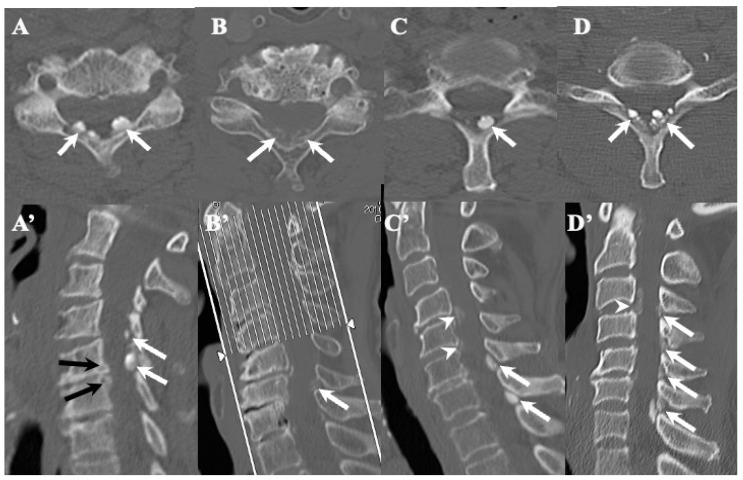
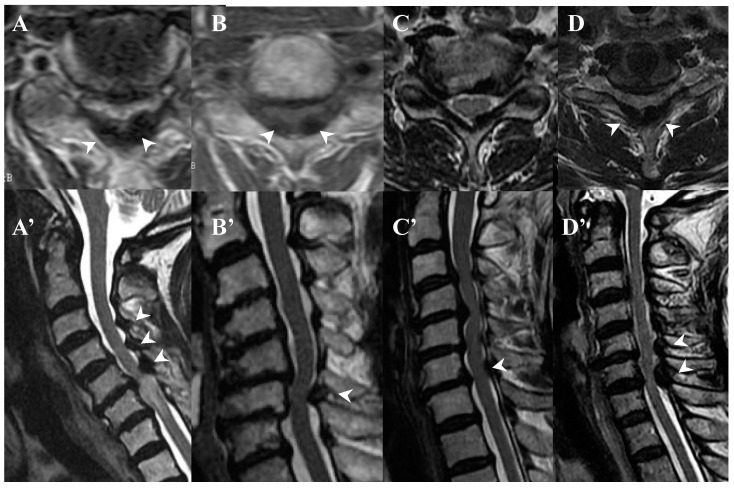
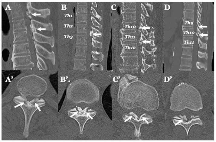
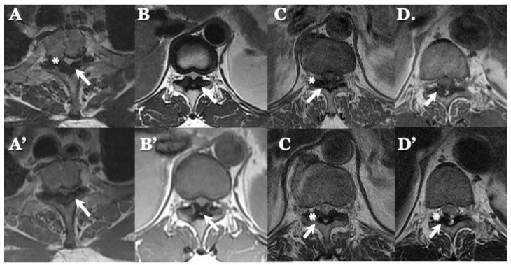
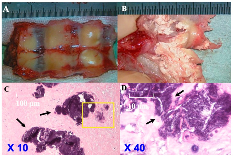
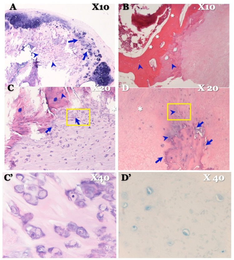
Results: In CLF, a computed tomography (CT) scan showed egg-shaped or speck-like calcification in the ligamentum flavum. Magnetic resonance (MR) imaging demonstrated spinal cord compression due to a thickened ligamentum flavum, which appeared as a low-intensity mass. Pathological findings demonstrated fused islands of calcification resembling sand-like calcification. In OLF, CT showed beak-like ossification extending into the intervertebral foramen. MR imaging demonstrated spinal cord compression by a low-intensity mass. Pathological findings revealed laminar ossification of LF with chondrocytes near the calcification and laminar hyaline cartilage.
Conclusions: CLF and OLF appear to be distinct entities based on their clinical, neuroradiological, histopathological, and pathogenetic features. We suggest that the causes of CLF include both metabolic and dystrophic factors, while the pathogenesis of OLF is characterized by enchondral ossification induced by a genetic cascade triggered by shearing/tension stress.
Keywords: calcification of the ligamentum flavum; clinical feature; ossification of the ligamentum flavum; pathogenesis; radiological finding; surgical management.
Conflict of interest statement
The authors report no conflict of interest concerning the materials or methods used in this study or the findings specified in this paper.
Figures

Similar articles
-
Multiple-level ossification of the ligamentum flavum in the cervical spine combined with calcification of the cervical ligamentum flavum and posterior atlanto-axial membrane.Eur Spine J. 2013 May;22 Suppl 3(Suppl 3):S416-20. doi: 10.1007/s00586-012-2521-7. Epub 2012 Oct 6. Eur Spine J. 2013. PMID: 23053758 Free PMC article.
-
The short-term outcomes of minimally invasive decompression surgery in patients with lumbar ossification or calcification of the ligamentum flavum.J Neurosurg Spine. 2020 Nov 6;34(2):203-210. doi: 10.3171/2020.6.SPINE20946. Print 2021 Feb 1. J Neurosurg Spine. 2020. PMID: 33157534
-
[Symptomatic cervical stenosis due to calcification of the ligamentum flavum after mild cervical trauma].Neurocirugia (Astur). 2007 Apr;18(2):141-6. doi: 10.1016/s1130-1473(07)70302-0. Neurocirugia (Astur). 2007. PMID: 17497062 Spanish.
-
Pathophysiology of Calcification and Ossification of the Ligamentum Flavum in the Cervical Spine.Neurosurg Clin N Am. 2018 Jan;29(1):47-54. doi: 10.1016/j.nec.2017.09.016. Neurosurg Clin N Am. 2018. PMID: 29173435 Review.
-
Cervical myelopathy resulting from combined ossification of the ligamentum flavum and posterior longitudinal ligament: report of two cases and literature review.Spine J. 2013 Jan;13(1):e1-6. doi: 10.1016/j.spinee.2012.10.038. Epub 2012 Dec 21. Spine J. 2013. PMID: 23266147 Review.
Cited by
-
Pathogenesis of thoracic ossification of the ligamentum flavum.Front Pharmacol. 2024 Oct 31;15:1496297. doi: 10.3389/fphar.2024.1496297. eCollection 2024. Front Pharmacol. 2024. PMID: 39545059 Free PMC article. Review.
References
Publication types
LinkOut - more resources
Full Text Sources
