

Outer Retinal and Choroidal Changes in Adolescents with Long-Lasting Type 1 Diabetes
- PMID: 38202235
- PMCID: PMC10779656
- DOI: 10.3390/jcm13010229
Outer Retinal and Choroidal Changes in Adolescents with Long-Lasting Type 1 Diabetes
Abstract
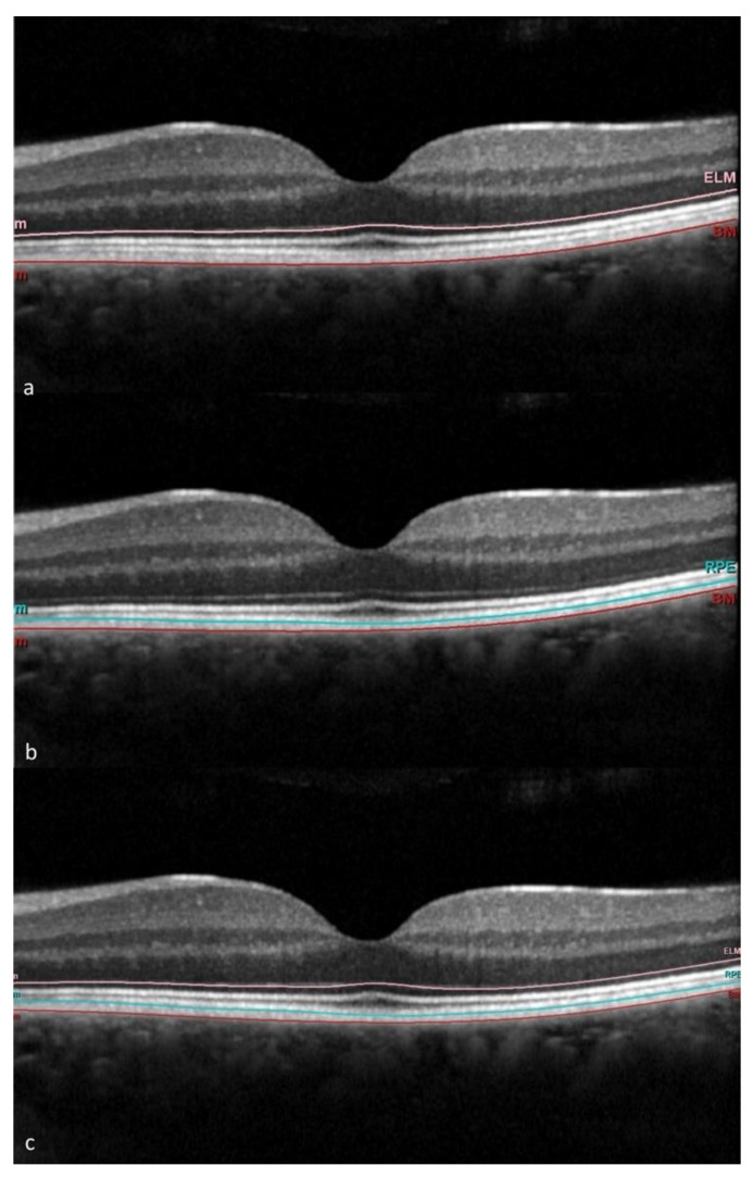
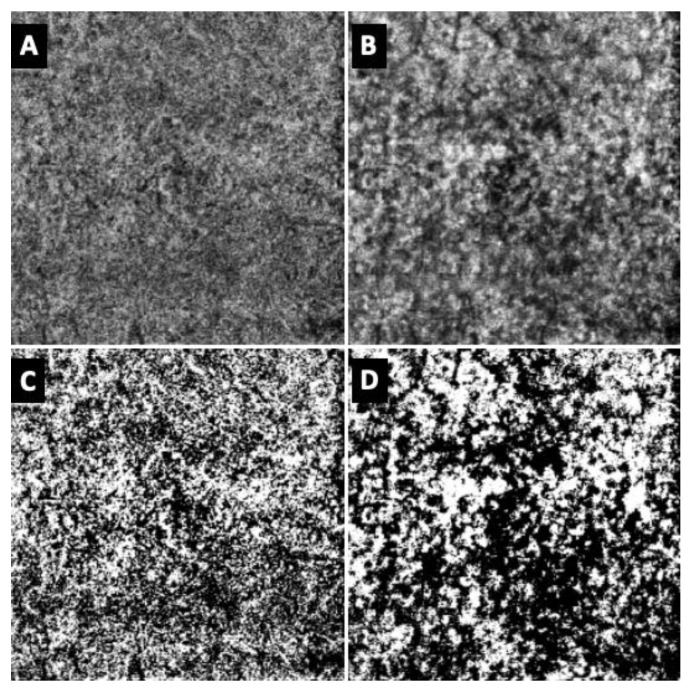
This study aimed to assess outer retinal layer (ORL), retinal pigment epithelium (RPE), choroid (Ch) and choriocapillaris (CC) modifications in adolescents with long-lasting (>10 years) type 1 diabetes (T1D) without (noDR) or with diabetic retinopathy (DR). ORL and RPE thickness were measured at optical coherence tomography (OCT) macular scans. Vascular parameters of Ch and CC were quantified after elaboration of macular OCT-angiography (OCTA) images. Insulin dose and auxological and metabolic parameters were correlated with OCT and OCTA findings in patients. ORL thickness was higher in DR eyes than in noDR and healthy controls (HC), and RPE thickness was higher in noDR and DR eyes than in HC, with statistical significance for some sectors in noDR versus HC. No OCTA parameters of CC and Ch differed among groups, and no significant correlation was observed with auxological and metabolic parameters. In conclusion, ORL and RPE were both increased in adolescents with long-lasting T1D. Such changes were not associated with insulin dose and glycemia control, nor to any choroid or choriocapillaris flow change clinically detectable at OCTA, and they could be potential imaging biomarkers of disease progression.
Keywords: OCT; OCT angiography; adolescents; choriocapillaris; choroid; continuous glucose monitoring; diabetic retinopathy; outer retina; retinal pigment epithelium; type 1 diabetes.
Conflict of interest statement
The authors declare no conflicts of interest.
Figures
Similar articles
-
Retinal Microvascular and Neuronal Changes Are Also Present, Even If Differently, in Adolescents with Type 1 Diabetes without Clinical Diabetic Retinopathy.J Clin Med. 2022 Jul 8;11(14):3982. doi: 10.3390/jcm11143982. J Clin Med. 2022. PMID: 35887746 Free PMC article.
-
Central and peripheral changes in the retina and choroid in patients with diabetes mellitus without clinical diabetic retinopathy assessed by ultra-wide-field optical coherence tomography angiography.Front Public Health. 2023 Jun 13;11:1194320. doi: 10.3389/fpubh.2023.1194320. eCollection 2023. Front Public Health. 2023. PMID: 37383256 Free PMC article.
-
The thickness and volume of the choroid, outer retinal layers and retinal pigment epithelium layer changes in patients with diabetic retinopathy.Int J Ophthalmol. 2018 Dec 18;11(12):1957-1962. doi: 10.18240/ijo.2018.12.14. eCollection 2018. Int J Ophthalmol. 2018. PMID: 30588430 Free PMC article.
-
Imaging-based Assessment of Choriocapillaris: A Comprehensive Review.Semin Ophthalmol. 2023 Jul;38(5):405-426. doi: 10.1080/08820538.2022.2109939. Epub 2022 Aug 18. Semin Ophthalmol. 2023. PMID: 35982638 Review.
-
[Pathophysiology of macular diseases--morphology and function].Nippon Ganka Gakkai Zasshi. 2011 Mar;115(3):238-74; discussion 275. Nippon Ganka Gakkai Zasshi. 2011. PMID: 21476310 Review. Japanese.
Cited by
-
Cerebral Small Vessel Disease Outperforms Brain Atrophy as an Imaging Biomarker in Diabetic Retinopathy.J Diabetes. 2025 Feb;17(2):e70058. doi: 10.1111/1753-0407.70058. J Diabetes. 2025. PMID: 39968694 Free PMC article.
-
New marker for the detection of pre-retinopathy in patients with type 1 diabetes mellitus: systemic immuno-inflammation index.BMC Ophthalmol. 2025 May 19;25(1):296. doi: 10.1186/s12886-025-04138-0. BMC Ophthalmol. 2025. PMID: 40389932 Free PMC article.
References
-
- Pilotto E., Torresin T., Leonardi F., Gutierrez De Rubalcava Doblas J., Midena G., Moretti C., Midena E. Retinal Microvascular and Neuronal Changes Are Also Present, Even If Differently, in Adolescents with Type 1 Diabetes without Clinical Diabetic Retinopathy. J. Clin. Med. 2022;11:3982. doi: 10.3390/jcm11143982. - DOI - PMC - PubMed
LinkOut - more resources
Full Text Sources
