. 2023 Dec 21;13(1):31.
doi: 10.3390/plants13010031.

In Vivo Phosphorylation of the Cytosolic Glucose-6-Phosphate Dehydrogenase Isozyme G6PD6 in Phosphate-Resupplied Arabidopsis thaliana Suspension Cells and Seedlings

Affiliations

In Vivo Phosphorylation of the Cytosolic Glucose-6-Phosphate Dehydrogenase Isozyme G6PD6 in Phosphate-Resupplied Arabidopsis thaliana Suspension Cells and Seedlings

Milena A Smith et al. Plants (Basel). .

Abstract

Glucose-6-phosphate dehydrogenase (G6PD) catalyzes the first committed step of the oxidative pentose phosphate pathway (OPPP). Our recent phosphoproteomics study revealed that the cytosolic G6PD6 isozyme became hyperphosphorylated at Ser12, Thr13 and Ser18, 48 h following phosphate (Pi) resupply to Pi-starved (-Pi) Arabidopsis thaliana cell cultures. The aim of the present study was to assess whether G6PD6 phosphorylation also occurs in shoots or roots following Pi resupply to -Pi Arabidopsis seedlings, and to investigate its relationship with G6PD activity. Interrogation of phosphoproteomic databases indicated that N-terminal, multi-site phosphorylation of G6PD6 and its orthologs is quite prevalent. However, the functions of these phosphorylation events remain unknown. Immunoblotting with an anti-(pSer18 phosphosite-specific G6PD6) antibody confirmed that G6PD6 from Pi-resupplied, but not -Pi, Arabidopsis cell cultures or seedlings (i.e., roots) was phosphorylated at Ser18; this correlated with a significant increase in extractable G6PD activity, and biomass accumulation. Peptide kinase assays of Pi-resupplied cell culture extracts indicated that G6PD6 phosphorylation at Ser18 is catalyzed by a Ca2+-dependent protein kinase (CDPK), which correlates with the 'CDPK-like' targeting motif that flanks Ser18. Our results support the hypothesis that N-terminal phosphorylation activates G6PD6 to enhance OPPP flux and thus the production of reducing power (i.e., NADPH) and C-skeletons needed to establish the rapid resumption of growth that ensures Pi-resupply to -Pi Arabidopsis.

Keywords: CDPK; glucose-6-phosphate dehydrogenase; oxidative pentose–phosphate pathway; phosphate nutrition; post-translational modification; protein phosphorylation.


Conflict of interest statement

The authors declare no conflict of interest.

Figures

Figure 1
Figure 1
Overview of the oxidative pentose phosphate pathway. The OPPP integrates with the upper portion of the glycolytic pathway, providing a bypass for G6P oxidation to triose-phosphates while generating reducing power in the form of NADPH, and anabolic precursors such as ribose-5-phosphate and erythrose-4-phosphate required for nucleotide synthesis and the shikimate pathway, respectively. Abbreviations: 6PGL, 6-phosphogluconolactonase; 6PGDH, 6-phosphogluconate dehydrogenase; RPI, ribose 5-phosphate isomerase; RPE, ribulose 5-phosphate epimerase; TKT, transketolase; TAL, transaldolase; SHI, sedoheptulose 7-phosphate isomerase; SH17BPase, sedoheptulose 1,7-biphosphatase; SHPK, sedoheptulokinase; HK, hexokinase; GPI, glucose phosphate isomerase; PFK, phosphofructokinase; FBA, fructose biphosphate aldolase; TPI, triosephosphate isomerase; GAPDH, glyceraldehyde 3-phosphate dehydrogenase; PK, pyruvate kinase. Figure created with Biorender.com.
Figure 2
Figure 2
Multiple sequence alignment of Arabidopsis G6PD6’s N-terminus with N-termini of other G6PDs. G6PD6 phosphosites that were mapped via LC-MS/MS following 48 h of Pi resupply to –Pi Arabidopsis cell cultures [18] are colored in red and underlined, other experimentally determined phosphosites (Table 1) are colored in red, and corresponding conserved residues within other G6PDs are colored in blue. Arabidopsis G6PD6 is marked with an arrow. N-termini sequences are highlighted as follows: green = Arabidopsis G6PDp; pink = plant G6PDc; yellow = animal G6PD; blue = fungal G6PD; purple = bacterial G6PD. The abbreviations are as follows: An, Aspergillus niger (common mold); At, Arabidopsis thaliana (thale cress); Ce, Caenorhabditis elegans (nematode); Cv, Chlorella vulgaris (green alga); Ec, Escherichia coli; Gm, Glycine max (soybean); Hs, Homo sapiens (human); Hv, Hordeum vulgare (barley); Mm, Mus musculus (mouse); Np, Nostoc punctiforme (cyanobacterium); Os, Oryza sativa (rice); Pp, Physcomitrium patens (earthmoss); Rn, Rattus norvegicus (rat); Sl, Solanum lycopersicum (tomato); St, Solanum tuberosum (potato). Protein accession numbers are listed in Supplementary Table S1.
Figure 3
Figure 3
Motif analysis of phosphorylated Arabidopsis G6PD6 residues. Sequence alignment of amino acids flanking G6PD6 Ser12, Thr13, and Ser18 residues that were phosphorylated 48 h following Pi resupply to –Pi Arabidopsis cell cultures [18]; basic and hydrophobic residues are colored red and blue, respectively. The optimal CDPK/SnRK recognition motif is located above the alignment to highlight residues of interest, where φ represents a hydrophobic amino acid, B represents a basic amino acid, and X represents any amino acid.
Figure 4
Figure 4
Structural model of Arabidopsis G6PD6. The predicted G6PD6 protein model (AF-Q9FJI5-F1) was obtained from AlphaFold and annotated in the SWISS-MODEL workspace. Based on pairwise sequence alignment with human G6PD, conserved key residues that interact with G6P or NADP+ (deduced from crystallized structures of human G6PD) are annotated as follows: red = G6P (substrate); dark blue = NADP+ (coenzyme); magenta = NADP+ (structural) [43]. Phosphorylated residues are annotated in green. Domains were retrieved from InterPro (coenzyme (yellow) = IPR036291, β+α (light blue) = IPR022675) and binding sites are annotated accordingly.
Figure 5
Figure 5
Specificity of phosphosite-specific antibody raised against pSer18 of Arabidopsis G6PD6. (A) Sequence of phosphorylated synthetic peptide that was covalently coupled to keyhole limpet hemocyanin (KLH) and used for rabbit immunization. The sequence numbering represents amino acid position relative to G6PD6’s N-terminus. The Ser18 phosphorylation site is indicated. The peptide was synthesized with an extra N-terminal Cys residue to facilitate its conjugation to KLH. (B) Anti-pSer18 immune serum (±10 μg/mL of phospho- (P-) or dephospho- (deP-) peptide) was used to probe immunodot blots of varying amounts of the pSer18 P-peptide and corresponding deP-peptide.
Figure 6
Figure 6
G6PD6 is in vivo phosphorylated at Ser18, 48 h following Pi resupply to Pi-starved Arabidopsis suspension cells and seedlings. Clarified extracts from –Pi and 48 h Pi-resupplied (Pi-RS) cells or seedlings (shoots and roots) were subjected to SDS/PAGE followed by immunoblotting with anti-pSer18 (+10 μg/mL dephospho- (deP)-peptide) or anti-(pea G6PDc). Approximately 8 and 1 µg of protein were loaded into each lane of the anti-pSer18 and anti-(pea G6PDc) immunoblots, respectively.
Figure 7
Figure 7
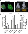
Impact of Pi deprivation (–Pi) and 48 h Pi resupply (Pi-RS) on appearance, G6PD activity, and biomass accumulation of Arabidopsis seedling roots and suspension cells. (A) Images are representative of at least five replicates (bar = 1 cm). (B) Dry weight values represent average gDW per flask of liquid cultured seedlings (roots) or suspension cells. All values represent means (±SE) of n ≥ 3 biological replicates; statistical significance was evaluated via a two-tailed, unpaired Student’s t-test (* p < 0.05, ** p < 0.01, *** p < 0.001, **** p < 0.0001).
Figure 8
Figure 8
Evidence that a CDPK catalyzes G6PD6 phosphorylation at Ser18. Reactions were incubated at 30 °C for 15 min with (+) and without (−) 0.5 mM ATP, and either 0.2 mM Ca2+ (+) or 5 mM EGTA (−) in a final volume of 10 µL. The reactions containing 0.4 mg/mL of synthetic dephospho- (deP-) peptide (corresponding to residues 10–24 of G6PD6; Figure 5A) were incubated with 4 µL of desalted clarified extract (equivalent to 36 µg of protein) from Pi-resupplied Arabidopsis cell cultures, and then dot-blotted with anti-pSer18 (+10 μg/mL deP-peptide) or anti-(pea G6PDc) (400 ng peptide/dot).

Similar articles

References

    1. Jiang Z., Wang M., Nicolas M., Ogé L., Pérez-Garcia M.-D., Crespel L., Li G., Ding Y., Le Gourrierec J., Grappin P., et al. Glucose-6-Phosphate Dehydrogenases: The Hidden Players of Plant Physiology. Int. J. Mol. Sci. 2022;23:16128. doi: 10.3390/ijms232416128. - DOI - PMC - PubMed
    1. Esposito S., Carfagna S., Massaro G., Vona V., Di Martino Rigano V. Glucose-6-phosphate dehydrogenase in barley roots: Kinetic properties and localisation of the isoforms. Planta. 2001;212:627–634. doi: 10.1007/s004250000443. - DOI - PubMed
    1. Esposito S., Massaro G., Vona V., Di Martino Rigano V., Carfagna S., Rigano C. Ammonium induction of a novel isoform of glucose-6P dehydrogenase in barley roots. Physiol. Plant. 2001;113:469–476. doi: 10.1034/j.1399-3054.2001.1130405.x. - DOI - PubMed
    1. Wakao S., Benning C. Genome-wide analysis of glucose-6-phosphate dehydrogenases in Arabidopsis. Plant J. 2005;41:243–256. doi: 10.1111/j.1365-313X.2004.02293.x. - DOI - PubMed
    1. Castiglia D., Cardi M., Landi S., Cafasso D., Esposito S. Expression and characterization of a cytosolic glucose 6 phosphate dehydrogenase isoform from barley (Hordeum vulgare) roots. Protein Expr. Purif. 2015;112:8–14. doi: 10.1016/j.pep.2015.03.016. - DOI - PubMed