Review
. 2023 Dec 22;29(1):89.
doi: 10.3390/molecules29010089.

"NO" Time in Fear Response: Possible Implication of Nitric-Oxide-Related Mechanisms in PTSD

Affiliations
Review

"NO" Time in Fear Response: Possible Implication of Nitric-Oxide-Related Mechanisms in PTSD

Mariana G Fronza et al. Molecules. .

Abstract

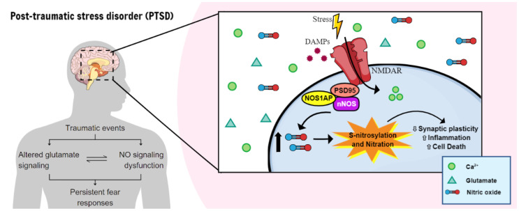
Post-traumatic stress disorder (PTSD) is a psychiatric condition characterized by persistent fear responses and altered neurotransmitter functioning due to traumatic experiences. Stress predominantly affects glutamate, a neurotransmitter crucial for synaptic plasticity and memory formation. Activation of the N-Methyl-D-Aspartate glutamate receptors (NMDAR) can trigger the formation of a complex comprising postsynaptic density protein-95 (PSD95), the neuronal nitric oxide synthase (nNOS), and its adaptor protein (NOS1AP). This complex is pivotal in activating nNOS and nitric oxide (NO) production, which, in turn, activates downstream pathways that modulate neuronal signaling, including synaptic plasticity/transmission, inflammation, and cell death. The involvement of nNOS and NOS1AP in the susceptibility of PTSD and its comorbidities has been widely shown. Therefore, understanding the interplay between stress, fear, and NO is essential for comprehending the maintenance and progression of PTSD, since NO is involved in fear acquisition and extinction processes. Moreover, NO induces post-translational modifications (PTMs), including S-nitrosylation and nitration, which alter protein function and structure for intracellular signaling. Although evidence suggests that NO influences synaptic plasticity and memory processing, the specific role of PTMs in the pathophysiology of PTSD remains unclear. This review highlights pathways modulated by NO that could be relevant to stress and PTSD.

Keywords: memory; nNOS; post-translational modifications.


Conflict of interest statement

The authors declare that the research was conducted in the absence of any commercial or financial relationships that could be construed as potential conflicts of interest.

Figures

Figure 1
Figure 1
The involvement of NO signaling in PTSD pathophysiology. Persistent fear responses and altered glutamate signaling, essential for synaptic plasticity and memory formation, are key features of PTSD, resulting from traumatic experiences. Activation of the NMDAR can trigger the formation of a complex comprising PSD95, nNOS, and NOS1AP, which is pivotal for the activation of nNOS and NO production, that, in turn, induce posttranslational modifications (PTMs), such as S-nitrosylation and nitration. Together, they alter protein function and structure for intracellular signaling, activating downstream pathways that modulate neuronal signaling, including synaptic plasticity/transmission, inflammation, and cell death.
Figure 2
Figure 2
Major products of alternatively spliced nNOS mRNAs: nNOS-α, β, μ and γ. The NOS1 gene comprises 29 exons, 28 introns, and these DNA elements are interspersed over a 250 Kb genome. All but exon 1 are translated to generate the most abundant isoform in the brain, nNOSα, a 150-kDa protein containing a PSD-95/discs large/ZO-1 homology domain (PDZ) that anchors this isoform to neuronal membranes through interactions with PSD95 and NMDA receptor. nNOSμ has a similar structure, but this variant contains a unique 102-base pair (34 amino acids) insert between the CaM and FMN binding domains and is majorly expressed in the heart and smooth muscle. nNOSβ translation is initiated at exon 1a generating a 136-kDa protein; meanwhile, the translation of nNOSγ within exon 5 generates a truncated 125 kDa isoform. Both nNOSβ and nNOSγ lack the PDZ domain and therefore are localized to the cytosolic fraction.* In vitro assays have shown that nNOSγ lacks significant catalytic activity, whereas nNOSβ possesses activity comparable to nNOSα. nNOS-α, β, and γ are majorly expressed in the brain [85].
Figure 3
Figure 3
Schematic of the involvement of NO in inflammatory and cell death pathways under stress conditions. In stressful situations, DAMPs like HMGB1 (mainly released by activated microglia and acting via TLR4 or RAGE; not shown) trigger the release of glutamate and the activation of NMDA-type glutamate receptors. These receptors are organized by PSD95, a protein containing multiple PDZ domains. PSD95 also facilitates the coupling of nNOS to NMDAR, enabling the direct activation of nNOS by the influx of Ca2+ and subsequent NO production. nNOS, through its PDZ domain, forms a binding interaction with COX-2 and the generated NO S-nitrosylates, ultimately leading to the production of prostaglandins (PGs), resulting in the production of chemokines and cytokines. Following exposure to stress, enhanced NO levels operate through various pathways: (1) Activation of JNK and p38, initiating a pro-apoptotic NO signal and resulting in the phosphorylation and accumulation of p53, activation of caspases, release of cytochrome c, and subsequent apoptosis. Under physiological conditions, XIAP, an E3 ubiquitin ligase, effectively inhibits caspases by targeting them for proteasomal degradation. However, under nitrosative conditions, NO inactivates the XIAP’s E3 ligase activity through S-nitrosylation, leading to caspase stabilization, and sensitization of neurons to apoptotic stimuli. Damaged mitochondria produce GSNO by transferring the nitrosyl group from cytochrome c’s heme iron to GSH. GSNO then translocates to various subcellular locations and participates in transnitrosylation, affecting interacting proteins such as NF-κB. (2) Production of ROS and RNS, responsible for S-nitrosylation of NF-kB, which translocates to the nucleus, leading to the production of iNOS and cytokines (e.g., pro-IL-1β, IL-6, and TNF-α). RNS also activates COX-2, increasing PGs release, and activation of NLRP3 inflammasome. (3) S-nitrosylation of GAPDH at Cys150 enables its interaction with Siah1, which escorts SNO-GAPDH to the nucleus, eliciting cell death. In the cytoplasm, SNO-GAPDH also interacts with GOSPEL, preventing its translocation to the nucleus, thereby inhibiting apoptosis. The competition between SNO-GOSPEL and Siah1 for SNO-GAPDH binding represents a regulatory mechanism that maintains cellular homeostasis in response to stressors. This balance may be disrupted in stress-related disorders such as PTSD. (4) Additionally, stress and DAMPs trigger the efflux of intracellular K+ through P2X7R, ultimately activating the NLRP3 inflammasome complex. This leads to the activation of caspase 1, which cleaves pro-IL-1β into IL-1β. One of the functions of IL-1β is to activate p38.
Figure 4
Figure 4
nNOS and the regulation of synaptic transmission and plasticity. After an action potential and influx of Ca2+ in the neuron, nNOS is activated through its domain CaM, producing NO. (1) In the docking step, NO can promote the S-nitrosylation of Sec1, inhibiting its binding to Munc-18 and thereby facilitating syntaxin 1 engagement with SNARE and fusion with the membrane. (2) After priming, nNOS might affect the disassembly of the SNARE complex by S-nitrosylating NSF, preventing its ATPase activation by SNAPs. Regarding postsynaptic transmission, S-nitrosylated thorase promotes the stabilization of the GluA2/GRIP1 complex and enhances AMPA endocytosis. On the other hand, thorase can activate NSF by transnitrosylation, promoting NSF binding on GluA2-PICK1, enhancing insertion of GluA2 at the synapse. NO can affect synaptic plasticity through the inhibition of TrkB activation by S-nitrosylation. Concomitantly, BDNF also upregulates the expression of nNOS and augments NO production, indicating that nNOS also plays a role in synaptic plasticity.

Similar articles

Cited by

References

    1. Chrousos G.P. Stress and disorders of the stress system. Nat. Rev. Endocrinol. 2009;5:374–381. doi: 10.1038/nrendo.2009.106. - DOI - PubMed
    1. Tseng Y.T., Schaefke B., Wei P., Wang L. Defensive responses: Behaviour, the brain and the body. Nat. Rev. Neurosci. 2023;24:655–671. doi: 10.1038/s41583-023-00736-3. - DOI - PubMed
    1. Parsons R.G., Ressler K.J. Implications of memory modulation for post-traumatic stress and fear disorders. Nat. Neurosci. 2013;16:146–153. doi: 10.1038/nn.3296. - DOI - PMC - PubMed
    1. Ressler K.J., Berretta S., Bolshakov V.Y., Rosso I.M., Meloni E.G., Rauch S.L., Carlezon W.A. Post-traumatic stress disorder: Clinical and translational neuroscience from cells to circuits. Nat. Rev. Neurol. 2022;18:273–288. doi: 10.1038/s41582-022-00635-8. - DOI - PMC - PubMed
    1. Bremner J.D., Staib L.H., Kaloupek D., Southwick S.M., Soufer R., Charney D.S. Neural correlates of exposure to traumatic pictures and sound in Vietnam combat veterans with and without posttraumatic stress disorder: A positron emission tomography study. Biol. Psychiatry. 1999;45:806–816. doi: 10.1016/S0006-3223(98)00297-2. - DOI - PMC - PubMed