. 2024 Jan 4;29(1):274.
doi: 10.3390/molecules29010274.

Pan-EGFR Inhibitor Dacomitinib Resensitizes Paclitaxel and Induces Apoptosis via Elevating Intracellular ROS Levels in Ovarian Cancer SKOV3-TR Cells

Affiliations

Pan-EGFR Inhibitor Dacomitinib Resensitizes Paclitaxel and Induces Apoptosis via Elevating Intracellular ROS Levels in Ovarian Cancer SKOV3-TR Cells

Ye Jin Lim et al. Molecules. .

Abstract

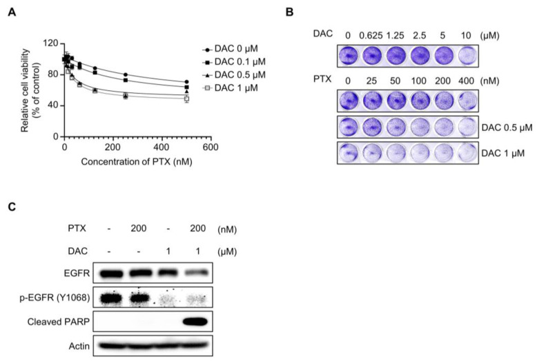
Paclitaxel is still used as a standard first-line treatment for ovarian cancer. Although paclitaxel is effective for many types of cancer, the emergence of chemoresistant cells represents a major challenge in chemotherapy. Our study aimed to analyze the cellular mechanism of dacomitinib, a pan-epidermal growth factor receptor (EGFR) inhibitor, which resensitized paclitaxel and induced cell cytotoxicity in paclitaxel-resistant ovarian cancer SKOV3-TR cells. We investigated the significant reduction in cell viability cotreated with dacomitinib and paclitaxel by WST-1 assay and flow cytometry analysis. Dacomitinib inhibited EGFR family proteins, including EGFR and HER2, as well as its downstream signaling proteins, including AKT, STAT3, ERK, and p38. In addition, dacomitinib inhibited the phosphorylation of Bad, and combination treatment with paclitaxel effectively suppressed the expression of Mcl-1. A 2'-7'-dichlorodihydrofluorescein diacetate (DCFH-DA) assay revealed a substantial elevation in cellular reactive oxygen species (ROS) levels in SKOV3-TR cells cotreated with dacomitinib and paclitaxel, which subsequently mediated cell cytotoxicity. Additionally, we confirmed that dacomitinib inhibits chemoresistance in paclitaxel-resistant ovarian cancer HeyA8-MDR cells. Collectively, our research indicated that dacomitinib effectively resensitized paclitaxel in SKOV3-TR cells by inhibiting EGFR signaling and elevating intracellular ROS levels.

Keywords: dacomitinib; ovarian cancer; paclitaxel-resistance; pan-EGFR inhibitor; reactive oxygen species.


Conflict of interest statement

The authors declare no conflicts of interest.

Figures

Figure 1
Figure 1
Cotreatment with dacomitinib and paclitaxel induced apoptosis in SKOV3-TR ovarian cancer cells. (A) SKOV3-TR cells were treated with various concentrations of dacomitinib (0, 0.1, 0.5, 1, 5, and 10 μM) for 48 h, and then LDH analysis was performed. (B) SKOV3-TR cells were cotreated with a combination of dacomitinib (0, 0.1, 0.5, and 1 µM) and serial dilutions of paclitaxel (0–500 nM) for 48 h. Cell viability was analyzed by WST-1 assay. (C) SKOV3-TR cells were cotreated with serial dilutions of paclitaxel (0–400 nM) and dacomitinib (0–10 μM) for 48 h, and then crystal violet assay was performed. (D) SKOV3-TR cells were subjected to treatments with DMSO (Mock), 200 nM paclitaxel, 1 µM dacomitinib, and a combination of 1 µM dacomitinib and 200 nM paclitaxel, each administered for 48 h. Subsequently, cellular observations were conducted under a microscope. (E) SKOV3-TR cells were subjected to treatments with 200 nM paclitaxel, 1 µM dacomitinib, and combination of 200 nM paclitaxel and 1 µM dacomitinib at various time intervals (0, 12, 24, and 48 h). Subsequently, the levels of cleaved PARP were analyzed by immunoblotting. (F) SKOV3-TR cells were treated with combination of dacomitinib (0, 0.1, 0.5, and 1 µM) and paclitaxel (0, 10, 100, and 200 nM) as indicated for 48 h, and then cleaved PARP was analyzed by immunoblotting. (G) SKOV3-TR cells were subjected to treatments with DMSO (Mock), 200 nM paclitaxel, 1 µM dacomitinib, and combination of 200 nM paclitaxel and 1 µM dacomitinib each for 48 h. Apoptotic cell death was analyzed by FACS analysis. Sub-G1 fraction (apoptotic cell fraction) was measured in percentages and shown as a graph. Actin was used as a loading control for each sample. The LDH assay data are presented as the mean percentage of control ± SD relative to the control. Values of *** p < 0.001 were considered statistically significant difference. DAC, dacomitinib; PTX, paclitaxel.
Figure 2
Figure 2
Dacomitinib suppressed EGFR and its downstream signaling in SKOV3-TR cells. (A) SKOV3-TR cells were subjected to treatments with 200 nM paclitaxel, 1 µM dacomitinib, and combination of 200 nM paclitaxel and 1 µM dacomitinib for 48 h. The expression of EGFR and HER2 and their phosphorylated forms (phosphorylated EGFR at Tyr1068 and phosphorylated HER2 at Tyr1248) were analyzed by immunoblotting. (B) Transcriptions of EGFR and HER2 were examined by RT-PCR. (C) SKOV3-TR cells were cotreated with dacomitinib (0, 0.1, 0.5, and 1 µM) and paclitaxel (0, 10, 100, and 200 nM) as indicated for 48 h. The expression levels of EGFR and HER2 and their phosphorylation (phosphorylated EGFR at Tyr1068 and phosphorylated HER2 at Tyr1248) were examined by immunoblotting. (D) SKOV3-TR cells were subjected to treatments with 200 nM paclitaxel, 1 µM dacomitinib, and combination of 200 nM paclitaxel and 1 µM dacomitinib, and then the expression levels of EGFR and HER2 and their phosphorylation status (phosphorylated EGFR at Tyr1068 and phosphorylated HER2 at Tyr1248) were examined at different time points (0, 0.5, 1.5, 3, and 6 h). (E) SKOV3-TR cells were treated with 200 nM paclitaxel, 1 µM dacomitinib, 1 µM geftinib, and 1 µM erlutinib in the presence or absence of 200 nM paclitaxel as indicated for 48 h and then the expression of EGFR and HER2 and their phosphorylation (phosphorylated EGFR at Tyr1068 and phosphorylated HER2 at Tyr1248) were examined by immunoblotting. (F) SKOV3-TR cells were subjected to treatments with 200 nM paclitaxel, 1 µM dacomitinib, and combination of 200 nM paclitaxel and 1 µM dacomitinib for 48 h. Then, the expression of AKT, STAT, ERK, and p38 and their phosphorylation levels (phosphorylated AKT at Ser473, phosphorylated STAT3 at Tyr705, phosphorylated STAT3 at Ser727, phosphorylated ERK at Thr202/Tyr204, and phosphorylated p38 at Thr180/Tyr182) were analyzed by immunoblotting. (G) Transcriptions of AKT, STAT3, ERK, and p38 were examined by RT-PCR. GAPDH was used as a loading control for the mRNAs in each sample. Actin was used as a loading control for each sample. PTX, paclitaxel; DAC, dacomitinib; GEF, gefitinib; ERL, erlotinib.
Figure 3
Figure 3
HER2 signaling did not affect the paclitaxel resensitization mechanism of dacomitinib in SKOV3-TR cells. (A) SKOV3-TR cells were treated with trastuzumab (10 µg/mL) for 48 h, and then the expression of EGFR and HER2 and their phosphorylation (phosphorylated EGFR at Tyr1068 and phosphorylated HER2 at Tyr1248) were examined by immunoblotting. (B) SKOV3-TR cells were cotreated with combination of a serial dilution of paclitaxel (0–500 nM) and trastuzumab (0, 10, 50, and 100 µg/mL) as indicated for 48 h. Cell viability analysis was performed by WST-1 assay. Actin was used as a loading control for each sample. TRA, trastuzumab.
Figure 4
Figure 4
Dacomitinib did not affect the expression and function of P-gp in SKOV3-TR cells. (A) SKOV3-TR cells were subjected to treatments with 200 nM paclitaxel, 1 µM dacomitinib, and combination of 200 nM paclitaxel and 1 µM dacomitinib for 48 h. Then, the expression levels of P-gp were examined by immunoblotting. (B) Transcription levels of MDR1 were analyzed by RT-PCR in SKOV3-TR cells treated with paclitaxel and dacomitinib as indicated. (C) SKOV3-TR cells (7 × 10⁵) were seeded and further incubated for 24 h. Then, cells were subjected to treatments with DMSO (Mock), 200 nM paclitaxel, 1 µM dacomitinib, and a combination of 200 nM paclitaxel and 1 µM dacomitinib for 24 h. Fluo-3/AM solution was added and further reacted at 37 °C for 1 h, and then the fluorescences were visualized using an inverted fluorescence microscope. SKOV3 cells, the parent cells of SKOV3-TR, were used as paclitaxel-sensitive controls. Actin was used as a loading control for each sample. GAPDH was used as a loading control for the mRNAs in each sample. PTX, paclitaxel; DAC, dacomitinib.
Figure 5
Figure 5
Dacomitinib suppressed the expression of Mcl-1 and the phosphorylation of Bad in paclitaxel-treated SKOV3-TR cells. (A) SKOV3-TR cells were subjected to treatments with 200 nM paclitaxel, 1 µM dacomitinib, and combination of 200 nM paclitaxel and 1 µM dacomitinib for 48 h. The expression levels of prosurvival Bcl-2 family proteins (Bcl-2, Bcl-xl, and Mcl-1), apoptosis effector Bcl-2 family protein (Bax), proapoptotic Bcl-2 family protein (Bad), and its phosphorylation (phospho-Bad at Ser155 residue) were examined by immunoblotting. The apoptotic mediators (caspase-3, cleaved caspase-3, and cleaved PARP) were also analyzed by immunoblotting. (B) Transcriptions of BCL-2, BCL-XL, BAX, and MCL-1 were analyzed by RT-PCR. Actin was used as a loading control for each sample. GAPDH was used as a loading control for the mRNAs in each sample. PTX, paclitaxel; DAC, dacomitinib.
Figure 6
Figure 6
Dacomitinib increased intracellular ROS levels in paclitaxel-treated SKOV3-TR cells. (A) SKOV3-TR cells were subjected to treatments with 200 nM paclitaxel, 1 µM dacomitinib, and combination of 200 nM paclitaxel and 1 µM dacomitinib, with or without 5 mM N-acetylcysteine for 48 h. Cells were additionally treated with 10 µM DCFH-DA and further incubated for 30 min at 37 °C. Intracellular ROS levels were analyzed by measuring fluorescence values at excitation wavelengths of 485 nm and emission wavelengths of 520 nm using a fluorescence microplate reader. Relative intracellular ROS levels were calculated as the percentage of fluorescence values in the treatment group compared to those in the control group. (B) SKOV3-TR cells were subjected to treatments with a serial dilution of paclitaxel (0–500 nM) with or without 5 mM N-acetylcysteine for 48 h, and then cell viability was examined by WST-1 assay. (C) SKOV3-TR cells were subjected to treatments with combination of serial dilutions of paclitaxel (0–500 nM) and 1 µM dacomitinib with or without 5 mM N-acetylcysteine for 48 h, and then cell viability was examined by WST-1 assay. (D) SKOV3-TR cells were subjected to treatments with combination of serial dilutions of paclitaxel (0–400 nM), 1 μM dacomitinib, and 5 mM N-acetylcysteine as indicated for 48 h and then stained with 0.2% crystal violet solution for 1 h at room temperature. The DCFH-DA assay data are presented as the mean percentage of control ± SD relative to the control. Values of ***, ### p < 0.001 were considered statistically significant. PTX, paclitaxel; DAC, dacomitinib; NAC, N-acetylcysteine.
Figure 7
Figure 7
Dacomitinib resensitized paclitaxel in HeyA8-MDR ovarian cancer cells. (A) HeyA8-MDR cells were cotreated with a serial dilution of paclitaxel (0–500 nM) and specific concentrations of dacomitinib (0, 0.1, 0.5, and 1 µM) for 48 h. Cell viability was analyzed through WST-1 assay. (B) HeyA8-MDR cells were cotreated with serial dilutions of paclitaxel (0–400 nM) and dacomitinib (0–10 μM) for 48 h and then stained with 0.2% crystal violet solution for 1 h at room temperature. (C) HeyA8-MDR cells were subjected to treatments with paclitaxel (200 nM), dacomitinib (1 µM), and combination of paclitaxel (200 nM) and dacomitinib (1 µM) for 48 h. Immunoblotting was used to validate the expression levels of EGFR and its phosphorylation status (phosphorylated EGFR at Y1068). Actin was used as a loading control for each sample. PTX, paclitaxel; DAC, dacomitinib.

Similar articles

References

    1. Jayson G.C., Kohn E.C., Kitchener H.C., Ledermann J.A. Ovarian cancer. Lancet. 2014;384:1376–1388. doi: 10.1016/S0140-6736(13)62146-7. - DOI - PubMed
    1. Reid B.M., Permuth J.B., Sellers T.A. Epidemiology of ovarian cancer: A review. Cancer Biol. Med. 2017;14:9–32. doi: 10.20892/j.issn.2095-3941.2016.0084. - DOI - PMC - PubMed
    1. Stewart C., Ralyea C., Lockwood S. Ovarian Cancer: An Integrated Review. Semin. Oncol. Nurs. 2019;35:151–156. doi: 10.1016/j.soncn.2019.02.001. - DOI - PubMed
    1. Matulonis U.A., Penson R.T., Domchek S.M., Kaufman B., Shapira-Frommer R., Audeh M.W., Kaye S., Molife L.R., Gelmon K.A., Robertson J.D., et al. Olaparib monotherapy in patients with advanced relapsed ovarian cancer and a germline BRCA1/2 mutation: A multistudy analysis of response rates and safety. Ann. Oncol. 2016;27:1013–1019. doi: 10.1093/annonc/mdw133. - DOI - PubMed
    1. Cortez A.J., Tudrej P., Kujawa K.A., Lisowska K.M. Advances in ovarian cancer therapy. Cancer Chemother. Pharmacol. 2018;81:17–38. doi: 10.1007/s00280-017-3501-8. - DOI - PMC - PubMed