. 2023 Dec 19;25(1):15.
doi: 10.3390/ijms25010015.

Release of Pro-Inflammatory/Angiogenic Factors by Retinal Microvascular Cells Is Mediated by Extracellular Vesicles Derived from M1-Activated Microglia

Affiliations

Release of Pro-Inflammatory/Angiogenic Factors by Retinal Microvascular Cells Is Mediated by Extracellular Vesicles Derived from M1-Activated Microglia

Elena Beltramo et al. Int J Mol Sci. .

Abstract

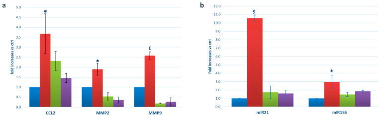
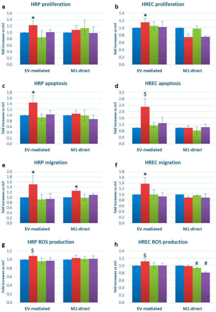
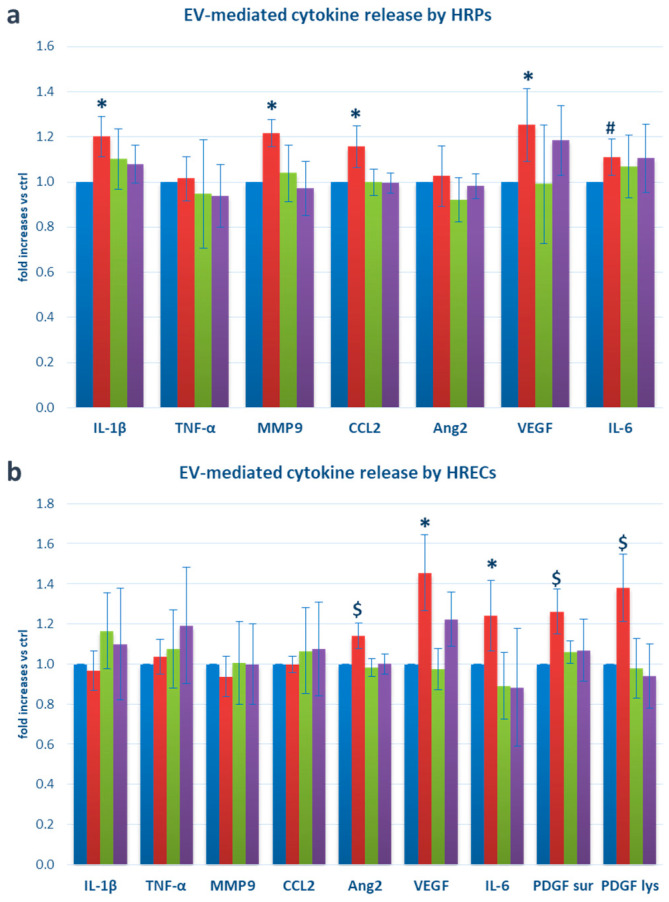
The interactions between the neuronal and vascular sides of the retina during diabetic retinopathy (DR) have gained increasing attention. Microglia is responsible for the immune response to inflammation inside the retina, which could be mediated by paracrine signals carried by extracellular vesicles (EVs). We aimed to characterize EVs released from immortalized human microglial cells in inflammation and investigate their effects on the retinal microvasculature and the anti-inflammatory potential of thiamine in this context. M1 pro-inflammatory polarization in microglia was induced through a cytokine cocktail. EVs were isolated from the supernatants, characterized, and used to stimulate human retinal endothelial cells (HRECs) and pericytes (HRPs). Microvascular cell functions and their release of pro-inflammatory/angiogenic factors were assessed. M1-derived EVs showed increased content of miR-21, miR-155, CCL2, MMP2, and MMP9, and enhanced apoptosis, proliferation, migration, and ROS production in HRPs and HRECs. IL-1β, IL-6, MMP9, CCL2, and VEGF release increased in HRPs exposed to M1-derived EVs, while HRECs showed augmented IL-6, Ang2, VEGF, and PDFG-B. Addition of thiamine to M1-microglial cultures reverted most of these effects. In conclusion, M1-derived EVs stimulate functional changes and secretion of pro-inflammatory/angiogenic molecules in microvascular cells, exacerbating inflammatory damage and retinopathy features. Thiamine added to microglia exerts anti-inflammatory effects.

Keywords: angiogenesis; diabetic retinopathy; endothelial cells; extracellular vesicles; inflammation; microglia; pericytes; thiamine.


Conflict of interest statement

The authors declare no conflict of interest. The funder had no role in the design of the study; in the collection, analyses, or interpretation of data; in the writing of the manuscript; or in the decision to publish the results.

Figures

Figure 1
Figure 1
EV expression of pro-inflammatory mRNAs (a) and miRNAs (b). Blue bars: EVs extracted from the supernatants of microglial cells cultured in physiological conditions (ctrl); red bars: EVs extracted from the supernatants of M1-activated microglial cells (M1); green bars: EVs extracted from the supernatants of microglial cells cultured in the presence of thiamine (T); purple bars: EVs extracted from the supernatants of microglial cells cultured in M1 conditions plus thiamine (M1+T). Mean ± SD of 5 independent experiments. * = p < 0.05 vs. ctrl and M1+T; £ = p <0.005 vs. ctrl and M1+T; $ = p < 0.001 vs. ctrl and M1+T.
Figure 2
Figure 2
Functional changes in HRPs and HRECs exposed to microglia-derived EVs: (a) HRP and (b) HREC proliferation; (c) HRP and (d) HREC apoptosis; (e) HRP and (f) HREC migration: (g) HRP and (h) HREC ROS production. Blue bars: cells exposed to EVs extracted from the supernatants of microglial cells cultured in physiological conditions (EV-mediated bars) or directly cultured in physiological conditions (M1-direct bars); red bars: cells exposed to EVs extracted from the supernatants of M1-activated microglial cells (EV-mediated bars) or directly cultured in the presence of the M1 cocktail (M1-direct); green bars: cells exposed to EVs extracted from the supernatants of microglial cells cultured with thiamine (EV-mediated bars) or directly cultured with thiamine (M1-direct bars); purple bars: cells exposed to EVs extracted from the supernatants of microglial cells cultured in M1 plus thiamine (EV-mediates bars) or directly with M1 plus thiamine (M1-direct bars). Mean ± SD of 5 independent experiments. * = p < 0.05 vs. ctrl and M1+T; $ = p < 0.005 vs. ctrl and M1+T; # = p < 0.05 vs. ctrl and M1.
Figure 3
Figure 3
Release of pro-inflammatory/pro-angiogenic factors by (a) HRPs and (b) HRECs following exposure to microglia-derived EVs. Blue bars: cells exposed to EVs extracted from the supernatants of microglial cells cultured in physiological conditions (ctrl); red bars: cells exposed to EVs extracted from the supernatants of M1-activated microglial cells (M1); green bars: cells exposed to EVs extracted from the supernatants of microglial cells cultured in the presence of thiamine (T); purple bars: cells exposed to EVs extracted from the supernatants of microglial cells cultured in M1 conditions plus thiamine (M1+T). Mean ± SD of 5 independent experiments. * = p < 0.05 vs. ctrl and M1+T; $ = p < 0.005 vs. ctrl and M1+T; # = p < 0.05 vs. ctrl.

References

    1. Teo Z., Tham Y., Yu M., Chee M., Rim T., Cheung N., Bikbov M., Wang Y., Tang Y., Lu Y., et al. Global Prevalence of Diabetic Retinopathy and Projection of Burden through 2045: Systematic Review and Meta-analysis. Ophthalmology. 2021;128:1580–1591. doi: 10.1016/j.ophtha.2021.04.027. - DOI - PubMed
    1. Simó R., Hernández C., European Consortium for the Early Treatment of Diabetic Retinopathy (EUROCONDOR) Neurodegeneration in the diabetic eye: New insights and therapeutic perspectives. Trends Endocrinol. Metab. 2014;25:23–33. doi: 10.1016/j.tem.2013.09.005. - DOI - PubMed
    1. Armulik A., Abramsson A., Betsholtz C. Endothelial/pericyte interactions. Circ. Res. 2005;97:512–523. doi: 10.1161/01.RES.0000182903.16652.d7. - DOI - PubMed
    1. Kettenmann H., Hanisch U.K., Noda M., Verkhratsky A. Physiology of microglia. Physiol. Rev. 2011;91:461–553. doi: 10.1152/physrev.00011.2010. - DOI - PubMed
    1. Arroba A.I., Valverde Á. Modulation of microglia in the retina: New insights into diabetic retinopathy. Acta Diabetol. 2017;54:527–533. doi: 10.1007/s00592-017-0984-z. - DOI - PubMed