Review
. 2023 Dec 19;25(1):33.
doi: 10.3390/ijms25010033.

Overview of Gene Expression Dynamics during Human Oogenesis/Folliculogenesis

Affiliations
Review

Overview of Gene Expression Dynamics during Human Oogenesis/Folliculogenesis

Bastien Ducreux et al. Int J Mol Sci. .

Abstract

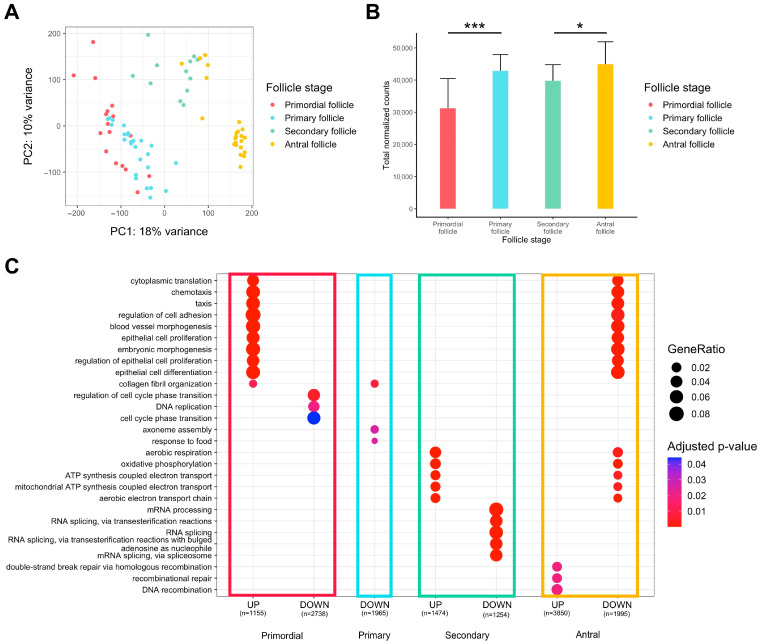
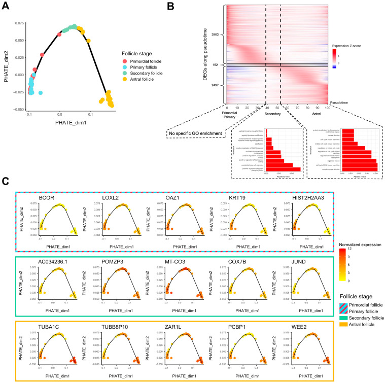
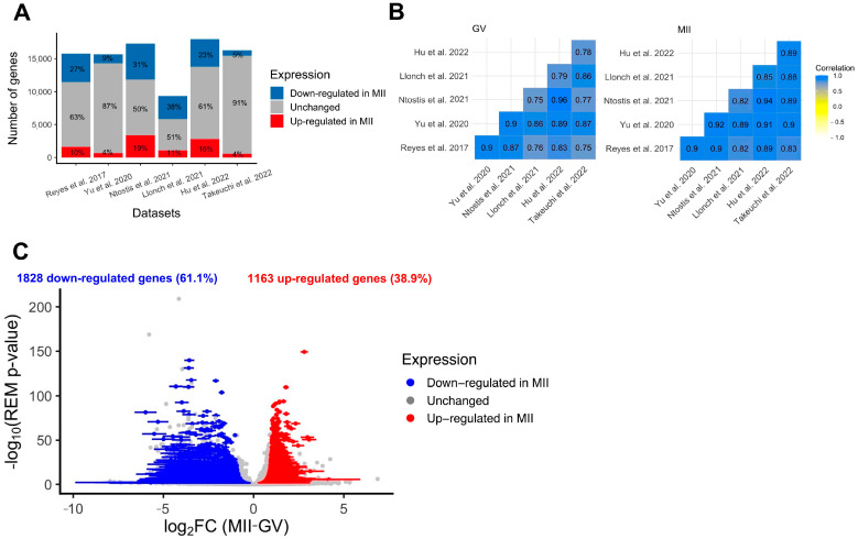
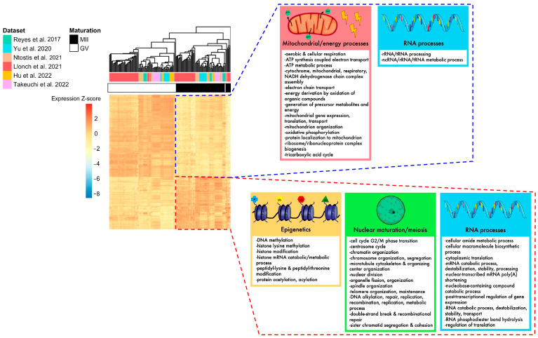
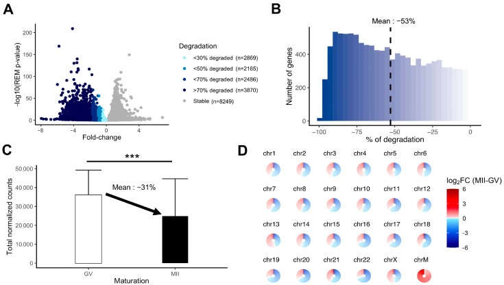
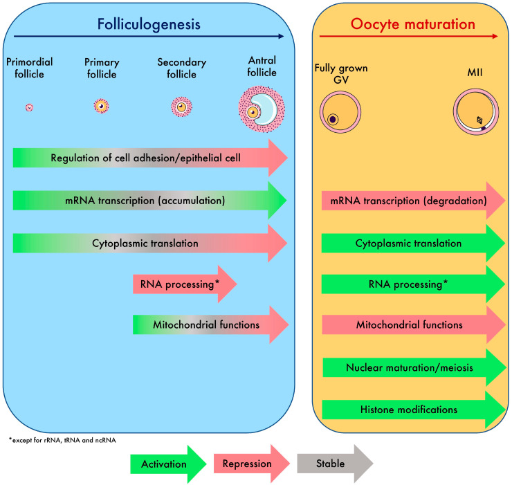
The oocyte transcriptome follows a tightly controlled dynamic that leads the oocyte to grow and mature. This succession of distinct transcriptional states determines embryonic development prior to embryonic genome activation. However, these oocyte maternal mRNA regulatory events have yet to be decoded in humans. We reanalyzed human single-oocyte RNA-seq datasets previously published in the literature to decrypt the transcriptomic reshuffles ensuring that the oocyte is fully competent. We applied trajectory analysis (pseudotime) and a meta-analysis and uncovered the fundamental transcriptomic requirements of the oocyte at any moment of oogenesis until reaching the metaphase II stage (MII). We identified a bunch of genes showing significant variation in expression from primordial-to-antral follicle oocyte development and characterized their temporal regulation and their biological relevance. We also revealed the selective regulation of specific transcripts during the germinal vesicle-to-MII transition. Transcripts associated with energy production and mitochondrial functions were extensively downregulated, while those associated with cytoplasmic translation, histone modification, meiotic processes, and RNA processes were conserved. From the genes identified in this study, some appeared as sensitive to environmental factors such as maternal age, polycystic ovary syndrome, cryoconservation, and in vitro maturation. In the future, the atlas of transcriptomic changes described in this study will enable more precise identification of the transcripts responsible for follicular growth and oocyte maturation failures.

Keywords: RNA-seq; folliculogenesis; maturation; oocyte; transcriptome.


Conflict of interest statement

The authors declare no conflict of interest.

Figures

Figure 1
Figure 1
Differential expression analysis of transcriptomic signature changes along the different stages of folliculogenesis (dataset from Zhang et al. (2018) [23]). (A) Principal component analysis of the whole expression dataset removing the 10% genes with the lowest variance. (B) Comparison of the normalized read counts total between the different follicle stages (significance assessed with a t-test, * p < 0.05, *** p < 0.005). (C) Comparative Gene Ontology enrichment analysis according to the up- or downregulation of transcripts between all four follicle stages. GeneRatio corresponds to the fraction of differentially expressed genes (DEGs) found in each Gene Ontology set.
Figure 2
Figure 2
Trajectory analysis of transcriptomic changes along folliculogenesis. (A) PHATE dimensionality reduction analysis of the whole expression dataset. (B) Heatmap of inferred expression of the most variable significant genes along folliculogenesis (6552 genes showing average expression changed by at least logFC > 1 within the oocyte developmental trajectory). Pseudotime is a metric that could be interpreted as a timing distance between one cell and its precursor cell and helps identify the ordering of cells along a lineage based on their gene expression profile. In this analysis, pseudotime 0 represents oocytes in the beginning of the window of the oocyte developmental trajectory (early follicles), while pseudotime 100 represents oocytes in the end of the window (antral follicles). Vertical lines delimit oocytes from the primordial/primary, secondary, and antral follicle groups. Horizontal lines delimit significantly differentially expressed genes along folliculogenesis reaching their peak expression during the primordial/primary (3903), secondary (152), and antral follicle groups (2497). (C) Variation of expression, for each follicle stage group, of the top 5 genes differentially expressed along folliculogenesis reaching their peak expression at this stage.
Figure 3
Figure 3
Differential expression analysis of genes during the GV-to-MII oocyte transition. (A) Results of individual differential expression analysis for the six datasets evaluated in this study. Numbers represent the percentage of genes upregulated, downregulated, or stable in MII versus GV oocytes. (B) Correlation of the normalized expression of all expressed genes for the six datasets evaluated in this study, respectively, for GV and MII oocytes. (C) Volcanoplot of the meta-analysis result for the six datasets evaluated in this study. A random effects model based on effect size (log2FC, difference of expression between MII and GV oocytes) and p-values from each dataset was applied. One gene is represented by one dot. A total of 2991 significant DEGs were identified (summary adjusted p-value from the random effect model < 0.05 and sign of log2FC consistent in all 6 studies). Blue dots represent significant DEGs downregulated in MII oocytes compared to GV. Similarly, red dots represent significant DEGs upregulated in MII oocytes compared to GV oocytes. The interval confidence of the summary log2FC is represented by the bars associated with the dots for each DEG [24,25,26,27,28,29].
Figure 4
Figure 4
Summary of biological pathways enriched for DEGs identified in the meta-analysis, according to their up- or downregulation. The heatmap of DEGs identified with the meta-analysis shows that MII and GV oocytes are correctly segregated according to their expression for these genes. Mitochondrial processes, energy processes, and some RNA processes were downregulated in MII oocytes (framed in blue), while nuclear maturation/meiosis, epigenetics, and some other RNA processes were upregulated in MII oocytes (framed in red) [24,25,26,27,28,29].
Figure 5
Figure 5
Measurement of the degradation of the maternal mRNA content in MII oocytes. (A) Volcanoplot of the meta-analysis result (MII versus GV) colored by the intensity of degradation between GV and MII stages (stable, <30% of degradation, <50% of degradation, <70% of degradation, or >70% of degradation). (B) Histogram focusing on the extent of degradation in downregulated transcripts in MII versus GV oocytes. (C) Comparison of total normalized counts between the GV and MII oocytes (significance assessed with a t-test, *** p < 0.005). (D) Assessment of degradation by chromosome. Each circle is divided into sections according to the number of genes on the chromosome and each section, representing one gene, is colored by the summary log2FC resulting from the meta-analysis result (MII versus GV) corresponding to the gene. ChrM: mitochondrial chromosome.
Figure 6
Figure 6
Summary of biological processes and global transcriptomic events driving oocyte growth and maturation identified in this study.

Similar articles

Cited by

References

    1. Bendsen E., Byskov A.G., Andersen C.Y., Westergaard L.G. Number of Germ Cells and Somatic Cells in Human Fetal Ovaries during the First Weeks after Sex Differentiation. Hum. Reprod. 2006;21:30–35. doi: 10.1093/humrep/dei280. - DOI - PubMed
    1. Tripathi A., Prem Kumar K.V., Chaube S.K. Meiotic Cell Cycle Arrest in Mammalian Oocytes. J. Cell Physiol. 2010;223:592–600. doi: 10.1002/jcp.22108. - DOI - PubMed
    1. Eppig J.J. Coordination of Nuclear and Cytoplasmic Oocyte Maturation in Eutherian Mammals. Reprod. Fertil. Dev. 1996;8:485–489. doi: 10.1071/RD9960485. - DOI - PubMed
    1. Watson A.J. Oocyte Cytoplasmic Maturation: A Key Mediator of Oocyte and Embryo Developmental Competence. J. Anim. Sci. 2007;85:E1–E3. doi: 10.2527/jas.2006-432. - DOI - PubMed
    1. Conti M., Franciosi F. Acquisition of Oocyte Competence to Develop as an Embryo: Integrated Nuclear and Cytoplasmic Events. Hum. Reprod. Update. 2018;24:245–266. doi: 10.1093/humupd/dmx040. - DOI - PMC - PubMed

LinkOut - more resources