. 2023 Dec 19;25(1):51.
doi: 10.3390/ijms25010051.

3'dNTP Binding Is Modulated during Primer Synthesis and Translesion by Human PrimPol

Affiliations

3'dNTP Binding Is Modulated during Primer Synthesis and Translesion by Human PrimPol

Cristina Velázquez-Ruiz et al. Int J Mol Sci. .

Abstract

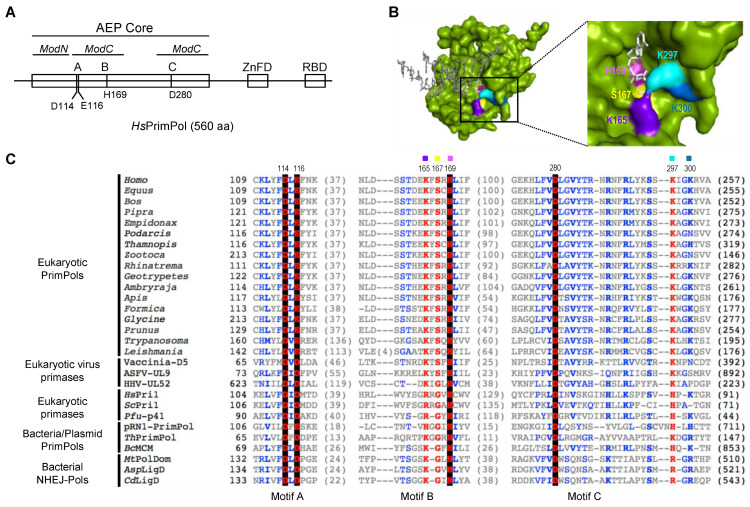
PrimPol is a DNA primase/polymerase from the Archaeo-Eukaryotic Primase (AEP) superfamily that enables the progression of stalled replication forks by synthesizing DNA primers ahead of blocking lesions or abnormal structures in the ssDNA template. PrimPol's active site is formed by three AEP-conserved motifs: A, B and C. Motifs A and C of human PrimPol (HsPrimPol) harbor the catalytic residues (Asp114, Glu116, Asp280) acting as metal ligands, whereas motif B includes highly conserved residues (Lys165, Ser167 and His169), which are postulated to stabilize 3' incoming deoxynucleotides (dNTPs). Additionally, other putative nucleotide ligands are situated close to motif C: Lys297, almost invariant in the whole AEP superfamily, and Lys300, specifically conserved in eukaryotic PrimPols. Here, we demonstrate that His169 is absolutely essential for 3'dNTP binding and, hence, for both primase and polymerase activities of HsPrimPol, whereas Ser167 and Lys297 are crucial for the dimer synthesis initiation step during priming, but dispensable for subsequent dNTP incorporation on growing primers. Conversely, the elimination of Lys165 does not affect the overall primase function; however, it is required for damage avoidance via primer-template realignments. Finally, Lys300 is identified as an extra anchor residue to stabilize the 3' incoming dNTP. Collectively, these results demonstrate that individual ligands modulate the stabilization of 3' incoming dNTPs to optimize DNA primer synthesis efficiency during initiation and primer maturation.

Keywords: 3′site dNTP ligands; AEP; AEP motif B; TLS; human PrimPol.


Conflict of interest statement

The authors declare no conflict of interest.

Figures

Figure 1
Figure 1
Highly conserved residues at motif B and close to motif C define the 3′ dNTP binding site in the AEP superfamily. (A) Scheme of human PrimPol domains. AEP core (in green), subdivided in ModN (aa 35 to 105) and ModC (aa 108–200 and 261–348), as proposed by Rechkoblit et al. (2016) [22]. The AEP core is shared among members of the AEP superfamily, and contains the conserved motifs A (red), B (blue) and C (red). A Zn-finger domain (ZnFD) is shown in purple and the RPA-binding domain (RBD) in orange found at C-terminal region. (B) Molecular surface of the AEP core of human PrimPol (green) in preternary complex with the 3′ incoming dNTP (white) and template/primer DNA (gray); as shown in the enlarged area, Lys165 (indigo), Ser167 (yellow), His169 (pink), Lys297 (cyan) and Lys300 (sky blue) are putative direct ligands of the 3′ incoming dNTP, forming the catalytic pocket together with the three invariant carboxylates (not shown for clarity). (C) Amino acid sequence alignment of the three conserved motifs A (shadowed in red), B (shadowed in blue) and C (shadowed in red) of different AEP enzymes, including Eukaryotic PrimPols from Homo sapiens (NP_001332824.1), Equus caballus (XP_023486679.1), Bos taurus (NP_001068956.1), Pipra filicauda (XP_027591218.1), Empidonax traillii (XP_027735658.1), Podarcis muralis (XP_028600037.1), Thamnophis elegans (XP_032080602.1), Zootoca vivipara (XP_034967959.1), Rhinatrema bivittatum (XP_029440727.1), Geotrypetes seraphini (XP_033799271.1), Amblyraja radiata (XP_032874431.1), Apis mellifera (XP_001121815.3), Formica exsecta (XP_029670346.1), Glycine soja (XP_028181145.1), Prunus avium (XP_021827012.1), Trypanosoma equiperdum (SCU71797.1), Leishmania donovani (XP_003863827.1), D5 primase from Vaccinia virus (Vaccinia-D5; P21010.1), UL9 helicase-containing protein from African Swine Fever Virus (ASFV-UL9; Q07183), UL52 primase subunit from Human herpesvirus 1 (HHV-UL52; P10236.1), small subunit of Homo sapiens primase (HsPri1; NP_000937.1), small subunit of Saccharomyces cerevisae primase (ScPri1; P10363.2), catalytic subunit of Pyrococcus furiosus primase (Pfu-p41; WP_011011222.1), Sulfolubus islandicus (pRN1) plasmid pRN1 ORF904 (pRN1-PrimPol; WP_010890202.1), PrimPol from Thermus thermophilus (ThPrimPol; WP_011173100.1), PrimPol-helicase from Bacillus cereus (BcMCM; WP_136983851.1), polymerase domain of Mycobacterium tuberculosis ligase D (MtPolDom; WP_003911307.1), ATP-dependent DNA ligase D from Arthrobacter sp. (AspLigD; TQK28754.1) and DNA ligase D from Clostridioides difficile (CdLigD; VTR08300.1). Non-conserved residues are in grey; conserved amino acids in blue; highly conserved in red and those invariant in black background. Selected HsPrimPol residues are indicated: Asp114, Glu116 and Asp280 (catalytic residues); Lys165, Ser167 and His169 (conserved residues within motif B); Lys297 and Lys300 (as potential interactors of 3′ incoming dNTPs).
Figure 2
Figure 2
Motif B residues and Lys297 are necessary to stabilize the 3′ incoming dNTP before priming. (A) Scheme of the mechanism of DNA priming by human PrimPol; (a) HsPrimPol modular organization: catalytic core in light gray and Zinc Finger domain in dark gray; (b) PrimPol DNA binding, which does not require the ZnFD; (c) dNTP (drawn as the base letter and 3 phosphates represented by blue balls) binding at the 3′elongation site that requires manganese ions and pairing with the templating base; (d) nucleotide binding at the 5′initiation site, which is configured by both the PrimPol core and the ZnFD; (e) dinucleotide formation coupled to PPi release; (f) primer elongation, coupled to separation of the PrimPol core and the ZnFD. (B) Binary complex (E:ssDNA) detected via EMSA using oligonucleotide 3′ T20-GTCC-T36 5′ (0.6 nM; 60-mer; [γ-32P]-labeled depicted as a red asterisk) and increasing concentrations of PrimPol WT or mutants (5, 20 and 80 nM). (C) Quantification (average of three different experiments) of the E:ssDNA complex using optical densitometry, expressed as percentage of the labeled ssDNA. (D) Binary (E:dGTP*) and preternary (E:dGTP*:ssDNA) complexes detected via EMSA using 400 nM of WT or mutants, 16 nM of [α32P]dGTP (α32P is indicated as a red ball while β and γ -phosphates in blue) and and 1 μM 3′ T20-GTCC-T36 5′ (unlabeled) as a template in the presence of Mn2+ (1 mM). Non-specific labeling of the oligonucleotide is indicated by a black asterisk.
Figure 3
Figure 3
His169 is essential for the initial step of PrimPol priming, which is further contributed by other 3′ dNTP ligands. (A) Primase assay of dimer formation was performed as indicated in Materials and Methods, by using as template the ssDNA oligonucleotide 3′ T20-GTCC-T36 5′ (1 μM), which contains a preferred priming sequence (GTC), WT or mutant HsPrimPols (100 nM), [α-32P]dGTP (16 nM; β and γ -phosphates are indicated with a blue ball and α32P with a red ball) as the 3′ site nucleotide, and ATP (1, 10, 100 μM) as the 5′ nucleotide. (B) Quantification of dimer synthesis formation (%) by mutants, relative to WT HsPrimPol (100%), averaged from three different experiments.
Figure 4
Figure 4
His169 is essential for dimer synthesis and primer extension, Ser167 and Lys297 are required to make dimers but not for primer extension, and Lys165, Ser167 and Lys297 are needed to form non-canonical extension products. (A) Dimer formation and further elongation by WT HsPrimPol or the indicated mutants (100 nM) using a template with the preferred priming site (3′ GTC) followed by a heterologous sequence, by sequentially adding [γ-32P]ATP (16 nM) (γ-32P is indicated as a red ball) as the 5′ nucleotide, and dGTP, dTTP and dCTP (100 μM) as 3′ incoming dNTPs, in the presence of 1 mM MnCl2. (B) Elongation of a synthetic mini-primer (3pAGT in blue with a blue box; 10 μM;) by WT HsPrimPol or the indicated mutants (100 nM) in the presence of dCTP, dTTP (10 μM), [α-32P]dGTP (16 nM; in red), and 1 mM MnCl2. Labeled dGMP insertion is indicated with a red ball. Non-canonical products are marked by asterisks.
Figure 5
Figure 5
Lys165, Ser167 and Lys297 are necessary to skip an abasic site. Synthetic primer (3pAGT) extension of WT HsPrimPol and mutants (100 nM) in the presence of either an undamaged template (part A) or a template containing an abasic (Ab) site (part B), using [α-32P]dGTP supplemented with dATP, dCTP or dTTP (100 μM), as indicated, and in the presence of 1 mM MnCl2. The upper autoradiograms show the position of different labeled products (indicated by colored asterisks), whereas the lower parts show schemes of their generation (by coloring the primer extension products congruently with the asterisks mentioned above).
Figure 6
Figure 6
His169 is indispensable for PrimPol polymerase activity, whereas other 3′ dNTP ligands have a secondary role. (A) Standing-start primer extension assay (standard DNA polymerization on a template/primer, labeled and indicated with a red asterisk) by WT HsPrimPol and 3′ incoming dNTP interactor mutants (50 nM), when using the indicated increasing concentrations of dNTPs, in the presence of 1 mM MnCl2. (B) Quantification of primer extension (%) of WT, K165A, S167A, K297A and K300A mutants when providing 1 μM dNTPs (average of three different experiments).
Figure 7
Figure 7
Structural analysis of human PrimPol 3′ incoming dNTP ligands. (A) (a): Spatial location of Lys165 (indigo); (b,c): interactions with 3′ incoming dNTP (white) and catalytic Glu116 (red); (d): mutant K165A (modeled). (B) (a): Spatial location of Ser167 (yellow); (b,c): interactions with Ser160 (orange), Glu116 (red), and 3′ site incoming dNTP (white); (d): mutant S167A (modeled). (C) (a): Spatial location of His169 (magenta); (b): interactions with catalytic Asp114 (red) and 3′ site incoming dNTP (white); modeled mutants H169A (c) and H169Y (d). (D) (a): Spatial location of Lys297 (cyan); (b,c): interactions with 3′ incoming dNTP (white) and Lys300 (sky blue); (d): mutant K297A (modeled). (E) (a): Spatial location of Lys300 (sky blue); (b): interactions with the 3′ incoming dNTP (white); (c): interactions with Lys297 (cyan) and Arg291 (dark blue); (d): mutant K300A (modeled). In all panels, green lines indicate hydrogen bonds while pink ones show steric clashes. Ca2+ as metal B in the catalytic center is shown as a sphere in grey. PDBid: 5L2X.

Similar articles

Cited by

References

    1. Frick D.N., Richardson C.C. DNA Primases. Annu. Rev. Biochem. 2001;70:39–80. doi: 10.1146/annurev.biochem.70.1.39. - DOI - PubMed
    1. Kuchta R.D., Stengel G. Mechanism and evolution of DNA primases. Biochim. Biophys. Acta (BBA) Proteins Proteom. 2010;1804:1180–1189. doi: 10.1016/j.bbapap.2009.06.011. - DOI - PMC - PubMed
    1. García-Gómez S., Reyes A., Martínez-Jiménez M.I., Chocrón E.S., Mourón S., Terrados G., Powell C., Salido E., Méndez J., Holt I.J., et al. PrimPol, an Archaic Primase/Polymerase Operating in Human Cells. Mol. Cell. 2013;52:541–553. doi: 10.1016/j.molcel.2013.09.025. - DOI - PMC - PubMed
    1. Wan L., Lou J., Xia Y., Su B., Liu T., Cui J., Sun Y., Lou H., Huang J. hPrimpol1/CCDC111 is a human DNA primase-polymerase required for the maintenance of genome integrity. Embo Rep. 2013;14:1104–1112. doi: 10.1038/embor.2013.159. - DOI - PMC - PubMed
    1. Bianchi J., Rudd S.G., Jozwiakowski S.K., Bailey L.J., Soura V., Taylor E., Stevanovic I., Green A.J., Stracker T.H., Lindsay H.D., et al. PrimPol Bypasses UV Photoproducts during Eukaryotic Chromosomal DNA Replication. Mol. Cell. 2013;52:566–573. doi: 10.1016/j.molcel.2013.10.035. - DOI - PMC - PubMed

LinkOut - more resources