Review
. 2023 Dec 21;25(1):124.
doi: 10.3390/ijms25010124.

Molecular Mechanisms of Neuroprotection by Ketone Bodies and Ketogenic Diet in Cerebral Ischemia and Neurodegenerative Diseases

Affiliations
Review

Molecular Mechanisms of Neuroprotection by Ketone Bodies and Ketogenic Diet in Cerebral Ischemia and Neurodegenerative Diseases

Jiwon Jang et al. Int J Mol Sci. .

Abstract

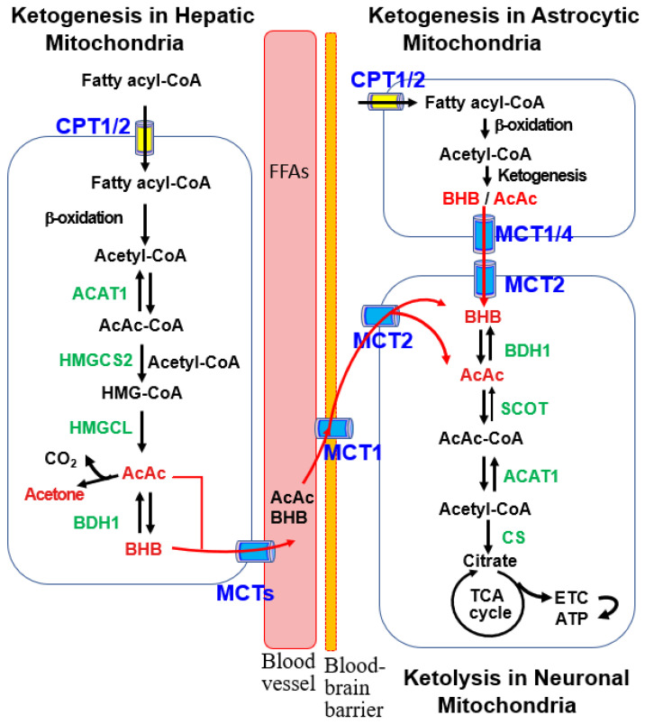
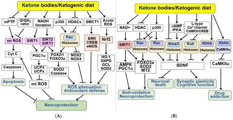
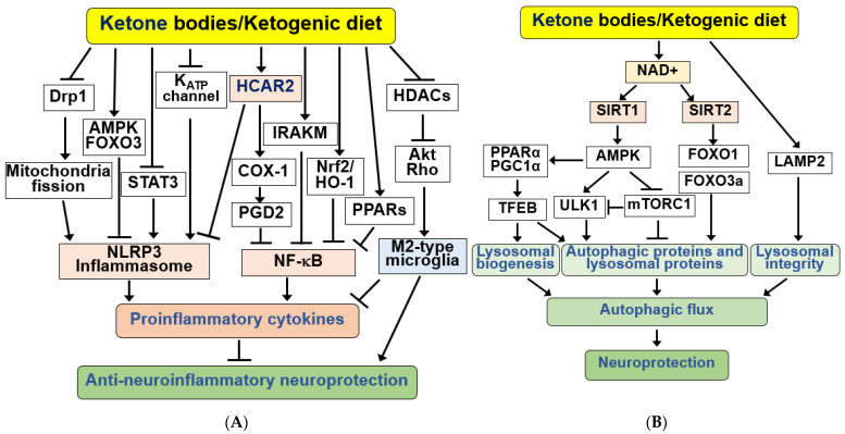
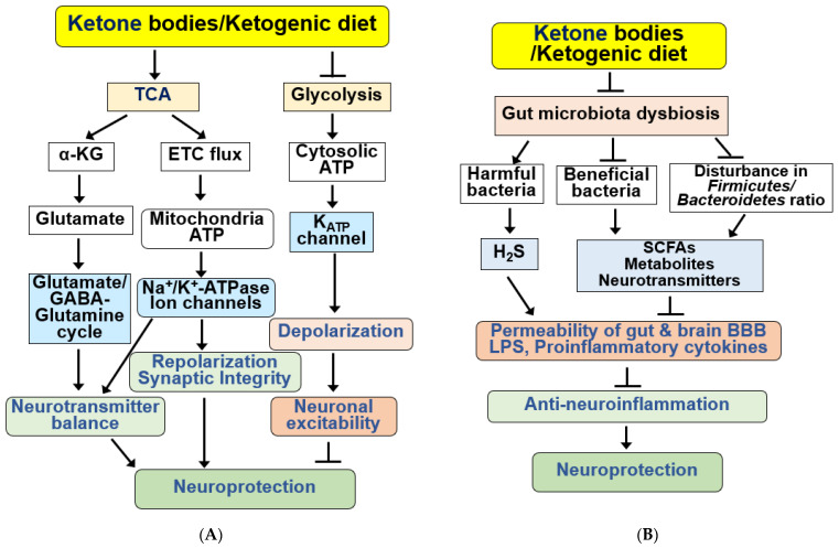
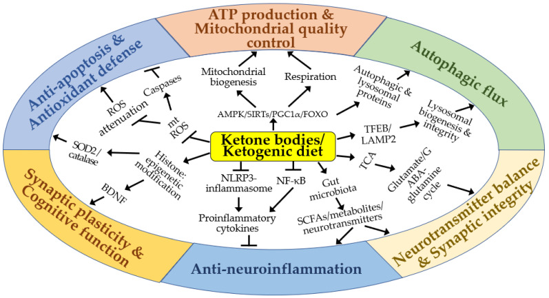
Ketone bodies (KBs), such as acetoacetate and β-hydroxybutyrate, serve as crucial alternative energy sources during glucose deficiency. KBs, generated through ketogenesis in the liver, are metabolized into acetyl-CoA in extrahepatic tissues, entering the tricarboxylic acid cycle and electron transport chain for ATP production. Reduced glucose metabolism and mitochondrial dysfunction correlate with increased neuronal death and brain damage during cerebral ischemia and neurodegeneration. Both KBs and the ketogenic diet (KD) demonstrate neuroprotective effects by orchestrating various cellular processes through metabolic and signaling functions. They enhance mitochondrial function, mitigate oxidative stress and apoptosis, and regulate epigenetic and post-translational modifications of histones and non-histone proteins. Additionally, KBs and KD contribute to reducing neuroinflammation and modulating autophagy, neurotransmission systems, and gut microbiome. This review aims to explore the current understanding of the molecular mechanisms underpinning the neuroprotective effects of KBs and KD against brain damage in cerebral ischemia and neurodegenerative diseases, including Alzheimer's disease and Parkinson's disease.

Keywords: Alzheimer’s disease; Parkinson’s disease; cerebral ischemia; ketogenic diet; ketone bodies; mitochondrial dysfunction; neurodegenerative disease; neuroinflammation; oxidative stress; β-hydroxybutyrate.


Conflict of interest statement

The authors declare no conflict of interest.

Figures

Figure 1
Figure 1
Ketogenic and ketolytic pathways in the liver and brain cells. Ketogenesis primarily occurs in the mitochondria of hepatocytes with acetyl-CoA produced by β-oxidation of fatty acyl-CoA. This process proceeds through stepwise reactions catalyzed by acetoacetyl-CoA thiolase 1 (ACAT1), 3-hydroxy-methylglutaryl-CoA synthase 2 (HMGCS2), 3-hydroxy-methylglutaryl-CoA lyase (HMGCL), and β-hydroxybutyrate dehydrogenase 1 (BDH1). The resulting ketone bodies (KBs), acetoacetate (AcAc) and β-hydroxybutyrate (BHB), are released into circulating blood via monocarboxylic acid transporters (MCTs). After uptake into the brain via MCT1, ketolysis occurs in the mitochondria of brain neurons, where BHB oxidation to acetyl-CoA is catalyzed by BDH1, succinyl-CoA:3-oxoacid-CoA transferase (SCOT), and ACAT1. After conversion of acetyl-CoA to citrate by citrate synthase (CS), ATP is generated through the tricarboxylic acid (TCA) cycle and the electron transport chain (ETC). KBs are also produced in the mitochondria of brain astrocytes, as described in the hepatic ketogenic pathway, and are provided to neurons as an energy source.
Figure 2
Figure 2
Neuroprotective effects of KBs and KD by providing energy source and improving mitochondrial dysfunction in the brain. (A) KBs serve as energy fuel for the brain. KBs promote ATP production by decreasing the NAD+/NADH ratio and increasing the coenzyme Q/QH2 ratio and ΔG’ of ATP hydrolysis. KBs exert neuroprotective effects through its use as an energy fuel. (B) KBs exert beneficial effects by improving mitochondrial function. KBs and KD improve mitochondrial dysfunction by increasing mitochondrial biogenesis through SIRT1/2/3, AMPK, PGC1α, and FOXO1/3a pathways after increasing NAD+ production. KBs and KD also increase mitochondrial respiration through PGC1α, complex I/II. The signaling pathways mediated by KBs and KD improve mitochondrial quality control and increase ATP production, resulting in neuroprotection. All arrows indicate activation.
Figure 3
Figure 3
Signaling functions of KBs and KD for anti-apoptosis, antioxidant defense, and epigenetic and post-translational modifications. (A) KBs exert beneficial effects by inducing anti-apoptotic and antioxidant defenses. KBs and KD protect neurons from apoptosis by inhibiting the mitochondrial permeability transition pore (mPTP), mt ROS production, and caspase activation. KBs and KD induce antioxidant defenses through the SIRT1/2/3, PGC1α, FOXO1/3a, and ERK/CREB/eNOS pathways and acetylation of histones, resulting in stress resistance and neuroprotection. The antioxidant Nrf2 signaling pathway is activated via acute ROS. (B) KBs and KD exhibit their beneficial effects through epigenetic and post-translational modifications of histones and non-histone proteins. KBs exert their neuroprotective effects through the SIRT1/AMPK/PGC1α pathway in a NAD+-dependent manner. Additionally, KBs regulate HDAC and p300 to promote acetylation at lysine 27 of histone H3 (H3K27Ac, Kac). KBs increase brain-derived neurotrophic factor (BDNF) expression by inducing acetylation (Kac), increasing trimethylation at lysine 4 (H3K4me3, Kme3), decreasing trimethylation of H3K27 (H3K27me3, Kme3), reducing ubiquitination at lysine 119 of histone H2A (H2AK119ub, Kub), and increasing β-hydroxybutyrylation on lysine 9 of histone H3 (H3K9bhb, Kbhb). KBs also increase the Kbhb of CaMKIIα, thereby reducing CaMKIIα activity and drug addiction. All arrows indicate activation and truncated cut lines indicate inhibition.
Figure 4
Figure 4
Signaling functions of KBs and KD for anti-neuroinflammation and autophagic flux. (A) KBs exert anti-inflammatory responses by activating hydroxycarboxylic acid receptor 2 (HCAR2), which inhibits nuclear factor kappa B (NF-κB) and NOD-like receptor protein 3 (NLRP3) inflammasomes. KBs and KD inhibit the NLRP3 inflammasome through KATP channels, Drp1, AMPK/FOXO3, and STAT3 pathways, and NF-κB through COX-1/PGD2, IRAKM, PPARs, and Nrf2/HO-1 pathways. KBs and KD also suppress inflammation through Akt/RhoGTPase-mediated ramification via HDAC inhibition and M2 microglial polarization. (B) KBs and KD induce autophagy and lysosomal proteins through SIRT1/2, AMPK, ULK1, mTORC1, and FOXO1/3a pathways and maintain lysosomal integrity through LAMP2. These changes increase autophagic flux, resulting in increased neuroprotection. All arrows indicate activation and truncated cut lines indicate inhibition.
Figure 5
Figure 5
Functions of KBs and KD for the regulation of neurotransmission systems and gut microbiota. (A) KBs and KD exert various beneficial effects on the regulation of neurotransmission systems. KBs/KD produce α-ketoglutarate (α-KG) through the TCA cycle and regulate neurotransmitter balance by regulating the glutamate–GABA/glutamate–glutamine cycle. KBs and KD also produce mitochondrial ATP and regulate synaptic integrity through ion channels and Na+/K+-ATPase. KBs and KD inhibit neuronal excitability through hyperpolarization by inhibiting glycolysis, reducing cytoplasmic ATP levels, and opening KATP channels. KBs and KD regulate sympathetic function through FFAR3 and N-type Ca2+ channels and norepinephrine release. (B) KBs and KD improve gut microbiota dysbiosis, increasing beneficial bacteria and reducing harmful proinflammatory bacteria. KB and KD also reverse disturbance in the ratio of Firmicutes to Bacteroidetes. Bacteria-derived SCFAs, metabolites, neurotransmitters, H2S, and LPS regulate the permeability of the gut-brain BBB and the production of proinflammatory cytokines. These alterations by KBs and KD result in anti-neuroinflammatory neuroprotection. All arrows indicate activation and truncated cut lines indicate inhibition.
Figure 6
Figure 6
A schematic diagram of the molecular mechanisms by which ketone bodies and ketogenic diet may exert neuroprotective effects in vitro and in vivo. Arrows and truncated lines indicate activation and inhibition, respectively.

References

    1. Cahill G.F., Jr. Fuel metabolism in starvation. Annu. Rev. Nutr. 2006;26:1–22. doi: 10.1146/annurev.nutr.26.061505.111258. - DOI - PubMed
    1. Puchalska P., Crawford P.A. Metabolic and Signaling Roles of Ketone Bodies in Health and Disease. Annu. Rev. Nutr. 2021;41:49–77. doi: 10.1146/annurev-nutr-111120-111518. - DOI - PMC - PubMed
    1. Pietrzak D., Kasperek K., Rekawek P., Piatkowska-Chmiel I. The Therapeutic Role of Ketogenic Diet in Neurological Disorders. Nutrients. 2022;14:1952. doi: 10.3390/nu14091952. - DOI - PMC - PubMed
    1. Watanabe M., Tozzi R., Risi R., Tuccinardi D., Mariani S., Basciani S., Spera G., Lubrano C., Gnessi L. Beneficial effects of the ketogenic diet on nonalcoholic fatty liver disease: A comprehensive review of the literature. Obes. Rev. 2020;21:e13024. doi: 10.1111/obr.13024. - DOI - PMC - PubMed
    1. Wlodarek D. Role of Ketogenic Diets in Neurodegenerative Diseases (Alzheimer’s Disease and Parkinson’s Disease) Nutrients. 2019;11:169. doi: 10.3390/nu11010169. - DOI - PMC - PubMed