. 2023 Dec 21;25(1):161.
doi: 10.3390/ijms25010161.

A Cancer-Specific Monoclonal Antibody against Podocalyxin Exerted Antitumor Activities in Pancreatic Cancer Xenografts

Affiliations

A Cancer-Specific Monoclonal Antibody against Podocalyxin Exerted Antitumor Activities in Pancreatic Cancer Xenografts

Hiroyuki Suzuki et al. Int J Mol Sci. .

Abstract

Podocalyxin (PODXL) overexpression is associated with poor clinical outcomes in various tumors. PODXL is involved in tumor malignant progression through the promotion of invasiveness and metastasis. Therefore, PODXL is considered a promising target of monoclonal antibody (mAb)-based therapy. However, PODXL also plays an essential role in normal cells, such as vascular and lymphatic endothelial cells. Therefore, cancer specificity or selectivity is required to reduce adverse effects on normal cells. Here, we developed an anti-PODXL cancer-specific mAb (CasMab), PcMab-6 (IgG1, kappa), by immunizing mice with a soluble PODXL ectodomain derived from a glioblastoma LN229 cell. PcMab-6 reacted with the PODXL-positive LN229 cells but not with PODXL-knockout LN229 cells in flow cytometry. Importantly, PcMab-6 recognized pancreatic ductal adenocarcinoma (PDAC) cell lines (MIA PaCa-2, Capan-2, and PK-45H) but did not react with normal lymphatic endothelial cells (LECs). In contrast, one of the non-CasMabs, PcMab-47, showed high reactivity to both the PDAC cell lines and LECs. Next, we engineered PcMab-6 into a mouse IgG2a-type (PcMab-6-mG2a) and a humanized IgG1-type (humPcMab-6) mAb and further produced the core fucose-deficient types (PcMab-6-mG2a-f and humPcMab-6-f, respectively) to potentiate the antibody-dependent cellular cytotoxicity (ADCC). Both PcMab-6-mG2a-f and humPcMab-6-f exerted ADCC and complement-dependent cellular cytotoxicity in the presence of effector cells and complements, respectively. In the PDAC xenograft model, both PcMab-6-mG2a-f and humPcMab-6-f exhibited potent antitumor effects. These results indicated that humPcMab-6-f could apply to antibody-based therapy against PODXL-expressing pancreatic cancers.

Keywords: PODXL; cancer-specific monoclonal antibody; defucosylated antibody; pancreatic cancer; podocalyxin.


Conflict of interest statement

The authors declare no conflict of interest.

Figures
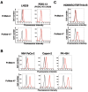
Figure 1
Figure 1
The reactivity of anti-PODXL mAbs (PcMab-6 and PcMab-47) against cancer and normal cells using flow cytometry. (A) LN229 and PODXL-KO LN229 (PDIS-13) cells were treated with 10 µg/mL of PcMab-6 (red line), PcMab-47 (red line), or blocking buffer (black line). (B) Pancreatic cancer MIA PaCa-2, Capan-2, and PK-45H cells were treated with 10 µg/mL of PcMab-6 (red line), PcMab-47 (red line), or blocking buffer (black line). (C) Lymphatic endothelial HDMVEC/TERT164-B cells were treated with 10 µg/mL of PcMab-6 (red line), PcMab-47 (red line), or blocking buffer (black line). Then, cells were treated with Alexa Fluor 488-conjugated anti-mouse IgG.
Figure 2
Figure 2
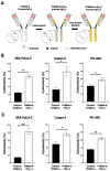
ADCC and CDC via PcMab-6-mG2a-f. (A) The VH cDNA of PcMab-6 (mouse IgG1) and the CH of mouse IgG2a were cloned into a vector. The VL and CL cDNAs of PcMab-6 were cloned into another vector. To generate a core-fucose-deficient mouse IgG2a mAb (PcMab-6-mG2a-f), the antibody expression vectors were transfected into BINDS-09 (Fut8-knocked-out ExpiCHO-S cells). (B) ADCC induced by PcMab-6-mG2a-f or control mouse IgG2a (mIgG2a) against MIA PaCa-2, Capan-2, and PK-45H cells. (C) CDC induced by PcMab-6-mG2a-f or control mIgG2a against MIA PaCa-2, Capan-2, and PK-45H cells. Values are shown as mean ± SEM. Asterisks indicate statistical significance (** p < 0.01, * p < 0.05; Welch’s t-test).
Figure 3
Figure 3
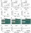
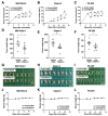
Antitumor activity of PcMab-6-mG2a-f against pancreatic cancer xenografts. (AC) MIA PaCa-2 (A), Capan-2 (B), and PK-45H (C) cells were subcutaneously injected into BALB/c nude mice (day 0). In total, 100 μg of PcMab-6-mG2a-f or control mouse IgG (mIgG) were intraperitoneally injected into each mouse on day 4 (PK-45H) or day 7 (MIA PaCa-2 and Capan-2). Additional antibodies were injected on days 11 and 18 (PK-45H) or days 14 and 21 (MIA PaCa-2 and Capan-2). The tumor volume is represented as the mean ± SEM. ** p < 0.01, * p < 0.05 (ANOVA and Sidak’s multiple comparisons test). (DF) The mice were euthanized on day 25 (PK-45H) or day 28 (MIA PaCa-2 and Capan-2) after cell implantation. The tumor weights of MIA PaCa-2 (D), Capan-2 (E), and PK-45H (F) xenografts were measured. Values are presented as the mean ± SEM. ** p < 0.01, * p < 0.05 (Welch’s t-test). (GI) MIA PaCa-2 (G), Capan-2 (H), and PK-45H (I) xenograft tumors (scale bar, 1 cm). (JL) Body weights of MIA PaCa-2 (J), Capan-2 (K), and PK-45H (L) xenograft-bearing mice treated with control mIgG or PcMab-6-mG2a-f. n.s., not significant.
Figure 4
Figure 4
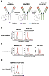
Establishment of the humanized and defucosylated antibody humPcMab-6-f. (A) The complementarity determining region (CDR) of PcMab-6 VH, the frame sequences of VH in human Ig, and the CH of human IgG1 were cloned into a vector. The CDR of PcMab-6 VL, the frame sequences of VL in human Ig, and the CL of the human kappa chain were cloned into another vector. To generate a core-fucose-deficient form (humPcMab-6-f), the antibody expression vectors were transfected into BINDS-09 cells. (B) LN229 and PODXL-KO LN229 (PDIS-13) cells were treated with 10 µg/mL of humPcMab-6-f (red line) or blocking buffer (black line). (C) MIA PaCa-2, Capan-2, and PK-45H cells were treated with 10 µg/mL of humPcMab-6-f (red line) or blocking buffer (black line). (D) HDMVEC/TERT164-B cells were treated with 10 µg/mL of humPcMab-6-f (red line) or blocking buffer (black line). Then, cells were treated with FITC-conjugated anti-human IgG.
Figure 5
Figure 5
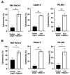
ADCC and CDC caused by humPcMab-6-f against pancreatic cancers. (A) ADCC induced by humPcMab-6-f or control human IgG (hIgG) against MIA PaCa-2, Capan-2, and PK-45H cells. (B) CDC induced by humPcMab-6-f or control hIgG against MIA PaCa-2, Capan-2, and PK-45H cells. Values are shown as mean ± SEM. Asterisks indicate statistical significance (** p < 0.01, * p < 0.05; Welch’s t-test).
Figure 6
Figure 6
Antitumor activity of humPcMab-6-f against pancreatic cancer xenografts. (AC) MIA PaCa-2 (A), Capan-2 (B), and PK-45H (C) cells were subcutaneously injected into BALB/c nude mice (day 0). In total, 100 μg of humPcMab-6-f or control hIgG were intraperitoneally injected into each mouse on day 7. Additional antibodies were injected on day 14. Human NK cells were also injected around the tumors on days 7 and 14. The tumor volume is represented as the mean ± SEM. ** p < 0.01, * p < 0.05 (ANOVA and Sidak’s multiple comparisons test). (DF) The mice were euthanized on day 21 after cell implantation. The tumor weights of the MIA PaCa-2 (D), Capan-2 (E), and PK-45H (F) xenografts were measured. Values are presented as the mean ± SEM. ** p < 0.01, * p < 0.05 (Welch’s t-test). (GI) MIA PaCa-2 (G), Capan-2 (H), and PK-45H (I) xenograft tumors (scale bar, 1 cm). (JL) Body weights of MIA PaCa-2 (J), Capan-2 (K), and PK-45H (L) xenograft-bearing mice treated with control hIgG or humPcMab-6-f. n.s., not significant.

References

    1. Doyonnas R., Nielsen J.S., Chelliah S., Drew E., Hara T., Miyajima A., McNagny K.M. Podocalyxin is a CD34-related marker of murine hematopoietic stem cells and embryonic erythroid cells. Blood. 2005;105:4170–4178. doi: 10.1182/blood-2004-10-4077. - DOI - PubMed
    1. Nielsen J.S., McNagny K.M. Novel functions of the CD34 family. J. Cell Sci. 2008;121:3683–3692. doi: 10.1242/jcs.037507. - DOI - PubMed
    1. Choo A.B., Tan H.L., Ang S.N., Fong W.J., Chin A., Lo J., Zheng L., Hentze H., Philp R.J., Oh S.K., et al. Selection against undifferentiated human embryonic stem cells by a cytotoxic antibody recognizing podocalyxin-like protein-1. Stem Cells. 2008;26:1454–1463. doi: 10.1634/stemcells.2007-0576. - DOI - PubMed
    1. McNagny K.M., Pettersson I., Rossi F., Flamme I., Shevchenko A., Mann M., Graf T. Thrombomucin, a novel cell surface protein that defines thrombocytes and multipotent hematopoietic progenitors. J. Cell Biol. 1997;138:1395–1407. doi: 10.1083/jcb.138.6.1395. - DOI - PMC - PubMed
    1. Kriehuber E., Breiteneder-Geleff S., Groeger M., Soleiman A., Schoppmann S.F., Stingl G., Kerjaschki D., Maurer D. Isolation and characterization of dermal lymphatic and blood endothelial cells reveal stable and functionally specialized cell lineages. J. Exp. Med. 2001;194:797–808. doi: 10.1084/jem.194.6.797. - DOI - PMC - PubMed