Comparative Study
. 2023 Dec 21;25(1):166.
doi: 10.3390/ijms25010166.

Comparative Analysis of Cyclization Techniques in Stapled Peptides: Structural Insights into Protein-Protein Interactions in a SARS-CoV-2 Spike RBD/hACE2 Model System

Affiliations
Comparative Study

Comparative Analysis of Cyclization Techniques in Stapled Peptides: Structural Insights into Protein-Protein Interactions in a SARS-CoV-2 Spike RBD/hACE2 Model System

Sára Ferková et al. Int J Mol Sci. .

Abstract

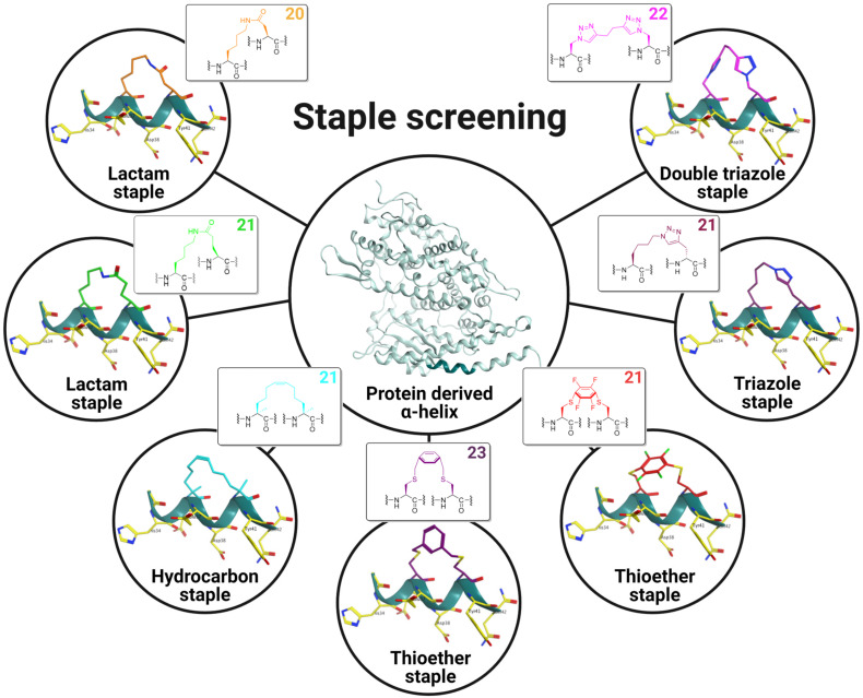
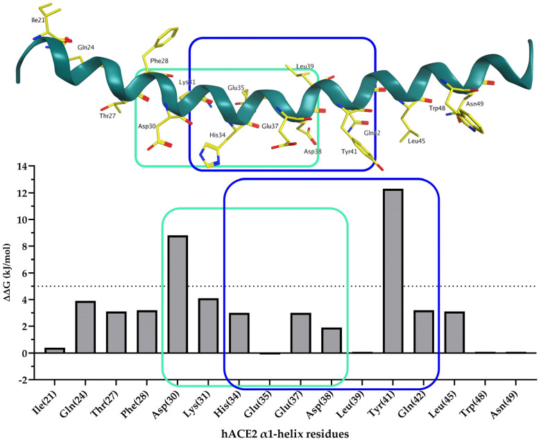
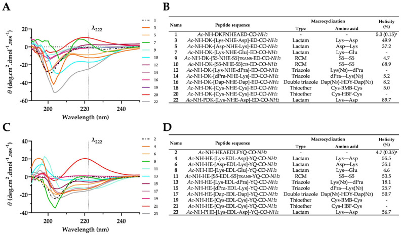
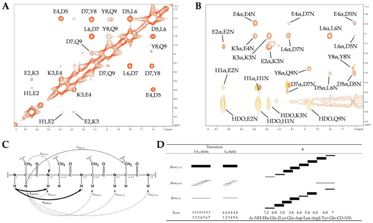
Medicinal chemistry is constantly searching for new approaches to develop more effective and targeted therapeutic molecules. The design of peptidomimetics is a promising emerging strategy that is aimed at developing peptides that mimic or modulate the biological activity of proteins. Among these, stapled peptides stand out for their unique ability to stabilize highly frequent helical motifs, but they have failed to be systematically reported. Here, we exploit chemically diverse helix-inducing i, i + 4 constraints-lactam, hydrocarbon, triazole, double triazole and thioether-on two distinct short sequences derived from the N-terminal peptidase domain of hACE2 upon structural characterization and in silico alanine scan. Our overall objective was to provide a sequence-independent comparison of α-helix-inducing staples using circular dichroism (CD) and nuclear magnetic resonance (NMR) spectroscopy. We identified a 9-mer lactam stapled peptide derived from the hACE2 sequence (His34-Gln42) capable of reaching its maximal helicity of 55% with antiviral activity in bioreporter- and pseudovirus-based inhibition assays. To the best of our knowledge, this study is the first comprehensive investigation comparing several cyclization methods with the goal of generating stapled peptides and correlating their secondary structures with PPI inhibitions using a highly topical model system (i.e., the interaction of SARS-CoV-2 Spike RBD with hACE2).

Keywords: SARS-CoV-2; circular dichroism (CD); nuclear magnetic resonance (NMR); peptidomimetics; protein–protein interaction (PPI).


Conflict of interest statement

The authors declare no conflict of interest.

Figures

Figure 1
Figure 1
Staple screening performed on a short hACE2-derived α-helical sequence (His34-Gln42) (shown in pine green) via i, i + 4 side-to-side chain cyclizations by lactamization, olefin ring-closing metathesis (RCM), S-alkylation, S-arylation and copper(I)-catalyzed Huisgen 1,3-dipolar azide-alkyne cycloaddition (CuAAC). Ball-and-stick model staple representations are shown in the insets and macrocycle sizes are indicated in the upper-righthand corner. Residues from hACE2 involved in the SARS-CoV-2 S RBD/hACE2 interaction are shown as yellow sticks labeled with three-letter codes.
Figure 2
Figure 2
In silico alanine scan (BudeAlaScan a) results for α1-helix (Ser19-Thr52) of hACE2 taken from the crystal structure of SARS-CoV-2 S RBD bound with hACE2 (PDB 6M0J). Residues from hACE2 involved in the SARS-CoV-2 S RBD/hACE2 interface are shown as yellow sticks labeled with three-letter codes. Selected hACE2-derived sequences (Asp30-Asp38) and (His34-Gln42) are outlined in light green and blue, respectively. a BudeAlaScan is an online software (version 1.0) available at https://pragmaticproteindesign.bio.ed.ac.uk/balas/ (accessed on 7 June 2023); it is only applicable to proteins consisting of natural amino acids.
Figure 3
Figure 3
Data generated from Far-UV CD spectra for hACE2-derived peptides measured at a concentration of 100 µM in 10 mM sodium phosphate buffer at pH 7.4 and 25 °C. (A) Far-UV CD spectra of hACE2 (Asp30-Asp38)-derived linear and i, i + 4 staple peptides in molar ellipticity per residue. (B) Table listing the derivatives of 1 with a focus on the macrocyclization technique and measure-based calculated helicity (%) via CD. (C) CD spectra of hACE2 (His34-Gln42)-derived linear and i, i + 4 staple peptides in molar ellipticity per residue. (D) Table listing the derivatives of 2 with a focus on the macrocyclization technique and measure-based calculated helicity (%) via CD. HDY 1,5-Hexadiyne; BMB 1,4-Bis(bromomethyl)benzene; HFB hexafluorobenzene. a Predicted helicity (%) using AGADIR online software available at http://agadir.crg.es (accessed on 17 April 2023); it is only applicable to peptides consisting of natural amino acids.
Figure 4
Figure 4
Secondary chemical shifts a of (A) CαH and (B) NH protons for peptide 4. Secondary chemical shifts of linker-formed residues have been highlighted with an orange-colored outline. a Random-coil chemical shifts for 20 common amino acids followed by alanine were measured using a peptide with free N- and C-termini at pH 5.0 and 25 °C.
Figure 5
Figure 5
Characteristic short- and medium-range sequential NOEs for an α-helix assessment. (A) The dNN and (B) dαN regions of a NOESY spectrum recorded for peptide 4. (C) Graphical illustration of sequential and medium-range 1H-1H distances in a peptide sequence. (D) Schematic representation of NOESY patterns involving NH and CαH protons observed in a NOESY spectrum recorded for α-helix compared with peptide 4. The horizontal lines of various lengths indicate NOE connectivities between protons of peptide sequences; the thicknesses of the lines is proportional to the observed strong, medium, and weak NOEs signal intensities. * The protons could not be assigned unambiguously.
Figure 6
Figure 6
The SARS-CoV-2 Spike RBD/hACE2 inhibition assessment involving a bioluminescence-based bioreporter assay and a pseudovirus-based entry inhibition assay. (A) Split-luciferase bioreporter assay demonstrating the disruptor capacity of peptides 1, 2, linear 4, 4, linear 11 and 11. Asterisks indicate a statistically significant difference between the RLUs measured for SmBiT-ACE2 + LgBiT-RBD and the neutralizing Ab control or an individual peptide. (B) Antiviral activity of peptides linear 4, 4, linear 11 and 11 was assessed using a SARS-CoV-2 pseudovirus carrying a fluorescent reporter gene and HEK-293T-hACE2 cells transfected with TMPRSS2. The fluorescence data (RFUs) were converted into percentages via normalization with the infection-free control (Dulbecco’s Eagle Medium; DMEM) set as 0%; we furthermore considered that the pseudovirus-only samples represented maximum infection (100%) using GraphPad Prism software (version 9.3.1). Both experiments were repeated in triplicate three times, and the data are expressed as means ± standard error of the mean (SEM) (error bars). The means of more than two groups were compared using one-way ANOVA with Tukey’s multiple comparison correction. For all analyses, **** p < 0.0001; ** 0.0017 p 0.0025; n.s., not significant.
Figure 7
Figure 7
Plasma stability measured in rat plasma over 24 h of incubation at 37 °C. (A) The proteolytic stability of hACE2 (Asp30-Asp38)-derived peptides recorded as a function of degraded peptide over 24 h. (B) The proteolytic stability of hACE2 (His34-Gln42)-derived peptides recorded as a function of degraded peptide over 24 h. The data are plotted as means and SEMs of duplicate independent experiments. The percentage of residual peptide was monitored using UPLC-MS. All of the experiments were repeated three times.

Similar articles

Cited by

References

    1. Lan J., Ge J., Yu J., Shan S., Zhou H., Fan S., Zhang Q., Shi X., Wang Q., Zhang L., et al. Structure of the SARS-CoV-2 Spike Receptor-Binding Domain Bound to the ACE2 Receptor. Nature. 2020;581:215–220. doi: 10.1038/s41586-020-2180-5. - DOI - PubMed
    1. Wang Q., Zhang Y., Wu L., Niu S., Song C., Zhang Z., Lu G., Qiao C., Hu Y., Yuen K.Y., et al. Structural and Functional Basis of SARS-CoV-2 Entry by Using Human ACE2. Cell. 2020;181:894–904.e9. doi: 10.1016/j.cell.2020.03.045. - DOI - PMC - PubMed
    1. Wang Y., Liu M., Gao J. Enhanced Receptor Binding of SARS-CoV-2 through networks of Hydrogen-Bonding and hydrophobic Interactions. Proc. Natl. Acad. Sci. USA. 2020;117:13967–13974. doi: 10.1073/pnas.2008209117. - DOI - PMC - PubMed
    1. Jones S., Thornton J.M. Review Principles of Protein-Protein Interactions. Proc. Natl. Acad. Sci. USA. 1996;93:13–20. doi: 10.1073/pnas.93.1.13. - DOI - PMC - PubMed
    1. Smith M.C., Gestwicki J.E. Features of Protein-Protein Interactions That Translate into Potent Inhibitors: Topology, Surface Area and Affinity. Expert Rev. Mol. Med. 2012;14:e16. doi: 10.1017/erm.2012.10. - DOI - PMC - PubMed

Publication types