. 2023 Dec 23;25(1):269.
doi: 10.3390/ijms25010269.

The Potential of Congo Red Supplied Aggregates of Multitargeted Tyrosine Kinase Inhibitor (Sorafenib, BAY-43-9006) in Enhancing Therapeutic Impact on Bladder Cancer

Affiliations

The Potential of Congo Red Supplied Aggregates of Multitargeted Tyrosine Kinase Inhibitor (Sorafenib, BAY-43-9006) in Enhancing Therapeutic Impact on Bladder Cancer

Małgorzata Lasota et al. Int J Mol Sci. .

Abstract

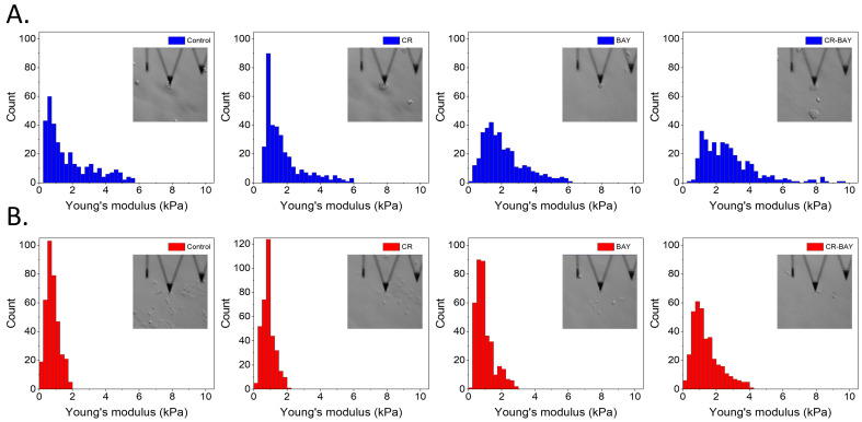
Bladder cancer is a common malignancy associated with high recurrence rates and potential progression to invasive forms. Sorafenib, a multi-targeted tyrosine kinase inhibitor, has shown promise in anti-cancer therapy, but its cytotoxicity to normal cells and aggregation in solution limits its clinical application. To address these challenges, we investigated the formation of supramolecular aggregates of sorafenib with Congo red (CR), a bis-azo dye known for its supramolecular interaction. We analyzed different mole ratios of CR-sorafenib aggregates and evaluated their effects on bladder cancer cells of varying levels of malignancy. In addition, we also evaluated the effect of the test compounds on normal uroepithelial cells. Our results demonstrated that sorafenib inhibits the proliferation of bladder cancer cells and induces apoptosis in a dose-dependent manner. However, high concentrations of sorafenib also showed cytotoxicity to normal uroepithelial cells. In contrast, the CR-BAY aggregates exhibited reduced cytotoxicity to normal cells while maintaining anti-cancer activity. The aggregates inhibited cancer cell migration and invasion, suggesting their potential for metastasis prevention. Dynamic light scattering and UV-VIS measurements confirmed the formation of stable co-aggregates with distinctive spectral properties. These CR-sorafenib aggregates may provide a promising approach to targeted therapy with reduced cytotoxicity and improved stability for drug delivery in bladder cancer treatment. This work shows that the drug-excipient aggregates proposed and described so far, as Congo red-sorafenib, can be a real step forward in anti-cancer therapies.

Keywords: Congo red; bladder cancer; multidisciplinary treatment; personalized medicine; tyrosine kinases inhibitor.


Conflict of interest statement

The authors declare no conflict of interest.

Figures

Figure 1
Figure 1
Effect of sorafenib alone and in aggregates with Congo red on bladder cells (RT4 and T24). The dose-dependent effect of sorafenib alone and in aggregates with Congo red on the viability of RH30 and RD cells after 48 h incubation (A,C,E). Statistical significance between non-treated and treated samples was evaluated using ANOVA with Dunnett post-test: ns—non-significant (p > 0.05) in comparison with a control sample (without investigated compounds); * 0.01 < p < 0.05, ** 0.001 < p < 0.01, ***—p < 0.001 Growth inhibition curve in standard culture conditions (B,D,F).
Figure 2
Figure 2
The effect of sorafenib on apoptosis and necrosis of RT4 and T24 cells. The images show flow cytometry analysis of Annexin-V and PI staining presented on a dot-plot graph. Graphic representation of four cell states: alive—the lower left square; cells undergoing necrosis—the upper left square; cells in early apoptosis—the right lower square; and cells in late apoptosis the upper right square. RT4 cells were incubated with BAY (at the IC50 concentration) and CR-BAY 5:1 (A). T24 cells were incubated with BAY (at the IC50) and CR-BAY 5:1 (B). Cumulative bar charts show the inter-relation between the state of RT4 cells after 48 h and 72 h exposure to BAY and CR-BAY 5:1 (C). Cumulative bar charts show the inter-relation between the state of T24 cells after 48 h and 72 h exposure to BAY and CR-BAY 5:1 (D). Statistical significance between investigated samples was evaluated using a Mann–Whitney test (only statistically significant data were marked): * 0.01 < p < 0.05, ** 0.001 < p < 0.01).
Figure 3
Figure 3
Effect of sorafenib alone and in aggregates with Congo red on human uroepithelial cells (SV-HUC-1). Growth inhibition curve (A). The dose-dependent effect of sorafenib alone and in aggregates with Congo red (CR60-BAY, CR-BAY 5:1) on the viability of SV-HUC-1 cells after 24 h incubation, Gray shading indicates negative values. The lines indicate the IC50 value (B). The cytotoxic influence of BAY-43-9006 and in aggregate with Congo red (5:1) by LDH test (results presented as % LDH released relative to control). Statistical significance between non-treated and treated samples was evaluated using ANNOVA with Dunnett post-test: ns—non-significant (p > 0.05) in comparison with a control sample (without BAY or CR-BAY); * 0.01 < p < 0.05, ** 0.001 < p < 0.01, *** p < 0.001 (C). The effect of BAY-43-9006 and in aggregate with Congo red (5:1) on apoptosis and necrosis of human uroepithelial cells (D,E).
Figure 4
Figure 4
Effect of sorafenib alone and in aggregates with Congo red on the motility of bladder cancer cell lines. Imaging under a 4× objective (A) Exposure to sorafenib and aggregates CR-BAY inhibited migration of bladder cancer cell lines in a scratch assay observed after 96 h. (B) Percentage of scratch reduction after 96 h (n = 3). (C) In a chemotactic assay, cells incubated with BAY and CR-BAY showed reduced migration towards 10% FBS, n = 3. (D) Representative images of bladder tumor cell invasion by Matrigel to 10% FBS in vitro show reduced cell invasion capacity under the influence of sorafenib and aggregates CR-BAY. Imaging under a 10× objective (E) Percentage of invasive cells after passing through Matrigel compared to the control, n = 4, Statistical analysis: ns—non-significant (p > 0.05), * p < 0.05. Data in graphs are represented as mean +/− SD.
Figure 5
Figure 5
Histograms of the Young’s modulus values for RT4 (A) and T24 (B) cells. Inset images show cells during nanomechanical analysis.
Figure 6
Figure 6
Representative confocal microscopy images of the RT4 cells. Scale bar of all images represent 20 µm.
Figure 7
Figure 7
Representative confocal microscopy images of the T24 cells. Scale bar of all images represent 20 µm.
Figure 8
Figure 8
The effect of investigated compounds on the p-AKT, AKT, p-ERK, and ERK protein levels in the RT4 cell line after 3 and 6 h of incubation with inhibitors (at the IC50 concentration). Results show representative Western blot images with immunodetection and densitometric evaluation of the bands (AD), #-expression of protein was not detected. Statistical significance between non-treated and treated samples was evaluated using ANOVA with Dunnett post-test: ns—non-significant (p > 0.05), * 0.01 < p < 0.05.
Figure 9
Figure 9
The effect of investigated compounds on the p-AKT, AKT, p-ERK, and ERK protein level in the T24 cell line after 3 and 6 h of incubation with inhibitors (at the IC50 concentration). Results show representative Western blot images with immunodetection and densitometric evaluation of the bands (AD), #—expression of protein was not detected. Statistical significance between non-treated and treated samples was evaluated using ANOVA with Dunnett post-test: ns—non-significant (p > 0.05), * 0.01 < p < 0.05.
Figure 10
Figure 10
Analysis of aggregates formation. (A) Electrophoretic separation in 1% agar gel of samples is shown. The samples include (1) CR, (2) CR-BAY aggregates at a ratio of 5:1 and (3) 2-1, (4) BAY (as control) at a ratio of 5-1, and (5) 2-1. The CR concentration among all samples was kept constant at 40 µM. The gel is presented as a paper blot on the left panel and, after development, with potassium permanganate on the right panel. (B) UV-VIS spectra for selected CR-BAY aggregates ratios and varying BAY concentrations. The dashed line indicates the theoretical absorbance when BAY (40 µM) is added to CR (40 µM). (C) The absorbance at maxima wavelengths (270 nm in the upper panel and 484 nm in the bottom panel) is plotted for CR-BAY aggregates against changing concentrations of BAY. (D) DLS measurements, the size distribution by number prevalence for selected samples is depicted. The line profiles represent BAY at different concentrations, while the filled curves represent aggregates of CR and BAY at different ratios. (E) DLS measurements of BAY and CR-BAY series. The average size with standard deviation is plotted against the concentration of two substances, sorafenib (upper panel) and Congo red (CR), with a constant amount of sorafenib (8 µM, bottom panel).

Similar articles

Cited by

References

    1. Lobo N., Afferi L., Moschini M., Mostafid H., Porten S., Psutka S.P., Gupta S., Smith A.B., Williams S.B., Lotan Y. Epidemiology, Screening, and Prevention of Bladder Cancer. Eur. Urol. Oncol. 2022;5:628–639. doi: 10.1016/j.euo.2022.10.003. - DOI - PubMed
    1. Powles T., Bellmunt J., Comperat E., De Santis M., Huddart R., Loriot Y., Necchi A., Valderrama B.P., Ravaud A., Shariat S.F., et al. Bladder cancer: ESMO Clinical Practice Guideline for diagnosis, treatment and follow-up. Ann. Oncol. 2022;33:244–258. doi: 10.1016/j.annonc.2021.11.012. - DOI - PubMed
    1. Dobruch J., Oszczudłowski M. Bladder Cancer: Current Challenges and Future Directions. Medicina. 2021;57:749. doi: 10.3390/medicina57080749. - DOI - PMC - PubMed
    1. Zhu S., Yu W., Yang X., Wu C., Cheng F. Traditional Classification and Novel Subtyping Systems for Bladder Cancer. Front. Oncol. 2020;10:102. doi: 10.3389/fonc.2020.00102. - DOI - PMC - PubMed
    1. Lenis A.T., Lec P.M., Chamie K. Bladder cancer a review. J. Am. Med. Assoc. 2020;324:1980–1991. doi: 10.1001/jama.2020.17598. - DOI - PubMed