. 2023 Dec 25;25(1):298.
doi: 10.3390/ijms25010298.

Nickel Challenge In Vitro Affects CD38 and HLA-DR Expression in T Cell Subpopulations from the Blood of Patients with Nickel Allergy

Affiliations

Nickel Challenge In Vitro Affects CD38 and HLA-DR Expression in T Cell Subpopulations from the Blood of Patients with Nickel Allergy

Metin Artuc et al. Int J Mol Sci. .

Abstract

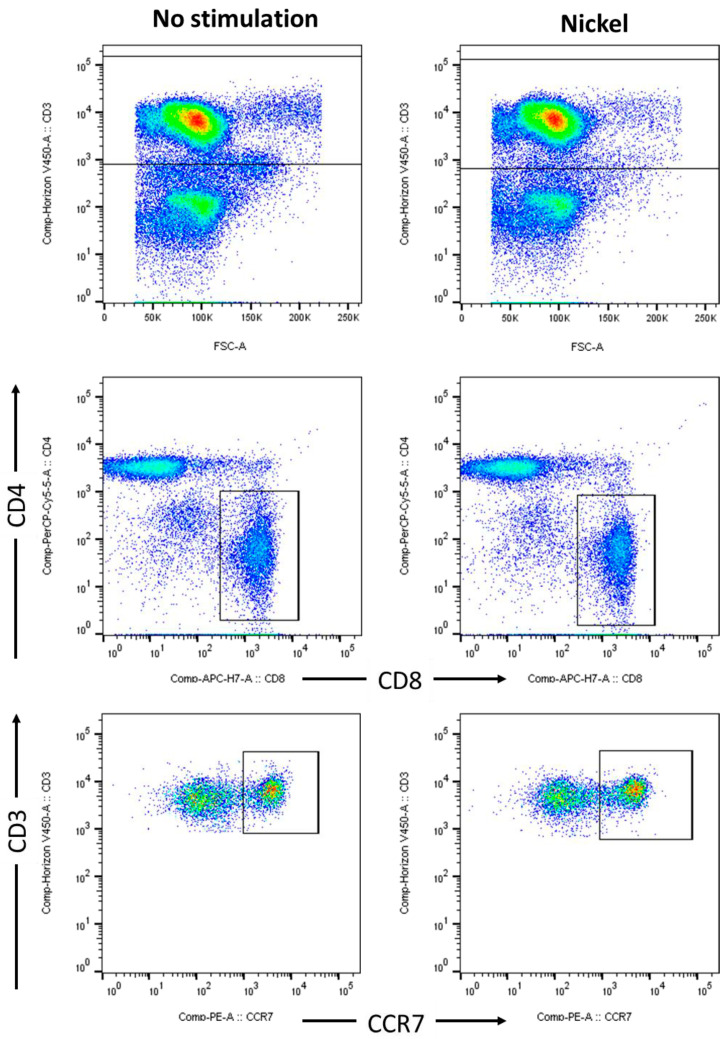
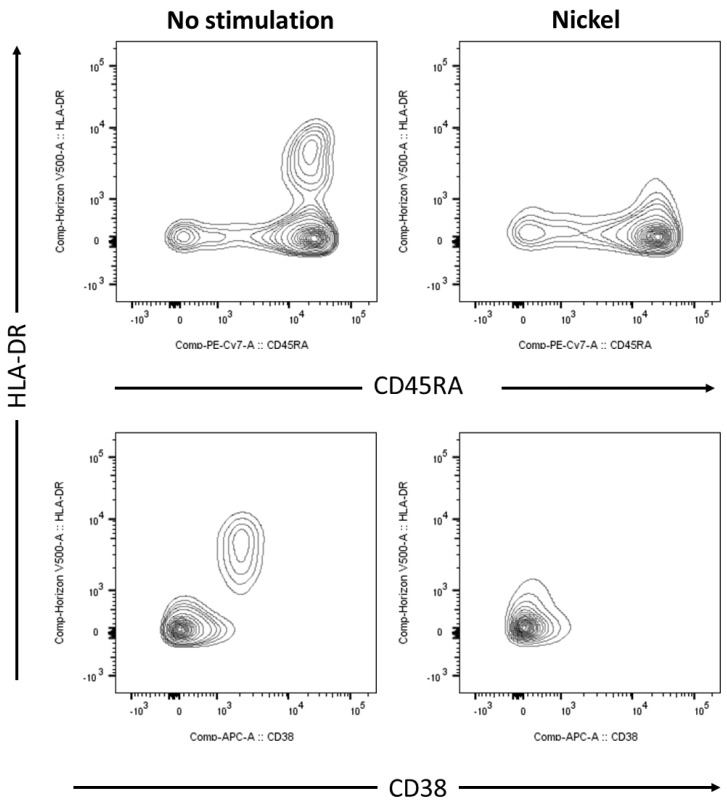
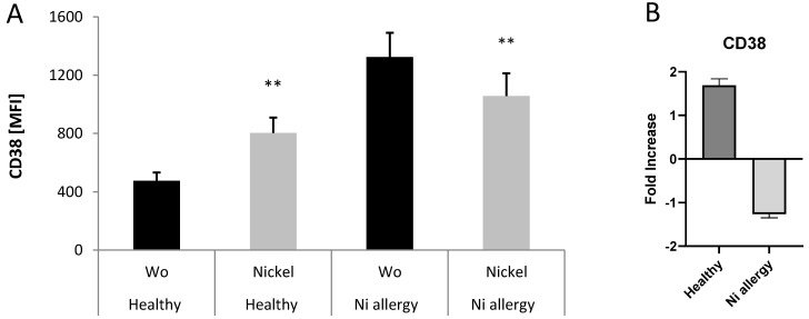
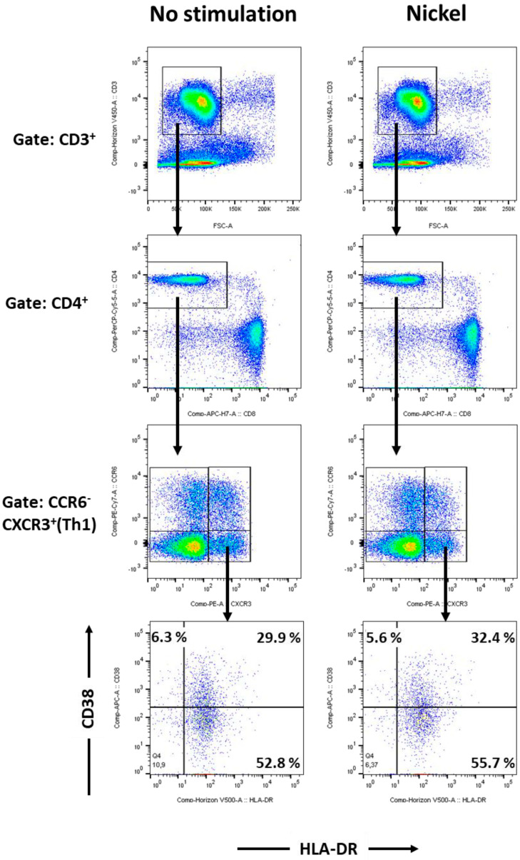
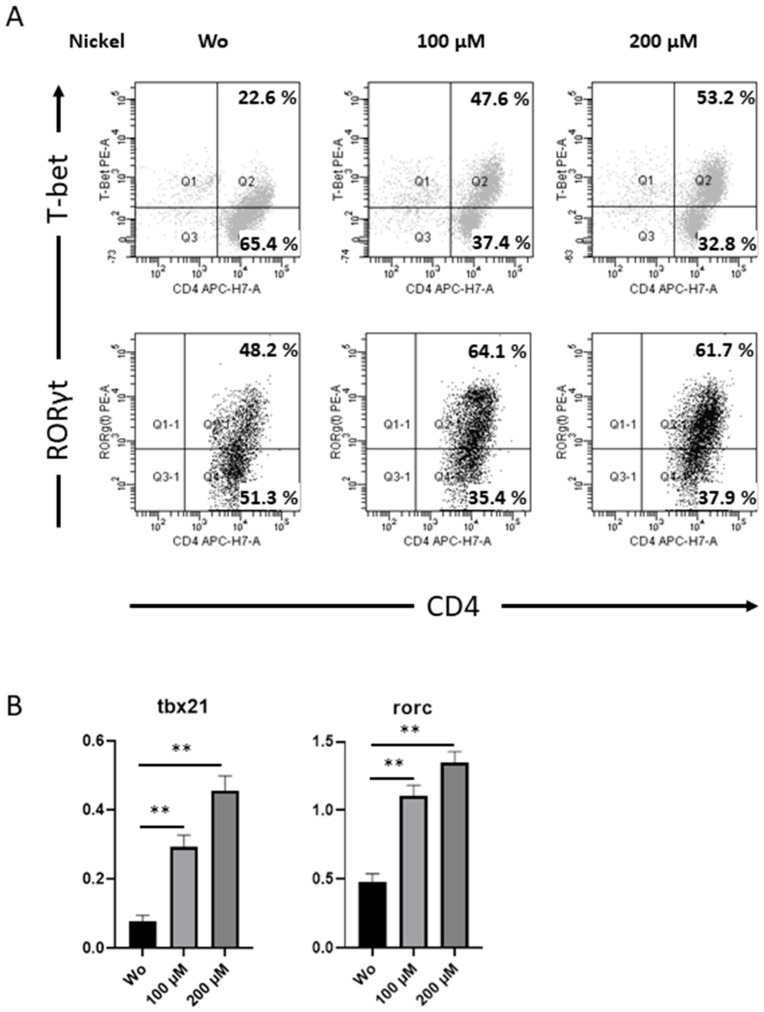
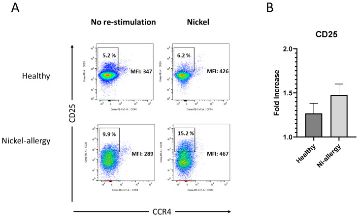
Nickel allergy is a major health problem and shows clinical manifestation of contact eczema. The response of specific lymphocyte subpopulations in sensitized patients after new challenge to nickel has until now not been studied in detail. To evaluate if nickel-based elicitation reaction could be objectively identified by multi-parametric flow cytometry, immunophenotyping of specific T cells was applied. White blood cells from 7 patients (4 positive in patch test, 3 negative) were challenged by nickel and in vitro short-term culture. Standardized antibody-dye combinations, specific for T helper(h)1, Th17 and cytotoxic T cell activation, were selected according to the recommendations of Stanford Human Immune Monitoring Center. In cytotoxic CD8+CCR7+CD45RA+ T cells from patients suffering from nickel allergy, CD38 and HLA-DR were elevated comparing to healthy donors. After challenge to nickel in vitro both markers decreased in CD8+CCR7+CD45RA+ T cells but found up-regulated in CD4+CCR7+CD45RA+CCR6-CXCR3+Th1 cells. Intracellular expression of T-bet and RORγt further indicated Th1 and Th17 cells. Finally, CD4+CD25+CCR4- T cells increased after challenge with nickel in PBMCs of patients with nickel allergy. Flow cytometry based quantification of T cell markers might be used as a specific and reliable method to detect chemical induced skin sensitization and confirm diagnostic patch testing in the clinics.

Keywords: T helper cells; cytotoxic T cells; flow cytometry; lymphocytes; nickel allergy; phenotyping.


Conflict of interest statement

The authors declare no conflict of interest.

Figures

Figure 1
Figure 1
Cytotoxic T cells: Gating strategy and immunophenotyping of nickel re-stimulated PBMCs from nickel allergic patients. Flow cytometric analyses of PBMCs from human peripheral blood, stimulated for 48 h by 200 µM NiSO4 (right panel) or control (left panel). According to the recommendations of Stanford Human Immune Monitoring Center, naïve and central memory T cells were stained and discriminated by CD3 (upper row), CD4 vs CD8 (middle row) and CCR7 (lower row). FACS analyses show dot plots of one donor, representing results from a total of n = 4 patients suffering from nickel allergy; PBMC, peripheral blood mononuclear cells, NiSO4, nickel(II)sulfate.
Figure 2
Figure 2
Expression of HLA-DR and CD38 on nickel re-stimulated cytotoxic T cells from blood of patients with nickel allergy. Flow cytometric analyses of PBMCs from human peripheral blood, stimulated for 48 h by 200 µM NiSO4 (right panel) or control (left panel). Total population of PBMCs was gated on viable (L2310 negative), CD3+CD8+CCR7+ cells and were further analyzed for co-expression of HLA-DR and CD38 using a T cell panel as described in materials and methods. FACS analyses show contour plots of one donor, representing results from a total of n = 4 patients suffering from nickel allergy; PBMC, peripheral blood mononuclear cells, NiSO4, nickel(II)sulfate.
Figure 3
Figure 3
Expression of CD38 on nickel re-stimulated CD3+CD8+CCR7+ T cells from blood of healthy donors and patients suffering from nickel allergy (A). Bars represent mean values + SD of mean fluorescence intensities (MFI) for CD38 measurement on PBMCs from three healthy donors and four patients suffering from nickel allergy. PBMCs from both groups were stimulated for 48 h by 200 µM NiSO4 or left untreated (Wo) and analyzed using T cell panel as described in materials and methods. Differences of untreated cells to cells stimulated by nickel were analyzed for statistical significance (**, 0.001 < p < 0.01) in both groups. Bars in (B) indicate analyses of the means for fold increase CD38 in nickel treated cells comparing to unstimulated cells in healthy donors and patients with Ni allergy.
Figure 4
Figure 4
T helper 1 cells: Gating strategy and immunophenotyping of nickel re-stimulated PBMCs from patients suffering from nickel allergy. Flow cytometric analyses of PBMCs from human peripheral blood, stimulated for 48 h by 200 µM NiSO4 (right panel) or control (left panel). T helper cells were stained using a Th1/Th2/Th17 panel as described in materials and methods and discriminated by CD3 (upper row), CD4 vs CD8 (middle row) and chemokine receptors CCR6 and CXCR3. T helper 1 cells were identified as CD3+CD4+CCR6CXCR3+ cells and analysed for CD38 and HLA-DR expression. FACS analyses show dot plots of one donor, representing results from a total of n = 4 patients suffering from nickel allergy; PBMC, peripheral blood mononuclear cells, NiSO4, nickel(II)sulfate.
Figure 5
Figure 5
Expression of Th-specific transcriptions factors in viable CD4+ Th cells. T helper cells, isolated from PBMCs of human peripheral blood, stimulated for 48 h by 200 µM NiSO4 (right panel) or control (left panel) were co-stained with mAbs against T-bet and RORγt. Intracellular FACS analyses show dot plots of one donor, representing results from a total of n = 4 patients suffering from nickel allergy (A). mRNA expression for tbx21 and rorc was measured for donors as in (A) by quantitative PCR. Analysis was calculated after normalization to β-actin (B); PBMC, peripheral blood mononuclear cells, NiSO4, nickel(II)sulfate (**, 0.001 < p < 0.01).
Figure 6
Figure 6
Activated T cells: Gating strategy and immunophenotyping of nickel re-stimulated PBMCs from patients suffering from nickel allergy. Flow cytometric analyses of PBMCs from human peripheral blood (A), stimulated for 48 h by 200 µM NiSO4 (right panel) or control (left panel). Th cells were stained using a T reg panel as described in materials and methods and discriminated by gates on viable CD3+, CD4+ and 127 low cells. FACS analyses show expression of CD25 vs CCR4 of one donor, representing results from a total of n = 4 patients suffering from nickel allergy and n = 3 healthy donors. Bars in (B) indicate analyses of the means for fold increase CD25 in nickel treated cells comparing to unstimulated cells in healthy donors and patients with Ni allergy; PBMC, peripheral blood mononuclear cells, NiSO4, nickel(II)sulfate.

Similar articles

References

    1. Yu W., Freeland D.M.H., Nadeau K.C. Food allergy: Immune mechanisms, diagnosis and immunotherapy. Nat. Rev. Immunol. 2016;16:751–765. doi: 10.1038/nri.2016.111. - DOI - PMC - PubMed
    1. Kupper T.S., Fuhlbrigge R.C. Immune surveillance in the skin: Mechanisms and clinical consequences. Nat. Rev. Immunol. 2004;4:211–222. doi: 10.1038/nri1310. - DOI - PMC - PubMed
    1. Peiser M., Tralau T., Heidler J., Api A.M., Arts J.H., Basketter D.A., English J., Diepgen T.L., Fuhlbrigge R.C., Gaspari A.A., et al. Allergic contact dermatitis: Epidemiology, molecular mechanisms, in vitro methods and regulatory aspects. Current knowledge assembled at an international workshop at BfR, Germany. Cell. Mol. Life Sci. 2012;69:763–781. doi: 10.1007/s00018-011-0846-8. - DOI - PMC - PubMed
    1. Abbas A.K., Murphy K.M., Sher A. Functional diversity of helper T lymphocytes. Nature. 1996;383:787–793. doi: 10.1038/383787a0. - DOI - PubMed
    1. O’Shea J.J., Paul W.E. Mechanisms underlying lineage commitment and plasticity of helper CD4+ T cells. Science. 2010;327:1098–1102. doi: 10.1126/science.1178334. - DOI - PMC - PubMed