. 2023 Dec 25;25(1):313.
doi: 10.3390/ijms25010313.

Serpin-4 Negatively Regulates Prophenoloxidase Activation and Antimicrobial Peptide Synthesis in the Silkworm, Bombyx mori

Affiliations

Serpin-4 Negatively Regulates Prophenoloxidase Activation and Antimicrobial Peptide Synthesis in the Silkworm, Bombyx mori

Xingtao Qie et al. Int J Mol Sci. .

Abstract

The prophenoloxidase (PPO) activation and Toll antimicrobial peptide synthesis pathways are two critical immune responses in the insect immune system. The activation of these pathways is mediated by the cascade of serine proteases, which is negatively regulated by serpins. In this study, we identified a typical serpin, BmSerpin-4, in silkworms, whose expression was dramatically up-regulated in the fat body and hemocytes after bacterial infections. The pre-injection of recombinant BmSerpin-4 remarkably decreased the antibacterial activity of the hemolymph and the expression of the antimicrobial peptides (AMPs) gloverin-3, cecropin-D, cecropin-E, and moricin in the fat body under Micrococcus luteus and Yersinia pseudotuberculosis serotype O: 3 (YP III) infection. Meanwhile, the inhibition of systemic melanization, PO activity, and PPO activation by BmSerpin-4 was also observed. Hemolymph proteinase 1 (HP1), serine protease 2 (SP2), HP6, and SP21 were predicted as the candidate target serine proteases for BmSerpin-4 through the analysis of residues adjacent to the scissile bond and comparisons of orthologous genes in Manduca sexta. This suggests that HP1, SP2, HP6, and SP21 might be essential in the activation of the serine protease cascade in both the Toll and PPO pathways in silkworms. Our study provided a comprehensive characterization of BmSerpin-4 and clues for the further dissection of silkworm PPO and Toll activation signaling.

Keywords: Bombyx mori; antimicrobial peptide; immune system; prophenoloxidase; serpin.


Conflict of interest statement

The authors declare no conflicts of interest.

Figures

Figure 1
Figure 1
The functional domain (A), three-dimensional structure (B), and phylogenetic assays of BmSerpin-4 (C). (A) A predicted signal peptide that consists of the first 17 residues and a serpin domain (34–402 aa) was found in the amino acid sequence of BmSerpin-4 (NP_001037090). A reactive center loop with the predicted P1-P1′ scissile bond (R-I) is located in the C terminal (360–379 aa) of the serpin domain. (B) The tertiary structure of mature BmSerpin-4 was retrieved and visualized from AlphaFold protein structure database (https://alphafold.com (accessed on 2 May 2023)). BmSerpin-4 has the typical structure of three (A–C) β-sheets and nine α-helical linkers, and the A and C β-sheets are linked by the reactive center loop. The different colors in the diagram represent the corresponding credibility, as indicated on the right. (C) The phylogram was built by a neighbor-joining method using MEGA 5.10 after aligning the selected serpin protein sequences of B. mori, M. Sexta, and D. melanogaster using ClustalW. Bm serpin4: NP_001037090; Ms serpin4: AAS68503.1; Bm serpin5: AAS68506.1; Ms serpin5: AAS68507.1; Bm serpin7: NP_001139701; Ms serpin9: AYK02794.1; Bm serpin8: NP_001139702; Ms serpin77Ba:XP_030032247.2; Bm serpin31: NP_001139722; Ms serpinB5: XP_030040571; Bm serpin12: NP_001036857; Ms serpin12: AYK02795.1; Dm serpin100A: NP_651818.1; Bm serpin10: NP_001139703; Dm serpin27A: NP_001260143.1; Bm serpin3: XP_037867815; Ms serpin3: AAO21505.1; Dm serpin31A: NP_609341.1; Dm serpin35F: NP_001036553.1; Dm serpin43Ab: NP_001027395.1; Dm serpin75F: NP_001036614.1; Dm serpin43Ad: NP_610261.1; Bm serpin29: NP_001139720; Bm serpin26: NP_001139718; Bm serpin19: NP_001139712; Bm serpin23: NP_001139716; Bm serpin30: NP_001139721; Bm serpin15: NP_001139707; Bm serpin20: NP_001139713; Bm serpin25: NP_001139717; Bm serpin22: NP_001139715; Bm serpin16: NP_001139708; Bm serpin18: NP_001139711; Dm serpin85F: NP_649965.2; Bm serpin34: NP_001129364; Dm serpin28F: NP_524957.2; Dm serpin38F: NP_001286112.1; Dm serpin42Dd: NP_001163067.1; Dm serpin28B: NP_001260209.1; Dm serpin47C:NP_001163116.1; Dm serpin42Da: NP_524955.2; Dm serpin42Db: NP_001260746.1; Dm serpin42Dc: NP_001246154.1; Dm serpin43Aa: NP_524805.1; Dm serpin28Da: NP_001036345.1; Dm serpin28Db: NP_001356884.1; Dm serpin55B: NP_524953.1; Ms serpin13: AYK02793.1; Ms serpin28Dc: XP_030027527; Bm serpin13: NP_001139705; Dm serpin28Dc: NP_609172.1; Dm serpin88Ea: NP_524954.2; Dm serpin88Eb: NP_650427.1; Bm serpin6: NP_001103823; Ms serpin6: AAV91026.1; Bm serpin1: NP_001037305; Bm serpin1J:AAC47340; Ms serpin88Ea: XP_037300507; Bm serpin28: NP_001139719; Ms serpin2: AAB58491.1; Bm serpin2: NP_001037021; Bm serpin21: NP_001139714; Bm serpin9: NP_001037530; Dm serpin77Bb: NP_649206.1; Dm serpin77Bc: NP_001262103.1; Dm serpin77Ba: NP_001287128.1; Ms serpin7: ADM86478.1; Ms serpinB6: XP_030039875; Bm serpin32: NP_001139723; Bm serpin33: NP_001129363; Bm serpin14: NP_001139706.
Figure 2
Figure 2
The mRNA abundance of BmSerpin-4 in the immune tissues (A) and the expression of BmSerpin-4 in hemocytes (B) and fat body after bacterial infection (C). (A) The main immune tissues from the midgut, hemocytes, and fat body were collected to analyze the mRNA abundance of BmSerpin-4. Hemocyte (B) and fat body (C) samples were collected at 6, 12, and 24 hpi (hours post-infection) to analyze the transcription levels of BmSerpin-4 using q-PCR. The expression of BmSerpin-4 was normalized with IF4A gene of B. mori, and the values shown are the means (±SEM) of three independent biological experiments. (D) The protein levels of BmSerpin-4 in the hemolymph after infection by M. lutes and YP III at 6, 12, and 24 hpi were examined through Western blotting (WB). For (A), the relative expression of BmSerpin-4 in different tissues was compared to the expression in midgut, and the statistical differences between compared groups were denoted with asterisks. p-values were determined using Student’s t-test. ns: no significance; ** p < 0.01. For (B,C), the relative expression of the infection groups was compared to the expression of the control groups at each time point, and the statistical differences between the control groups and infection groups were denoted with asterisks. p-values were determined by one-way ANOVA, followed by Tukey’s multiple range test. * p < 0.05; ** p < 0.01; *** p < 0.001.
Figure 3
Figure 3
The expression and purification of recombinant Bmserpin-4. (A) Coomassie brilliant blue-stained SDS-PAGE. (B) Western blot probed with anti-His antibody. (C) Western blot probed with anti-BmSerpin-4 antibody. M—protein molecular mass markers; lane 1—total proteins from uninduced E. coli cells; lane 2—total proteins from induced E. coli cells; lane 3—soluble proteins from induced E. coli cells; lane 4—purified recombinant BmSerpin-4. The bands of recombinant Bmserpin-4 are indicated by the red arrows.
Figure 4
Figure 4
BmSerpin-4 inhibited systemic melanization. The anatomic melanization of tissues and plasma after pre-injection of 10 µg of BSA following approximately 2 × 10⁸ dead YP III (A) or 1 × 10⁸ dead M. luteus infection (C). The anatomic melanization of tissues and plasma after pre-injection of 10 µg of BmSerpin-4 following approximately 2 × 10⁸ dead YP III (B) or 1 × 10⁸ dead M. luteus (D) infection. The samples were collected and observed 12 h after the injection of bacterial cells.
Figure 5
Figure 5
BmSerpin-4 blocked the PO activity and spontaneous melanization of the hemolymph. The spontaneous melanization of the hemolymph collected from the larvae pre-injected with BSA or BmSerpin-4 was observed. The PO activity of the hemolymph from the larvae pre-injected with BSA or BmSerpin-4 and then activated with or without pathogen-associated molecular patterns (PAMPs, including curdlan, LPS, and PG) was measured. The values shown are the means (±SEM) of three independent experiments. The statistical differences between the compared groups are denoted with asterisks. p-values were determined using Student’s t-test. * p < 0.05; *** p < 0.001; **** p < 0.0001.
Figure 6
Figure 6
BmSerpin-4 inhibited the PPO activation of the hemolymph. Western blotting shows the clipping/activation of PPO1 (left panel) and PPO2 (right panel) and PO complex formation in the hemolymph pre-injected with BSA or BmSerpin-4 following curdlan (from A. faecalis) (A), LPS (from P. ingivalis) (B), PG (from M. luteus) (C), and PG (from E. coli) (D) activation. The blue arrows indicate the PO complexes in the plasma; the black arrows indicate PPO1 and PPO2; the red arrows indicate PO1 and PO2. M—protein molecular mass markers; 1, 5, 25—the hemolymph samples were incubated with the PAMPs for 1, 5, 25 minutes respectively.
Figure 7
Figure 7
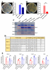
The production of AMPs was negatively regulated by BmSerpin-4. (A) The inhibition zone against YP III using the hemolymph collected from larvae pre-injected with BSA or BmSerpin-4 and then infected with YP III. (B) The inhibition zone against M. luteus using the hemolymph collected from the larvae pre-injected with BSA or BmSerpin-4 and then infected with M. luteus. For (A) and (B), the hemolymph collected from larvae injected with BSA was used as negative control. The antibacterial activity of the hemolymph was determined by the diameter of inhibition zone. (C) Plasma proteins were separated using tricine-SDS-PAGE and visualized with Coomassie brilliant blue staining. Note that only the lower part of the gel is shown. The boxes indicate the bands in the gel induced by bacteria. (D) The mass spectrometry results of the protein bands that were excised from the gel in (C). (E) The mRNA abundance of antimicrobial peptide genes gloverin3 (Glov 3), cecropin-D (Cec D), cecropin-E (Cec E), and moricin (Mor) in the fat body collected from the larvae pre-injected with BSA or BmSerpin-4 and then infected with YP III or M. luteus. The fat body collected from larvae injected with BSA was used as control. Each dot in the graph represents an individual experiment. For (AC), the values shown are the means (±SEM) of three independent experiments. The statistical differences between the compared groups are denoted with asterisks. p-values were determined using Student’s t-test. * p < 0.05; ** p < 0.01.
Figure 8
Figure 8
The prediction of the candidate target serine proteases for BmSerpin-4. (A) Comparison of activation site sequences between BmSerpin-4/MsSerpin-4 and proteases from B. mori, D. melanogaster, M. sexta, and T. molitor. PAP—prophenoloxidase-activating protease; SPE—Spätzle-processing enzyme; SAE—SPE-activating enzyme. The P1-P1′ scissile bond is indicated by an arrow. The yellow background indicates MsSerpin-4 and its corresponding identified target serine proteases, as well as BmSerpin-4 and the predicted candidate target serine proteases for BmSerpin-4. Each yellow background frame indicates a corresponding orthology between the serpins or serine proteases of B. mori and M. sexta. (B) A model for regulation of PPO activation and Toll pathway cascade in B. mori by BmSerpin-4. Arrows indicate activation of downstream components or steps. Dashed arrows indicate potentially more than one step. *: activated/clipped.

Similar articles

Cited by

References

    1. Lemaitre B., Hoffmann J. The host defense of Drosophila melanogaster. Annu. Rev. Immunol. 2007;25:697–743. doi: 10.1146/annurev.immunol.25.022106.141615. - DOI - PubMed
    1. Govind S. Innate immunity in Drosophila: Pathogens and pathways. Insect Sci. 2008;15:29–43. doi: 10.1111/j.1744-7917.2008.00185.x. - DOI - PMC - PubMed
    1. Buchon N., Silverman N., Cherry S. Immunity in Drosophila melanogaster—From microbial recognition to whole-organism physiology. Nat. Rev. Immunol. 2014;14:796–810. doi: 10.1038/nri3763. - DOI - PMC - PubMed
    1. Manniello M.D., Moretta A., Salvia R., Scieuzo C., Lucchetti D., Vogel H., Sgambato A., Falabella P. Insect antimicrobial peptides: Potential weapons to counteract the antibiotic resistance. Cell. Mol. Life. Sci. 2021;78:4259–4282. doi: 10.1007/s00018-021-03784-z. - DOI - PMC - PubMed
    1. Yu S., Luo F., Xu Y., Zhang Y., Jin L.H. Drosophila innate immunity involves multiple signaling pathways and coordinated communication between different tissues. Front. Immunol. 2022;13:905370. doi: 10.3389/fimmu.2022.905370. - DOI - PMC - PubMed

LinkOut - more resources