. 2023 Dec 25;25(1):316.
doi: 10.3390/ijms25010316.

Suppression of Neuroinflammation by Coffee Component Pyrocatechol via Inhibition of NF-κB in Microglia

Affiliations

Suppression of Neuroinflammation by Coffee Component Pyrocatechol via Inhibition of NF-κB in Microglia

Taisuke Murata et al. Int J Mol Sci. .

Abstract

According to numerous studies, it has been epidemiologically suggested that habitual coffee intake seems to prevent the onset of neurodegenerative diseases. In this study, we hypothesized that coffee consumption suppresses neuroinflammation, which is closely related to the development of neurodegenerative diseases. Using microglial BV-2 cells, we first found that the inflammatory responses induced by lipopolysaccharide (LPS) stimulation was diminished by both coffee and decaffeinated coffee through the inhibition of an inflammation-related transcription factor, nuclear factor-κB (NF-κB). Pyrocatechol, a component of roasted coffee produced by the thermal decomposition of chlorogenic acid, also exhibited anti-inflammatory activity by inhibiting the LPS-induced activation of NF-κB. Finally, in an inflammation model using mice injected with LPS into the cerebrum, we observed that intake of pyrocatechol as well as coffee decoctions drastically suppressed the accumulation of microglia and the expression of interleukin-6 (IL-6), tumor necrosis factor α (TNFα), CCL2, and CXCL1 in the inflammatory brain. These observations strongly encourage us to hypothesize that the anti-inflammatory activity of pyrocatechol as well as coffee decoction would be useful for the suppression of neurodegeneration and the prevention of the onsets of Alzheimer's (AD) and Perkinson's diseases (PD).

Keywords: NF-κB; coffee; microglia; neuroinflammation; pyrocatechol.


Conflict of interest statement

The authors declare no conflict of interest.

Figures

Figure 1
Figure 1
Coffee and decaffeinated coffee both inhibit LPS-induced NO production and expression of iNOS in BV-2 cells. (A,B) BV-2 cells were pretreated with coffee or decaffeinated coffee (Decaf) (0.31, 0.63, 1.25, 2.5, and 5% (v/v)) for 1 h followed by stimulation with LPS (1 μg/mL) for 12 h. (A) Cell viability was measured. (B) The amount of NO in the culture supernatant was measured. (C,D) BV-2 cells were pretreated with coffee or decaffeinated coffee (Decaf) (5% (v/v)) for 1 h followed by stimulation with LPS (1 μg/mL) for the indicated periods. (C) The expression of iNOS mRNA was analyzed by RT-PCR. (D) The expression of iNOS was examined by immunoblotting. The relative expression level of iNOS is shown in the graph. (AD) Values are the mean ± S.D. of three independent experiments. *** p < 0.001 indicates significant difference from control cells. ## p < 0.01 and ### p < 0.001 indicate significant differences from control cells treated with LPS.
Figure 2
Figure 2
Coffee and decaffeinated coffee both inhibit LPS-induced expression of inflammatory mediators in BV-2 cells. BV-2 cells were pretreated with coffee or decaffeinated coffee (Decaf) (5% (v/v)) for 1 h followed by stimulation with LPS (1 μg/mL) for the indicated periods. The mRNA expression of (A) IL-6, (B) TNFα, (C) CCL2, (D) CXCL1, and (E) COX-2 was analyzed by RT-PCR. Values are the mean ± S.D. of three independent experiments. * p < 0.05, ** p < 0.01, and *** p < 0.001 indicate significant differences from control cells. # p < 0.05, ## p < 0.01, and ### p < 0.001 indicate significant differences from control cells treated with LPS.
Figure 3
Figure 3
Coffee and decaffeinated coffee both inhibit the LPS-induced nuclear localization and transcriptional activation of NF-κB in BV-2 cells. (A) BV-2 cells were transfected with pNF-κB-Luc and pRL-TK as described in the Methods. Cells were pretreated with coffee or decaffeinated coffee (Decaf) decoctions (5% (v/v)) for 1 h followed by stimulation with LPS (1 μg/mL) for 8 h. Luciferase assay was performed. Values are the mean ± S.D. of three independent experiments. * and ** indicate p < 0.05 and p < 0.01, respectively. (B) BV2 cells were pretreated with coffee or decaffeinated coffee (Decaf) decoctions (5% (v/v)) for 1 h followed by stimulation with LPS (1 μg/mL) for indicated periods. The expression of IκBα was examined by immunoblotting. The relative expression level of IκBα is shown in the graphs. Values are the mean ± S.D. of three independent experiments. *** p < 0.001 indicates a significant difference from control cells. (C) BV-2 cells were pretreated with coffee or decaffeinated coffee (Decaf) decoctions (5% (v/v)) for 1 h followed by stimulation with LPS (1 μg/mL) for 30 min. The localization of NF-κB p65 was visualized with an antibody (green). DAPI (blue) was also applied to visualize nuclei. (Scale bar: 30 μm, magnification: ×200).
Figure 4
Figure 4
Coffee and decaffeinated coffee both have no effect on the LPS-induced phosphorylation of MAPK family in BV-2 cells. BV-2 cells were pretreated with coffee or decaffeinated coffee (Decaf) decoctions (5% (v/v)) for 1 h and were then stimulated with LPS (1 μg/mL) for the indicated periods. Immunoblotting was performed, and the relative phosphorylation level of ERK, p38, and JNK is shown in the graphs. Values are the mean ± S.D. of three independent experiments. * p < 0.05, ** p < 0.01, and *** p < 0.001 indicate significant differences from control cells.
Figure 5
Figure 5
The coffee ingredient, pyrocatechol, inhibits LPS-induced NO production and expression of iNOS in BV-2 cells. (A) The scheme shows the reaction producing pyrocatechol by roasting coffee beans. (B,C) BV-2 cells were pretreated with pyrocatechol (2.5, 5, and 10 μM) for 1 h followed by stimulation with LPS (1 μg/mL) for 12 h. (B) Cell viability was measured. (C) The amount of NO in the culture supernatant was measured. Values are the mean ± S.D. of three independent experiments. *** p < 0.001 indicates significant differences from control cells stimulated with LPS. (D,E) BV-2 cells were pretreated with pyrocatechol (5 μM) for 1 h followed by stimulation with LPS (1 μg/mL) for the indicated periods. (D) The expression of iNOS mRNA was analyzed by RT-PCR. (E) The expression of iNOS was examined by immunoblotting, and the relative expression level of iNOS is shown in the graph. * p < 0.05, ** p < 0.01, and *** p < 0.001 indicate significant differences from control cells. # p < 0.05, ## p < 0.01, and ### p < 0.001 indicate significant differences from control cells treated with LPS.
Figure 6
Figure 6
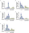
Pyrocatechol inhibits LPS-induced expression of inflammatory mediators in BV-2 cells. BV-2 cells were pretreated with pyrocatechol (2.5, 5, and 10 μM) for 1 h followed by stimulation with LPS (1 μg/mL) for 12 h. The mRNA expression of (A) IL-6, (B) TNFα, (C) CCL2, (D) CXCL, and (E) COX-2 was analyzed by RT-PCR. Values are the mean ± S.D. of three independent experiments. * p < 0.05, ** p < 0.01, and *** p < 0.001 indicate significant differences from control cells. # p < 0.05, ## p < 0.01, and ### p < 0.001 indicate significant differences from control cells treated with LPS.
Figure 7
Figure 7
Pyrocatechol inhibits the LPS-induced nuclear localization and transcriptional activation of NF-κB in BV-2 cells. (A) BV-2 cells were transfected with pNF-κB-Luc and pRL-TK as described in the Methods. Cells were pretreated with pyrocatechol (5 μM) for 1 h followed by stimulation with LPS (1 μg/mL) for 8 h and Luciferase assay was performed. Values are the mean ± S.D. of three independent experiments. * and ** indicate p < 0.05 and p < 0.01, respectively. (B) BV-2 cells were pretreated with pyrocatechol (5 μM) for 1 h followed by stimulation with LPS (1 μg/mL) for indicated periods. The expression of IκBα was examined by immunoblotting and the relative expression level of IκBα is shown in the graphs. Values are the mean ± S.D. of three independent experiments. ** p < 0.001 indicates significant difference from control cells. (C) BV-2 cells were pretreated with pyrocatechol (5 μM) for 1 h followed by a stimulation with LPS (1 μg/mL) for 30 min. The localization of NF-κB p65 was visualized with an antibody (green). DAPI (blue) was also applied to visualize nuclei. (Scale bar: 30 μm, magnification: ×200).
Figure 8
Figure 8
Pyrocatechol has no effect on the LPS-induced phosphorylation of the MAPK family in BV-2 cells. BV-2 cells were pretreated with pyrocatechol (5 μM) for 1 h and then stimulated with LPS (1 μg/mL) for the indicated periods. Immunoblotting was performed and the relative phosphorylation level of ERK, p38, and JNK is shown in the graphs. Values are the mean ± S.D. of three independent experiments. * p < 0.05, ** p < 0.01, and *** p < 0.001 indicate significant differences from control cells.
Figure 9
Figure 9
Coffee and decaffeinated coffee attenuate the LPS-induced accumulation of microglia and mRNA expression of IL-6, TNFα, CCL2, and CXCL1 in the murine brain. C57BL/6 mice (male, 4 weeks, n = 6) were given water, a 60% (v/v) coffee decoction, or 60% (v/v) decaffeinated coffee (Decaf) decoction for 4 weeks. (A) The body weight and food intake of each mouse were measured. (B) After 4 weeks of the intake of coffee or decaffeinated coffee (Decaf), LPS was injected into the right striatum of mice (n = 6). Seven days after the LPS injection, murine brain sections were prepared and two tissue sections obtained from six mice in each group were immunostained using anti-Iba1 antibody (Scale bar: 1 mm), and the luminance of each sample was measured by ImageJ. Data are presented as the mean ± SD of three independent experiments in the graph. *** indicates p < 0.001. (C) Six hours after the LPS injection, total RNA was extracted from murine brains. The mRNA expression of IL-6, TNFα, CCL2, and CXCL1 was analyzed by RT-PCR. Values are the mean ± S.D. of six independent experiments. *, **, and *** indicate p < 0.05, p < 0.01, and p < 0.001, respectively.
Figure 10
Figure 10
The intake of pyrocatechol attenuates the LPS-induced accumulation of microglia and mRNA expression of IL-6, TNFα, CCL2, and CXCL1 in the murine brain. C57BL/6 mice (male, 4 weeks, n = 6) were given water or 60 μM pyrocatechol for 4 weeks. (A) The body weight and food intake of each mouse were measured twice each week. (B) After 4 weeks of the intake of water or pyrocatechol, LPS was injected into the right striatum of the brain (n = 6). Seven days after the LPS injection, murine brain sections were prepared. Two tissue sections obtained from six mice in each group were immunostained using anti-Iba-1 atibody (Scale bar: 1 mm), and the luminance of each sample was measured by ImageJ. Data are presented as the mean ± SD of three independent experiments in the graph. *** indicates p < 0.05. (C) Six hours after the LPS injection, total RNA was extracted from murine brains. The mRNA expression of TNFα, IL-6, CCL2, and CXCL1 was analyzed by RT-PCR. Values are the mean ± S.D. of six independent experiments. ** and *** indicate p < 0.01 and p < 0.001, respectively.
Figure 11
Figure 11
Pyrocatechol, a coffee ingredient, suppresses neuroinflammation. Pyrocatechol, a coffee ingredient, suppresses neuroinflammation by inhibiting the activation of NF-κB in microglia. Therefore, the intake of coffee is expected to suppress the onset of various neurodegenerative diseases.

Similar articles

Cited by

References

    1. Saijo K., Glass C.K. Microglial cell origin and phenotypes in health and disease. Nat. Rev. Immunol. 2011;11:775–787. doi: 10.1038/nri3086. - DOI - PubMed
    1. Hoeffel G., Chen J., Lavin Y., Low D., Almeida F.F., See P., Beaudin A.E., Lum J., Low I., Forsberg E.C., et al. C-Myb (+) erythro-myeloid progenitor-derived fetal monocytes give rise to adult tissue-resident macrophages. Immunity. 2015;42:665–678. doi: 10.1016/j.immuni.2015.03.011. - DOI - PMC - PubMed
    1. Kreutzberg G.W. Microglia: A sensor for pathological events in the CNS. Trends Neurosci. 1996;19:312–318. doi: 10.1016/0166-2236(96)10049-7. - DOI - PubMed
    1. Laurenzi M.A., Arcuri C., Rossi R., Marconi P., Bocchini V. Effects of microenvironment on morphology and function of the microglial cell line BV-2. Neurochem. Res. 2001;26:1209–1216. doi: 10.1023/A:1013911205494. - DOI - PubMed
    1. Hanisch U.K. Microglia as a source and target of cytokines. Glia. 2002;40:140–155. doi: 10.1002/glia.10161. - DOI - PubMed