. 2023 Dec 26;25(1):329.
doi: 10.3390/ijms25010329.

Studying the Roles of the Renin-Angiotensin System in Accelerating the Disease of High-Fat-Diet-Induced Diabetic Nephropathy in a db/db and ACE2 Double-Gene-Knockout Mouse Model

Affiliations

Studying the Roles of the Renin-Angiotensin System in Accelerating the Disease of High-Fat-Diet-Induced Diabetic Nephropathy in a db/db and ACE2 Double-Gene-Knockout Mouse Model

Cheng-Yi Chen et al. Int J Mol Sci. .

Abstract

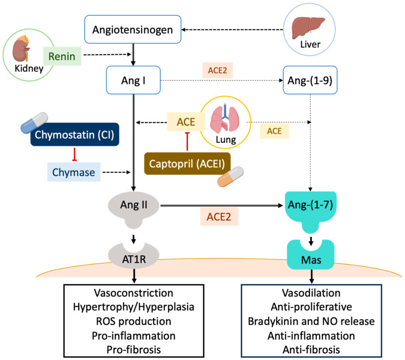
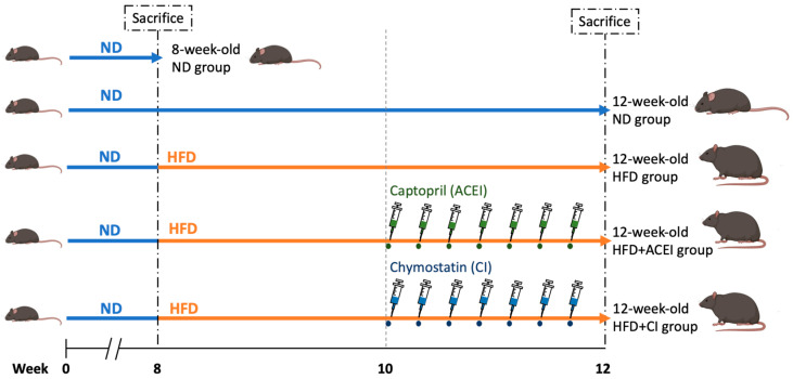
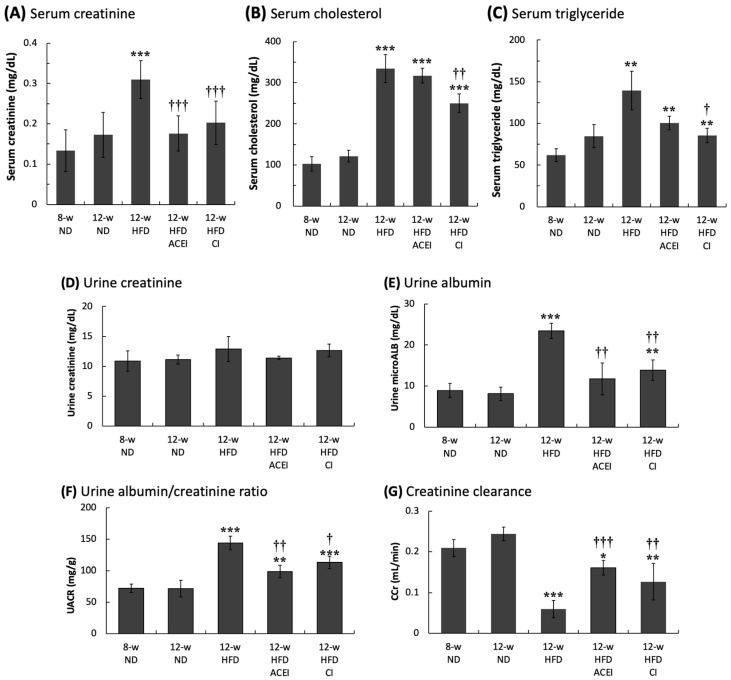
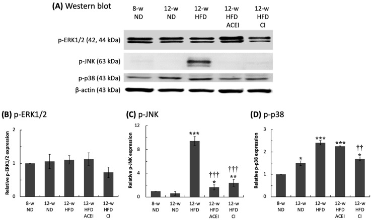
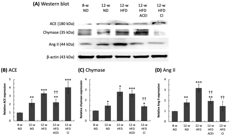
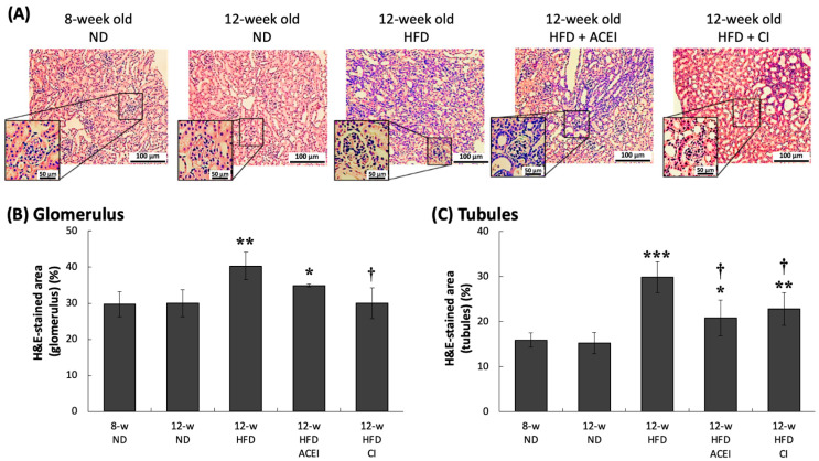
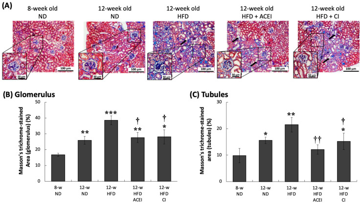
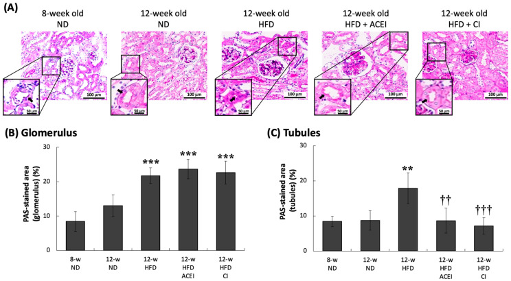
Diabetic nephropathy (DN) is a crucial metabolic health problem. The renin-angiotensin system (RAS) is well known to play an important role in DN. Abnormal RAS activity can cause the over-accumulation of angiotensin II (Ang II). Angiotensin-converting enzyme inhibitor (ACEI) administration has been proposed as a therapy, but previous studies have also indicated that chymase, the enzyme that hydrolyzes angiotensin I to Ang II in an ACE-independent pathway, may play an important role in the progression of DN. Therefore, this study established a model of severe DN progression in a db/db and ACE2 KO mouse model (db and ACE2 double-gene-knockout mice) to explore the roles of RAS factors in DNA and changes in their activity after short-term (only 4 weeks) feeding of a high-fat diet (HFD) to 8-week-old mice. The results indicate that FD-fed db/db and ACE2 KO mice fed an HFD represent a good model for investigating the role of RAS in DN. An HFD promotes the activation of MAPK, including p-JNK and p-p38, as well as the RAS signaling pathway, leading to renal damage in mice. Blocking Ang II/AT1R could alleviate the progression of DN after administration of ACEI or chymase inhibitor (CI). Both ACE and chymase are highly involved in Ang II generation in HFD-induced DN; therefore, ACEI and CI are potential treatments for DN.

Keywords: angiotensin converting enzyme II (ACE2); chymase; diabetic nephropathy; high-fat-diet; renin angiotensin system.


Conflict of interest statement

The authors declare no conflicts of interest.

Figures

Figure 1
Figure 1
Schematic representation of RAS cascade. The renin–angiotensin system (RAS) is a major regulatory system involved in blood pressure and water balance. The classical RAS is made up of a circulating endocrine system in which renin cleaves angiotensin I (Ang I). Inactive Ang I is then hydrolyzed by angiotensin-converting enzyme (ACE) and also by chymase, producing the octapeptide angiotensin II (Ang II). Ang II then binds to Ang II Type-I receptors (AT1R), which causes vasoconstriction and inflammation. Angiotensin-converting enzyme II (ACE2) cleaves the Ang II to generate the peptide angiotensin 1–7 (Ang 1–7), which acts via the Mas receptor, i.e., as Ang 1–7/Mas, to counteract the adverse effects of Ang II/AT1R. ACE and chyamse activity can be inhibited by an ACE inhibitor (ACEI; Captopril) and a chymase inhibitor (CI; Chymostatin), respectively [13,32].
Figure 2
Figure 2
Experimental design of feeding the HFD and therapeutic treatments in db/db and ACE2 KO mice. The mice were divided into five groups. One group of eight-week-old mice was sacrificed, and we sampled the blood and tissues for assay as the Control group. Next, db/db and ACE2 KO mice were fed a high-fat diet (HFD) for four weeks. After two weeks on the HFD, captopril (ACE inhibitor, ACEI) or chymostatin (chymase inhibitor, CI) were administered in a twice-daily dose of 10 mg/kg via intraperitoneal injection (i.p.). These mice were sacrificed at 12 weeks old, and we sampled the blood and tissues for further assay.
Figure 3
Figure 3
The serum and urinary biochemical parameters of db/db and ACE2 KO mice. Serum creatinine (A), total cholesterol (B) and triglyceride (C) were significantly increased in the HFD group. Both captopril (ACEI) and chymostatin (CI) treatments improved serum creatinine, but only chymostatin lowered the blood lipid level. Urine creatinine (D) showed no marked difference across groups. The high-fat diet (HFD) significantly increased the urine albumin (E) and urine albumin/creatinine ratio (UACR) (F) and significantly decreased the creatinine clearance (CCr) (G); these parameters were measured as a test of kidney function. Administration of ACEI or CI slowed the loss of kidney function. All parameters are expressed as the mean ± SD (n = 6) from each group. *, ** and *** indicate p < 0.05, p < 0.01 and p < 0.001, respectively, compared with the 12-week-old ND group. , †† and ††† indicate p < 0.05, p < 0.01 and p < 0.001, respectively, compared with the 12-week-old HFD group.
Figure 4
Figure 4
Relative expression of renal MAPK-related protein in db/db and ACE2 KO mice. The kidneys were sampled and protein was extracted for Western blotting assays (A). High-fat-diet (HFD) feeding did not affect the p-ERK1/2 level (B); however, significantly increased p-JNK (C) and p-p38 (D) levels were detected. Administration with captopril (ACEI) or chymostatin (CI) could slow down the abnormal MAPK activation. All parameters were expressed as the mean ± SD (n = 6) for each group. *, ** and *** indicate p < 0.05, p < 0.01 and p < 0.001, compared with the 8-w (8-week-old) ND group, respectively. †† and ††† indicate p < 0.01 and p < 0.001, compared with the 12-w (12-week-old) HFD group, respectively.
Figure 5
Figure 5
Relative levels of renal–RAS components in db/db and ACE2 KO mice. The kidneys were sampled, and proteins were extracted for Western blot assays (A). Feeding a high-fat diet (HFD) significantly accelerated abnormal activity in the renin–angiotensin system (RAS) by increasing ACE (B), chymase (C), and Ang II (D) levels. Administration of captopril (ACEI) and chymostatin significantly ameliorated the HFD-induced increase in Ang II levels. All parameters are expressed as the mean ± SD (n = 6) from each group. *, ** and *** indicate p < 0.05, p < 0.01 and p < 0.001, respectively, compared with the 8-w (8-week-old) ND group. †† indicates p < 0.01 compared with the 12-w (12-week-old) HFD group.
Figure 6
Figure 6
Hematoxylin and eosin (H&E)-stained kidney sections of db/db and ACE2 KO mice. Kidney sections from the mice were stained with H&E and then photographed for pathologic examination at 100× magnification (A). Captopril (ACEI) and chymostatin (CI) treatments decreased the high-fat-diet (HFD)-induced lymphocytic infiltration in the tubules (B,C). Glomerular hypertrophy was observed in the 12-week-old HFD group. All parameters are expressed as the mean ± SD for each group. *, ** and *** indicate p < 0.05, p < 0.01 and p < 0.001, respectively, compared with the 8-w (8-week-old) ND group. indicates p < 0.05 compared with the 12-w (12-week-old) HFD group (n = 3 for each group).
Figure 7
Figure 7
Masson’s trichrome-stained kidney sections of db/db and ACE2 KO mice. Kidney sections from the mice were stained with Masson’s trichrome and then photographed for pathologic verification at 100× magnification (A). Both the captopril (ACEI) and chymostatin (CI) treatments reduced the HFD-induced collagen accumulation in the glomerulus and tubules. Slightly greater glomerular injury was found in 12-w (12-week-old) ND groups (B,C). All HFD-fed groups showed greater evidence of missing or ruptured brush border and glomerular hypertrophy. All parameters are expressed as the mean ± SD from each group. *, ** and *** indicate p < 0.05, p < 0.01 and p < 0.001, respectively, compared with the 8-w (8-week-old) ND group. and †† indicate p < 0.05 and p < 0.01, respectively, compared with the 12-w (12-week-old) HFD group (n = 3 for each group).
Figure 8
Figure 8
PAS-stained kidney sections of db/db and ACE2 KO mice. Kidney sections from the mice were stained with PAS and then photographed for pathologic assessment at 200× magnification (A). Accumulations of PAS-positive matrix in the mesangium and severe tubular atrophy were observed in all of the HFD-fed groups. Administrating captopril (ACEI) or chymostatin (CI) ameliorated tubular atrophy but did not decrease mesangial matrix expansion (B,C). All parameters are expressed as the mean ± SD from each group. ** and *** indicate p < 0.01 and p < 0.001, respectively, compared with the 8-w (8-week-old) ND group. †† and ††† indicate p < 0.01 and p < 0.001, respectively, compared with the 12-w (12-week-old) HFD group (n = 3 for each group).

Similar articles

Cited by

References

    1. Al-Lawati J.A. Diabetes Mellitus: A Local and Global Public Health Emergency! Oman Med. J. 2017;32:177–179. doi: 10.5001/omj.2017.34. - DOI - PMC - PubMed
    1. Forouhi N.G., Wareham N.J. Epidemiology of diabetes. Medicine. 2014;42:698–702. doi: 10.1016/j.mpmed.2014.09.007. - DOI - PMC - PubMed
    1. Aldukhayel A. Prevalence of diabetic nephropathy among Type 2 diabetic patients in some of the Arab countries. Int. J. Health Sci. 2017;11:1–4. - PMC - PubMed
    1. Zhang X.X., Kong J., Yun K. Prevalence of Diabetic Nephropathy among Patients with Type 2 Diabetes Mellitus in China: A Meta-Analysis of Observational Studies. J. Diabetes Res. 2020;2020:2315607. doi: 10.1155/2020/2315607. - DOI - PMC - PubMed
    1. Stachura A., Khanna I., Krysiak P., Paskal W., Wlodarski P. Wound Healing Impairment in Type 2 Diabetes Model of Leptin-Deficient Mice-A Mechanistic Systematic Review. Int. J. Mol. Sci. 2022;23:8621. doi: 10.3390/ijms23158621. - DOI - PMC - PubMed